. 2023 May 29;24(11):9458.
doi: 10.3390/ijms24119458.

Plakophilin-3 Binds the Membrane and Filamentous Actin without Bundling F-Actin

Affiliations

Plakophilin-3 Binds the Membrane and Filamentous Actin without Bundling F-Actin

Jyoti Gupta et al. Int J Mol Sci. .

Abstract

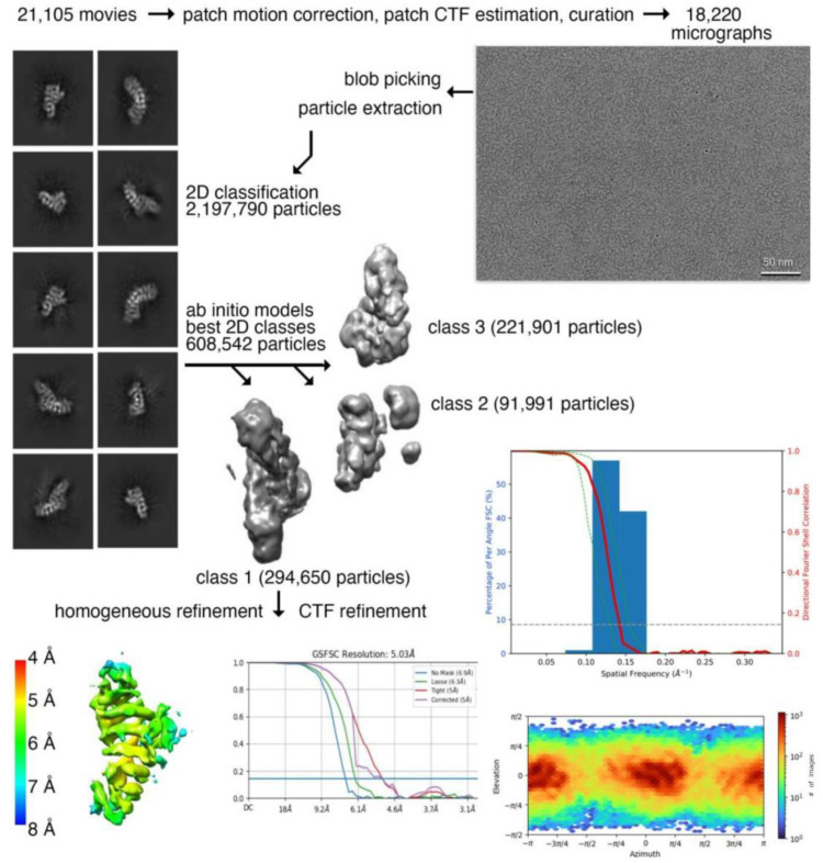
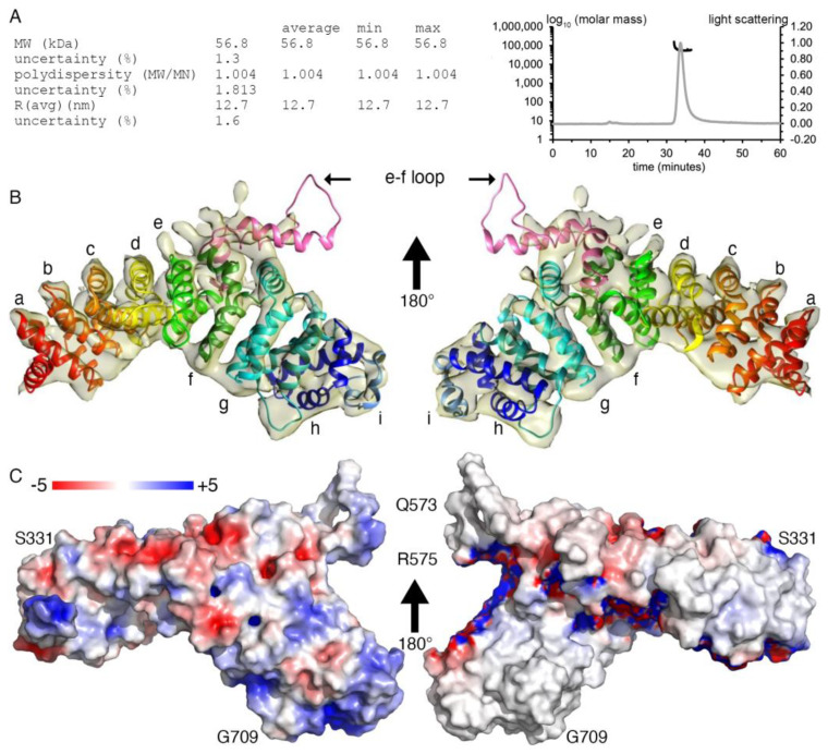
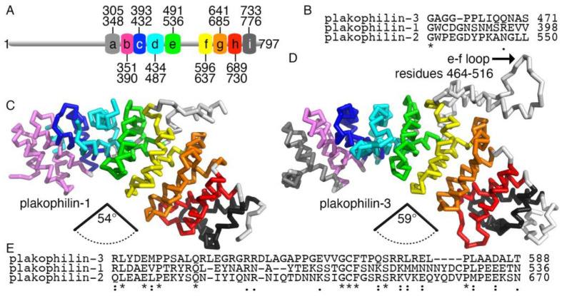
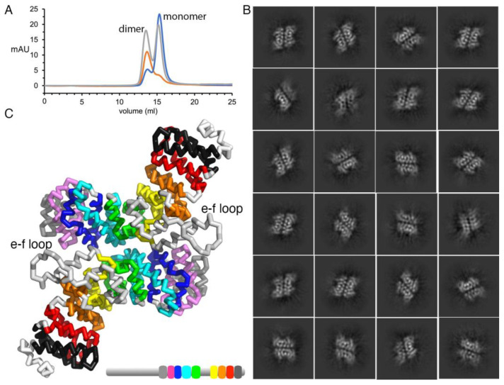
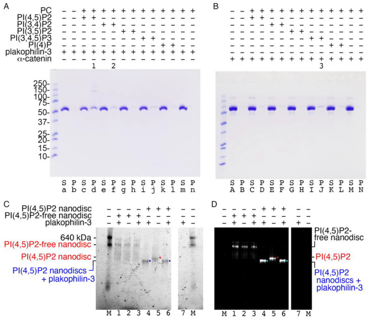
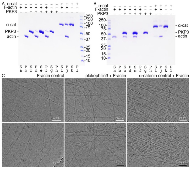
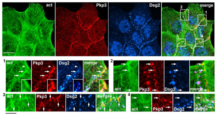
Plakophilin-3 is a ubiquitously expressed protein found widely in epithelial cells and is a critical component of desmosomes. The plakophilin-3 carboxy-terminal domain harbors nine armadillo repeat motifs with largely unknown functions. Here, we report the 5 Å cryogenic electron microscopy (cryoEM) structure of the armadillo repeat motif domain of plakophilin-3, one of the smaller cryoEM structures reported to date. We find that this domain is a monomer or homodimer in solution. In addition, using an in vitro actin co-sedimentation assay, we show that the armadillo repeat domain of plakophilin-3 directly interacts with F-actin. This feature, through direct interactions with actin filaments, could be responsible for the observed association of extra-desmosomal plakophilin-3 with the actin cytoskeleton directly attached to the adherens junctions in A431 epithelial cells. Further, we demonstrate, through lipid binding analyses, that plakophilin-3 can effectively be recruited to the plasma membrane through phosphatidylinositol-4,5-bisphosphate-mediated interactions. Collectively, we report on novel properties of plakophilin-3, which may be conserved throughout the plakophilin protein family and may be behind the roles of these proteins in cell-cell adhesion.

Keywords: actin cytoskeleton; armadillo; desmosomes; phosphatidylinositol 4,5-bisphosphate; plakophilin; plasma membrane.


Conflict of interest statement

The authors declare no conflict of interest.

Figures

Figure 1
Figure 1
CryoEM data processing workflow for plakophilin-3. Motion-corrected cryoEM micrograph for plakohilin-3 (top, right). Representative 2D class averages of monomeric plakophilin-3 used for 3D reconstruction (top, left). High-quality micrographs (18,220) were selected after motion correction and used for the selection of 2,197,790 particles. Multiple rounds of 2D classification were performed to remove noise, yielding 608,542 plakophilin-3 particles, which were subjected to ab initio classification. One out of three classes, class 1, containing 294,650 particles, revealed structural features for all nine armadillo repeat motifs of plakophilin-3 that were subjected to 3D refinement and contrast transfer function (CTF) refinement. (Middle, right) Directional Fourier shell correlation plot for plakophilin-3. The red line shows the global Fourier shell correlation (FSC), the green lines show the directional resolution spread values defined according to ±1 standard deviation from the mean of the directional resolutions, and the blue bars represent a histogram of 100 directional resolutions evenly sampled over the 3D FSC. A sphericity of 0.951 was determined at an FSC threshold of 0.5, which indicates significant isotropic angular distribution. (Bottom left) The resolution of the map was determined based on local resolution estimation in cryoSPARC. The orientation distribution plot obtained from cryoSPARC is shown at the bottom on the right.
Figure 2
Figure 2
CryoEM structure of the armadillo repeat motif domain of plakophilin-3. (A) The multi-angle light scattering size exclusion statistics obtained for plakophilin-3 are provided in the table (left). The profiles of the light scattering (gray) and the calculated molar mass (black) are as shown (right). (B) Back and front views of a cartoon drawing of our cryoEM plakophilin-3 structure (residues 305-797) and final Coulomb potential map. Each armadillo repeat motif is labeled (a–i), and the loop between the fifth (e) and sixth (f) armadillo repeat motifs is indicated. (C) Back and front views of the electrostatic surface potential representation of our plakophilin-3 cryoEM structure. The electrostatic potential gradient is from −5 to +5 kBT (red, negative; blue, positive), where kB is the Boltzmann constant, and T is the temperature. Some residues are labeled.
Figure 3
Figure 3
Plakophilin-3 is less compact than plakophilin-1. (A) Schematic domain structure of plakophilin-3 showing the nine armadillo repeat motifs and residue boundaries. (B) The insert in the fourth armadillo repeat motif is disordered in the plakophilin-1 structure and has a distinct sequence. (C) The human plakophilin-1 crystal structure (PDB entry 1xm9) [19] is more compact compared to (D) our plakophilin-3 cryoEM structure. In the plakophilin-1 structure, the angle between the Cα of residues 277, 428, and 586 is 54°. The angle between the equivalent Cα of residues 351, 501, and 668 in plakophilin-3 is 58.6°. (E) Amino acid sequence alignment of the three human plakophilin insert regions shows that plakophilin-3 is distinct in that region. The asterisk, colon, and period symbols below the sequence each represent identity, and high and low conservation, respectively.
Figure 4
Figure 4
Plakophilin-3 is a monomer and a dimer. (A) Size exclusion chromatogram for plakophilin-3. The apparent molecular weights for the plakophilin-3 monomer and dimer were found to be 60.25 kDa and 120.6 kDa, respectively, compared to the calculated molecular weights of 57.2 and 114.4 kDa. Initial plakophilin-3 chromatogram in gray is normalized. Fractions from the first peak that correspond to the dimer were pooled and reloaded, red. Fractions from the second peak that correspond to the monomer were pooled and reloaded, blue. (B) Representative 2D class averages of the particles for the plakophilin-3 dimer are shown. (C) The plakophilin-3 dimer model based on the 2D class averages suggests that the loop between armadillo repeat motifs e and f is near the dimer interface.
Figure 5
Figure 5
Plakophilin-3 binds to the membrane. (A) Plakophilin-3 binds to PI(4,5)P2-containing vesicles (lane d, indicated by “1”), which results in some dimerization, as determined by co-sedimentation. Plakophilin-3 remains in solution in the absence of lipids (lane a). Lane f (indicated by “2”) shows weak plakophilin-3 binding to PI(4,5)P2. (B) Our α-catenin control (residues 82-906) weakly binds PI(3,4,5)P2-containing vesicles (lane J, indicated by “3”), as reported [31]. S, supernatant; P, pellet; PC, phosphatidylcholine; PI(4,5)P2, phosphatidylinositol 4,5-bisphosphate; PI(3,4)P2, phosphatidylinositol 3,4-bisphosphate; PI(3,5)P2, phosphatidylinositol 3,5-bisphosphate; PI(3,4,5)P3, phosphatidylinositol 3,4,5-trisphosphate; PI(4)P, phosphatidylinositol 4-phosphate. Similar results were obtained from two independent experiments. (C,D) Native gel shift assay to detect the binding of plakophlin-3 to fluorescently labeled PI(4,5)P2-free and PI(4,5)P2-containing nanodiscs, as detected by (C) ultraviolet activation and (D) rhodamine fluorescence. Lanes 1–3, Plakophilin-3 showed no binding to the PI(4,5)P2-free nanodisc. Lanes 4–6, Binding of plakophilin-3 was observed for the PI(4,5)P2 nanodisc (blue or cyan dots, complex; red dots, unbound nanodiscs). Lanes 7, plakophilin-3 does not run into the native gel due to its basic pI; M, molecular weight markers. Data are representative of two independent experiments.
Figure 6
Figure 6
Plakophilin-3 binds filamentous actin without bundling F-actin. (A) High-speed centrifugation to detect F-actin binding. Plakophilin-3 co-sediments with F-actin (lanes d, f). Plakophilin-3 remains in solution in the absence of F-actin (lane g). Actin is fully polymerized (lane b). Our α-catenin control binds F-actin (lane j) and remains in solution in the absence of actin (lane k). Similar results were obtained from two independent experiments. (B) Low-speed centrifugation to detect F-actin bundling. Plakophilin-3 does not bundle F-actin (lane e). Purified actin (lane a), plakophilin-3 (lane g), and α-catenin (lane k) are soluble. Our α-catenin control bundles F-actin (lane j). (C) CryoEM micrographs. Left, F-actin alone. Middle, F-actin in the presence of plakophilin-3. Right, F-actin in the presence of α-catenin. Top and bottom panels are independent repeats.
Figure 7
Figure 7
Extra-desmosomal plakophilin-3 clusters are recruited to cell–cell junction-associated actin bundles. Triple-fluorescence microscopy of A431 cells stained for actin filaments (act, green), plakophilin-3 (Pkp3, red), and the desmosomal cadherin, desmoglein-2 (Dsg2, blue). The upper micrographs show a low magnification of a group of cells. Scale bar, 25 μm. Zoomed images of four representative cell-cell contact regions that are designated with dashed boxes (numbered 1 to 4) are in the bottom rows. Note that desmoglein-2 and plakophilin-3 are colocalized in desmosomes. In addition to desmosomes, each of the selected regions contains several plakophilin-3-positive clusters of different sizes (some of them are indicated with arrows), which are desmoglein-2-deficient but align along the actin-enriched structures. One of them (marked by the arrowhead in the region #1) is shown zoomed-in in the insets. Note that this large plakophilin-3 cluster incorporates several small desmosomes. Bars, 7 μm.
Figure 8
Figure 8
Extra-desmosomal plakophilin-3 clusters are associated with adherens junctions. (A) Triple-fluorescence microscopy of images A431 cells stained for plakophilin-3 (red) in combination with the constitutive proteins of adherens junction and desmosomes, β-catenin (green) and desmoplakin (blue), respectively. The upper micrographs show cells at a low magnification. Bar, 25 μm. Two representative cell–cell contact regions that are demarcated with dashed boxes (1 and 2) are shown zoomed-in in the bottom rows (bar, 7 μm) as single-stained images (β-catenin, plakophilin-3, and desmoplakin) in (B) or in three different combinations (β-catenin + desmoplakin; β-catenin + plakophilin-3; and merge) in (C). Note that both the desmoplakin-labeled desmosomes (some of them marked by arrows) and β-catenin-labeled adherens junctions (arrowheads) contain plakophilin-3. Additionally, note that the localizations of β-catenin and plakophilin-3 in the adherens junctions do not exactly match one another.

Similar articles

Cited by

References

    1. Bass-Zubek A.E., Godsel L.M., Delmar M., Green K.J. Plakophilins: Multifunctional scaffolds for adhesion and signaling. Curr. Opin. Cell Biol. 2009;21:708–716. doi: 10.1016/j.ceb.2009.07.002. - DOI - PMC - PubMed
    1. Neuber S., Muhmer M., Wratten D., Koch P.J., Moll R., Schmidt A. The desmosomal plaque proteins of the plakophilin family. Dermatol. Res. Pract. 2010;2010:101452. doi: 10.1155/2010/101452. - DOI - PMC - PubMed
    1. Hatzfeld M., Wolf A., Keil R. Plakophilins in desmosomal adhesion and signaling. Cell Commun. Adhes. 2014;21:25–42. doi: 10.3109/15419061.2013.876017. - DOI - PubMed
    1. Hofmann I. Plakophilins and their roles in diseased states. Cell Tissue Res. 2020;379:5–12. doi: 10.1007/s00441-019-03153-0. - DOI - PubMed
    1. Fujiwara M., Nagatomo A., Tsuda M., Obata S., Sakuma T., Yamamoto T., Suzuki S.T. Desmocollin-2 alone forms functional desmosomal plaques, with the plaque formation requiring the juxtamembrane region and plakophilins. J. Biochem. 2015;158:339–353. doi: 10.1093/jb/mvv048. - DOI - PubMed

LinkOut - more resources