. 2023 May 30;24(11):9474.
doi: 10.3390/ijms24119474.

A New Culture Model for Enhancing Estrogen Responsiveness in HR+ Breast Cancer Cells through Medium Replacement: Presumed Involvement of Autocrine Factors in Estrogen Resistance

Affiliations

A New Culture Model for Enhancing Estrogen Responsiveness in HR+ Breast Cancer Cells through Medium Replacement: Presumed Involvement of Autocrine Factors in Estrogen Resistance

Seok-Hoon Jang et al. Int J Mol Sci. .

Abstract

Hormone receptor-positive breast cancer (HR+ BC) cells depend on estrogen and its receptor, ER. Due to this dependence, endocrine therapy (ET) such as aromatase inhibitor (AI) treatment is now possible. However, ET resistance (ET-R) occurs frequently and is a priority in HR+ BC research. The effects of estrogen have typically been determined under a special culture condition, i.e., phenol red-free media supplemented with dextran-coated charcoal-stripped fetal bovine serum (CS-FBS). However, CS-FBS has some limitations, such as not being fully defined or ordinary. Therefore, we attempted to find new experimental conditions and related mechanisms to improve cellular estrogen responsiveness based on the standard culture medium supplemented with normal FBS and phenol red. The hypothesis of pleiotropic estrogen effects led to the discovery that T47D cells respond well to estrogen under low cell density and medium replacement. These conditions made ET less effective there. The fact that several BC cell culture supernatants reversed these findings implies that housekeeping autocrine factors regulate estrogen and ET responsiveness. Results reproduced in T47D subclone and MCF-7 cells highlight that these phenomena are general among HR+ BC cells. Our findings offer not only new insights into ET-R but also a new experimental model for future ET-R studies.

Keywords: aromatase inhibitor; autocrine factor; breast cancer; endocrine therapy; estrogen; medium replacement; resistance; responsiveness.


Conflict of interest statement

The authors declare no conflict of interest.

Figures

Figure 1
Figure 1
Growth curves of cell proliferation in normal fetal bovine serum (FBS) and dextran-coated charcoal-stripped FBS (CS-FBS) in the absence and presence of 17β-estradiol (E2) in two hormone receptor-positive breast cancer (HR+ BC) cell lines. MTT assay results at the relevant time after E2 treatment in MCF-7 (A) and T47D (B): cell seeding density, 0.05 × 10⁶ cells/mL; data, expressed as the mean ± standard deviation (SD) and normalized to the vehicle control (CTL) group of CS-FBS on Day 1; comparison using the two-way analysis of variance (ANOVA) test, based on each CTL at each day; * p < 0.05; ** p < 0.01; and *** p < 0.001.
Figure 2
Figure 2
Effectiveness of E2 and fulvestrant (Fulv) on cell proliferation based on cell density in T47D. MTT assay results: data, expressed as the mean ± SD and normalized to each CTL; basic comparison (above the graph), based on each CTL; additional comparison (line), between cell densities in each reagent; statistical analysis, Student’s t-test; * p < 0.05; and *** p < 0.001 (A). Phase-contrast microscopic images (40×) of the CTLs in A just before the MTT assay: scale bar, 500 μm; the figures show how much culture space T47D occupies at each cell density (B).
Figure 3
Figure 3
Effectiveness of E2, Fulv, and two representative chemotherapeutic agents on cell proliferation at two cell densities over time in T47D. MTT assay results for E2 and Fulv at cell seeding densities of 0.01 (A) and 0.05 (B) × 106 cells/mL for up to three days. MTT assay results for E2 and Fulv at a cell seeding density of 0.01 × 10⁶ cells/mL for up to five days (C). MTT assay results for doxorubicin (Doxo) and docetaxel (DT) at a cell seeding density of 0.01 × 10⁶ cells/mL for up to five days (D). Data, expressed as the mean ± SD and normalized to each CTL at each day; comparison (above or below the graph) using Student’s t-test, based on each CTL at each day; Doxo, 0.1 μM; DT, 1 nM; * p < 0.05; ** p < 0.01; and *** p < 0.001.
Figure 4
Figure 4
Effectiveness of E2 and Fulv on cell proliferation based on medium replacement, cell density, and FBS concentration in T47D. MTT assay results for E2 and Fulv under medium maintenance (MTN) and medium exchange (EXC) at cell seeding densities of 0.01 and 0.05 × 10⁶ cells/mL: additional comparison (line), between MTN and EXC in each reagent at each cell density (A). MTT assay results for E2 and Fulv based on FBS concentration: additional comparison (line), based on 10% FBS in each reagent (B). Data, expressed as the mean ± SD and normalized to each CTL; basic comparison (above the graph), based on each CTL; statistical analysis, Student’s t-test; * p < 0.05; ** p < 0.01; and *** p < 0.001.
Figure 5
Figure 5
Effects of EXC and BC cell culture supernatants on E2 and Fulv actions and cell proliferation in the medium supplemented without or with FBS in T47D. MTT assay results for E2 and Fulv under MTN and EXC in serum-free RPMI 1640 at cell seeding densities of 0.01 and 0.05 × 10⁶ cells/mL: additional comparison (line), between MTN and EXC in each reagent at each cell density (A). Cell proliferation rates of the CTLs in (A): data, re-normalized to MTN at 0.01; comparison (line), between MTN and EXC at each cell density (B). MTT assay results for E2 and Fulv when adding 1/5 volume (compared to the preexisting medium volume) of the supernatants under EXC in the 1% FBS medium: additional comparison (line), between the medium control and each supernatant in each reagent (C). Cell proliferation rates of the CTLs in (C): data, re-normalized to the medium control (D). Data, expressed as the mean ± SD; data in (A,C), normalized to each CTL; basic comparison (above the graph) in (A,C,D), based on each control group; statistical analysis, Student’s t-test; * p < 0.05; ** p < 0.01; and *** p < 0.001.
Figure 6
Figure 6
Effects of EXC, cell density, and BC cell culture supernatants on E2 and Fulv actions in single cell-derived T47D subclones or MCF-7. MTT assay results for E2 and Fulv under MTN and EXC in the T47D parental and subclone cells: cell seeding density, 0.02 × 10⁶ cells/mL; additional comparison (above the basic comparison mark), between MTN and EXC in each reagent in each subclone (A). MTT assay results for E2 and Fulv based on cell seeding density in MCF-7: additional comparison (line), based on 0.05 in each reagent (B). MTT assay results under MTN and EXC at cell seeding densities of 0.01 and 0.05 × 10⁶ cells/mL in MCF-7: additional comparison (line), between MTN and EXC in each reagent at each density (C). MTT assay results when adding 1/5 volume (compared to the preexisting medium volume) of the supernatants in MCF-7: additional comparison (line), between the medium control and each supernatant in each reagent (D). MTT assay results when adding 1/3 volume of MCF-7 culture supernatant in MCF-7: additional comparison (line), between the two groups in each reagent (E). Data, expressed as the mean ± SD and normalized to each CTL; basic comparison (above the graph), based on each CTL; statistical analysis, Student’s t-test; * p < 0.05; ** p < 0.01; and *** p < 0.001.
Figure 7
Figure 7
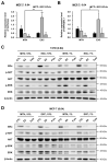
Effects of E2 and Fulv on the activity, target gene expression, or protein expression of the estrogen receptor (ER), protein kinase B (AKT), or extracellular signal-regulated kinase (ERK) under MTN and EXC in 10% or 1% FBS in MCF-7 or T47D. Results of the estrogen response element (ERE) reporter assay for the ER activity (A) and qRT-PCR for the expression of an ERα target gene (RAPGEFL1) (B) in MCF-7: data, expressed as the mean ± SD and normalized to CTL under MTN; basic comparison (above the graph), based on each CTL; additional comparison (line), between MTN and EXC in each reagent; statistical analysis, Student’s t-test; * p < 0.05; ** p < 0.01. Western blotting results in T47D (C) and MCF-7 (D). Cell seeding density, 0.04 × 10⁶ cells/mL; p-, phosphorylated.

References

    1. Sung H., Ferlay J., Siegel R.L., Laversanne M., Soerjomataram I., Jemal A., Bray F. Global Cancer Statistics 2020: GLOBOCAN Estimates of Incidence and Mortality Worldwide for 36 Cancers in 185 Countries. CA Cancer J. Clin. 2021;71:209–249. doi: 10.3322/caac.21660. - DOI - PubMed
    1. Dai X., Li T., Bai Z., Yang Y., Liu X., Zhan J., Shi B. Breast cancer intrinsic subtype classification, clinical use and future trends. Am. J. Cancer Res. 2015;5:2929–2943. - PMC - PubMed
    1. Cheang M.C., Chia S.K., Voduc D., Gao D., Leung S., Snider J., Watson M., Davies S., Bernard P.S., Parker J.S., et al. Ki67 index, HER2 status, and prognosis of patients with luminal B breast cancer. J. Natl. Cancer Inst. 2009;101:736–750. doi: 10.1093/jnci/djp082. - DOI - PMC - PubMed
    1. Porras L., Ismail H., Mader S. Positive Regulation of Estrogen Receptor Alpha in Breast Tumorigenesis. Cells. 2021;10:2966. doi: 10.3390/cells10112966. - DOI - PMC - PubMed
    1. Russo J., Russo I.H. The role of estrogen in the initiation of breast cancer. J. Steroid Biochem. Mol. Biol. 2006;102:89–96. doi: 10.1016/j.jsbmb.2006.09.004. - DOI - PMC - PubMed