. 2023 May 30;24(11):9481.
doi: 10.3390/ijms24119481.

Empagliflozin Ameliorates Bleomycin-Induced Pulmonary Fibrosis in Rats by Modulating Sesn2/AMPK/Nrf2 Signaling and Targeting Ferroptosis and Autophagy

Affiliations

Empagliflozin Ameliorates Bleomycin-Induced Pulmonary Fibrosis in Rats by Modulating Sesn2/AMPK/Nrf2 Signaling and Targeting Ferroptosis and Autophagy

Hemat El-Sayed El-Horany et al. Int J Mol Sci. .

Abstract

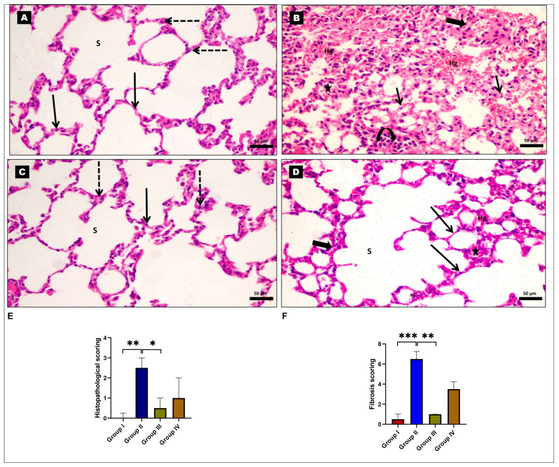
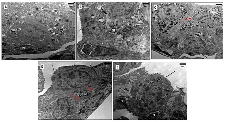
Pulmonary fibrosis (PF) is a life-threatening disorder that severely disrupts normal lung architecture and function, resulting in severe respiratory failure and death. It has no definite treatment. Empagliflozin (EMPA), a sodium-glucose cotransporter 2 (SGLT2) inhibitor, has protective potential in PF. However, the mechanisms underlying these effects require further elucidation. Therefore, this study aimed to evaluate the ameliorative effect of EMPA against bleomycin (BLM)-induced PF and the potential mechanisms. Twenty-four male Wister rats were randomly divided into four groups: control, BLM treated, EMPA treated, and EMPA+BLM treated. EMPA significantly improved the histopathological injuries illustrated by both hematoxylin and eosin and Masson's trichrome-stained lung tissue sections, as confirmed by electron microscopic examination. It significantly reduced the lung index, hydroxyproline content, and transforming growth factor β1 levels in the BLM rat model. It had an anti-inflammatory effect, as evidenced by a decrease in the inflammatory cytokines' tumor necrosis factor alpha and high mobility group box 1, inflammatory cell infiltration into the bronchoalveolar lavage fluid, and the CD68 immunoreaction. Furthermore, EMPA mitigated oxidative stress, DNA fragmentation, ferroptosis, and endoplasmic reticulum stress, as evidenced by the up-regulation of nuclear factor erythroid 2-related factor expression, heme oxygenase-1 activity, glutathione peroxidase 4 levels, and a decrease in C/EBP homologous protein levels. This protective potential could be explained on the basis of autophagy induction via up-regulating lung sestrin2 expression and the LC3 II immunoreaction observed in this study. Our findings indicated that EMPA protected against BLM-induced PF-associated cellular stress by enhancing autophagy and modulating sestrin2/adenosine monophosphate-activated protein kinase/nuclear factor erythroid 2-related factor 2/heme oxygenase 1 signaling.

Keywords: autophagy; bleomycin; empagliflozin; endoplasmic reticulum stress; ferroptosis; pulmonary fibrosis.


Conflict of interest statement

The authors declare no conflict of interest.

Figures

Figure 1
Figure 1
Representative photomicrographs of rat lung tissues stained with hematoxylin and eosin (H and E). (A) The control and (C) empagliflozin (EMPA)-treated groups show thin-walled alveoli (red arrows) and alveolar sacs (s). The thin-walled alveoli are made up of flat type I pneumocytes (black arrows) with densely pigmented nuclei and type II pneumocytes (dashed arrows) with big, rounded nuclei. (B) The bleomycin (BLM)-treated group reveals a disrupted architectural arrangement of the lung with markedly thickened alveolar septa (thick arrows) and extensive mononuclear cellular infiltration (curved arrows). Some alveoli appear collapsed (stars). Many of the alveolar epithelial lining cells exhibit vacuolar cytoplasmic necrosis with pyknotic nuclei (thin arrows). Severe interstitial hemorrhage (Hg) can be observed. (D) The EMPA+BLM-treated group shows many thin-walled alveoli (arrows) and alveolar sacs (S) with normal histology. However, there are still a few collapsed alveoli, minor alveolar wall thickening (thick arrows), and mild interstitial hemorrhage (Hg). Magnification is ×400; scale bar = 50 μm. (E) The histopathological and (F) fibrosis scoring among various studied groups (n = 6 rats in each group). * indicates a p < 0.05, ** indicates a p < 0.01, and *** indicates a p < 0.001.
Figure 2
Figure 2
Representative photomicrographs of rat lung tissues stained with Masson’s trichrome. (A,C) The control and empagliflozin (EMPA)-treated groups showed little collagen fibers in the alveolar walls and around the blood vessels (arrows). (B) The bleomycin (BLM)-treated group showed massive deposits of collagen fibers (arrows). (D) The EMPA+BLM-treated group demonstrated alveolar walls with minimal collagen fiber deposition (arrows) (×400; scale bar = 50 μm). (E) A morphometric analysis of the mean area percentage in the Masson trichrome-stained lung tissue sections from various studied groups (n = 6 rats in each group). * indicates a p < 0.05 and ** indicates a p < 0.01.
Figure 3
Figure 3
Effects of empagliflozin (EMPA) treatment on lung inflammatory status in a bleomycin (BLM)-induced pulmonary fibrosis model. Representative photomicrographs of CD68 immunohistochemical-stained lung sections. (A,C) The control and EMPA-treated groups show only a few positively stained macrophages (arrows) in the lung interalveolar septa. (B) The BLM-treated group exhibits an abundance of positively stained brown macrophages (arrows) in the interalveolar septa. (D) The EMPA + BLM-treated group shows a few positively stained brown macrophages (arrows) that are close to the control (×400, scale bar = 50 μm). (E) A morphometric analysis of the mean area percentage of CD68 immune reactivity; (F) the total leucocytic count; (G) neutrophil percentages; (H) lymphocyte percentages; (I) macrophage percentages; (J) bronchoalveolar lavage fluid (BALF) tumor necrosis factor-alpha (TNF-α); and (K) BALF high mobility group box 1 (HMBG1) among various studied groups (n = 6 rats in each group). Data are presented as mean and standard deviation (error bars) and analyzed using one-way analysis of variance (ANOVA) with Tukey’s post hoc test for multiple comparisons. ** indicates a p < 0.01 and *** indicates a p < 0.001.
Figure 4
Figure 4
Effects of empagliflozin treatment on lung fibrotic states in a bleomycin-induced pulmonary fibrosis model. (A) The lung index; (B) pulmonary transforming growth factor beta 1 (TGF-β1); and (C) pulmonary hydroxyproline levels in various studied groups (n = 6 rats in each group). Data are presented as mean and standard deviation (error bars) and analyzed using one-way analysis of variance (ANOVA) with Tukey’s post hoc test for multiple comparisons. *** indicates a p < 0.001.
Figure 5
Figure 5
Effect of empagliflozin treatment on redox status, ferroptosis, and endoplasmic reticulum stress in a bleomycin-induced pulmonary fibrosis model. (A) Pulmonary malondialdehyde (MDA); (B) 4-hydroxynonenal (4-HNE); (C) reduced glutathione (GSH); (D) glutathione peroxidase 4 (GPX4); (E) heme oxygenase 1 (HO-1); (F) Nrf2 relative expression; and (G) C/EBP homologous protein (CHOP) levels in various studied groups (n = 6 rats in each group). Data are presented as mean and standard deviation (error bars) and analyzed using one-way analysis of variance (ANOVA) with Tukey’s post hoc test for multiple comparisons. ** indicates a p < 0.01 and *** indicates a p < 0.001.
Figure 6
Figure 6
Effect of empagliflozin (EMPA) treatment on autophagy in a bleomycin (BLM)-induced pulmonary fibrosis model. Representative photomicrographs of LC3 II immunohistochemical-stained lung tissue sections showing (A,C) numerous cells with strong positive immunoreaction (arrows) in the form of brown cytoplasmic coloration in the control and EMPA-treated groups; (B) faint LC3 II immunoreaction (arrows) in the BLM-treated group; (D) a very strong LC3 II immunoreaction in some cells (arrows) with a weak LC3 II immunoreaction in few cells (dashed arrows) in the EMPA+BLM-treated group (×400, scale bar = 50 μm). (E) A morphometric analysis of the mean area percentage of LC3 II immune reactivity; (F) pulmonary Sesn2 relative expression levels; and (G) pulmonary adenosine monophosphate-activated protein kinase (AMPK) levels among various studied groups (n = 6 rats in each group). Data are presented as mean and standard deviation (error bars) and analyzed using one-way analysis of variance (ANOVA) with Tukey’s post hoc test for multiple comparisons. * indicates a p < 0.05, ** indicates a p < 0.01, and *** indicates a p < 0.001.
Figure 7
Figure 7
Electrophoretic pattern of DNA fragments. Lane 1: DNA marker (100–3000 bp); Lanes 2: the control group; Lanes 3 and 4: the bleomycin (BLM) group; Lanes 5: the empagliflozin (EMPA) group; and Lanes 6 and 7: the EMPA + BLM-treated group (n = 6 rats in each group).
Figure 8
Figure 8
Representative electron micrographs from lung sections of the studied groups. (A) The control group shows type I pneumocytes (P1) with elongated euchromatic nuclei surrounded by a thin rim of cytoplasm. Type II pneumocytes (P2) with rounded euchromatic nuclei and intact microvillous borders (arrows). Their cytoplasm contains multiple mitochondria (M) and characteristic lamellar bodies (LB) with evident lamellae. A marked deposition of collagen fibers (CL) is seen in the interstitial tissue. (B) The bleomycin (BLM)-treated group shows type II pneumocytes (P2) with irregularly shrunken nuclei (PK), disturbed mitochondria (M), empty lamellar bodies (LB), and detached microvilli (arrows). (C) The BLM-treated group shows many alveolar macrophages (AM) with characteristic indented nuclei, many lysosomes (LY), and pseudopodia (red arrow). In the interstitial tissues, there is a deposition of collagen fibrils (CL) and oedematous fluid (OD). Type II pneumocytes (P2) depict empty lamellar bodies (LB) and the loss of microvilli (black arrow). (D) The empagliflozin (EMPA)-treated group shows normal type II pneumocytes (P2) with distinguishing cytoplasmic lamellar bodies (LB), mitochondria (M), and intact microvilli (black arrow). Alveolar macrophages (AM) with lysosomes (LY) and pseudopodia (red arrow) could be seen. (E) The EMPA + BLM-treated group displays type II pneumocytes (P2) with numerous lamellar bodies (LB) filled with surfactant, intact microvilli (arrow), many mitochondria (M), and big euchromatic nuclei with visible nucleoli. Parts of typical type I pneumocytes (P1) are seen. However, there are few interstitial collagen fibers (CL) visible (magnification ×1500, scale bar = 2 μm).
Figure 9
Figure 9
Schematic presentation of the experimental design.

References

    1. Cheng H., Feng D., Li X., Gao L., Tang S., Liu W., Wu X., Yue S., Li C., Luo Z. Iron deposition-induced ferroptosis in alveolar type II cells promotes the development of pulmonary fibrosis. Biochim. Biophys. Acta (BBA)-Mol. Basis Dis. 2021;1867:166204. doi: 10.1016/j.bbadis.2021.166204. - DOI - PubMed
    1. Zhao Y., Yan Z., Liu Y., Zhang Y., Shi J., Li J., Ji F. Effectivity of mesenchymal stem cells for bleomycin-induced pulmonary fibrosis: A systematic review and implication for clinical application. Stem Cell Res. Ther. 2021;12:470. doi: 10.1186/s13287-021-02551-y. - DOI - PMC - PubMed
    1. Song S., Fu Z., Guan R., Zhao J., Yang P., Li Y., Yin H., Lai Y., Gong G., Zhao S. Intracellular hydroxyproline imprinting following resolution of bleomycin-induced pulmonary fibrosis. Eur. Respir. J. 2022;59:2100864. doi: 10.1183/13993003.00864-2021. - DOI - PMC - PubMed
    1. Zhu N., Zhang D., Wang W., Li X., Yang B., Song J., Zhao X., Huang B., Shi W., Lu R., et al. A Novel Coronavirus from Patients with Pneumonia in China, 2019. N. Engl. J. Med. 2020;382:727–733. doi: 10.1056/NEJMoa2001017. - DOI - PMC - PubMed
    1. Zhao Y., Lu J., Mao A., Zhang R., Guan S. Autophagy Inhibition Plays a Protective Role in Ferroptosis Induced by Alcohol via the p62–Keap1–Nrf2 Pathway. J. Agric. Food. Chem. 2021;69:9671–9683. doi: 10.1021/acs.jafc.1c03751. - DOI - PubMed

LinkOut - more resources