. 2023 May 30;24(11):9522.
doi: 10.3390/ijms24119522.

Chondrocyte Thrombomodulin Protects against Osteoarthritis

Affiliations

Chondrocyte Thrombomodulin Protects against Osteoarthritis

Lin Kang et al. Int J Mol Sci. .

Abstract

Osteoarthritis (OA) is a prevalent form of arthritis that affects over 32.5 million adults worldwide, causing significant cartilage damage and disability. Unfortunately, there are currently no effective treatments for OA, highlighting the need for novel therapeutic approaches. Thrombomodulin (TM), a glycoprotein expressed by chondrocytes and other cell types, has an unknown role in OA. Here, we investigated the function of TM in chondrocytes and OA using various methods, including recombinant TM (rTM), transgenic mice lacking the TM lectin-like domain (TMLeD/LeD), and a microRNA (miRNA) antagomir that increased TM expression. Results showed that chondrocyte-expressed TM and soluble TM [sTM, like recombinant TM domain 1 to 3 (rTMD123)] enhanced cell growth and migration, blocked interleukin-1β (IL-1β)-mediated signaling and protected against knee function and bone integrity loss in an anterior cruciate ligament transection (ACLT)-induced mouse model of OA. Conversely, TMLeD/LeD mice exhibited accelerated knee function loss, while treatment with rTMD123 protected against cartilage loss even one-week post-surgery. The administration of an miRNA antagomir (miR-up-TM) also increased TM expression and protected against cartilage damage in the OA model. These findings suggested that chondrocyte TM plays a crucial role in counteracting OA, and miR-up-TM may represent a promising therapeutic approach to protect against cartilage-related disorders.

Keywords: miRNA; osteoarthritis; thrombomodulin; transgenic mice.


Conflict of interest statement

The authors declare no conflict of interest.

Figures
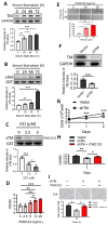
Figure 1
Figure 1
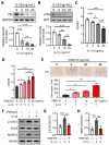
Shedding of the TM extracellular domain by RHBDL2 contributes to cell proliferation and migration in chondrocytes through its EGF-like domain. (A) Human chondrocytes (TC28a2) were incubated under serum starvation. Then, the cell lysates were collected at indicated time points for western blotting to evaluate the TM protein level. GAPDH was used as an internal loading control. (B) The concentrated conditioned media (CMs) from (A) were used to evaluate the protein level of soluble TM (sTM). Glutathione S-transferase (GST) was added as an internal control. (C) After incubation for 48 h, the harvested serum-free CMs with DCI (RHBDL2 inhibitor) were used to evaluate the sTM production. (D) The WST-1 cell proliferation assay was performed to evaluate the effect of recombinant TMD2-3 (rTMD23; TMD2 is an EGF-like repeat domain) on chondrocytes after treatment for 48 h. (E) rTMD123 exhibited a dose-dependent promotion of wound recovery in primary human articular chondrocytes (NHAC-kn). (F) TM protein level was significantly reduced by TM shRNA (shTM). (G) The growth curve of shTM-transfected TC28a2 cells in two days. (H) Effect of rTMD23 on shTM chondrocytes was assessed using MTT cell proliferation assay. (I) The transwell cell migration assay was used to evaluate the effect of rTMD23 on shTM chondrocytes. Scale bar: 100 μm. * p < 0.05; ** p < 0.01; *** p < 0.001. All experiments were repeated at least three times.
Figure 2
Figure 2
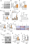
rTMD123 treatment attenuates IL-1β-reduced TM and sTM protein levels, chondrocyte proliferation, and migration by reducing STAT3 signaling and MMP13 expression. Human chondrocytes (TC28a2) were treated with IL-1β for 24 h. Then, the cell lysates and conditioned medium both were collected to evaluate the TM protein level (A) and sTM production (B). GST was used as an internal control. (C) The WST-1 cell proliferation assay was used to evaluate the effect of IL-1β on chondrocytes after treatment for 48 h. (D) rTMD123 reversed IL-1β-reduced cell proliferation in a dose-dependent manner. (E) The transwell cell migration assay was performed to assess the effect of rTMD123 on IL-1β-inhibited cell migration. Scale bar: 100 μm. (FH) IL-1β-mediated STAT3 signaling and MMP13 were arrested by treatment with rTMD123. * p < 0.05; ** p < 0.01; *** p < 0.001.
Figure 3
Figure 3
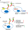
rTMD123 treatment protects knee functions in ACLT-induced OA mice by increasing articular cartilage TM expression and reducing MMP 13 level. (A) Illustrated experimental design. The green arrowhead indicates the ACLT surgery was performed. The “+” indicates the injection of rTMD123. The black arrows indicate mice undergoing tests to assess knee function. (B) Weight-bearing distribution test. (C) Results of treadmill test at week 4. n = 5 in each group. Mice were sacrificed at week 4 after the ACLT surgery, and knee joint samples were collected for IHC staining (D,E) and western blotting analysis (F) to evaluate the expression of TM and MMP 13. The red-brown color represents signal-positive cells. (G,H) Quantitative results of (F). Scale bar: 50 μm. * p < 0.05; ** p < 0.01; *** p < 0.001.
Figure 4
Figure 4
Accelerated knee joint function deterioration due to deletion of TM lectin-like domain after ACLT surgery could be improved by rTMD123. (A) Graph showing the difference of TM protein structure between TMwt/wt and TMLeD/LeD. (B) Weight-bearing distribution test. (C) Treadmill test. Results of the weight distribution test (D) and treadmill test (E) two weeks after ACLT surgery with or without rTMD123 (100 μg/kg) injection. (F) After surgery and treatment with rTMD123 for four weeks, knee sections were stained with safranin O and toluidine blue to evaluate the cartilage area (G) and OARSI score (H). Scale bar: 50 μm. ** p < 0.01; *** p < 0.001.
Figure 5
Figure 5
Delayed administration of rTMD123 one week after ACLT surgery still protected knee function in mice. (A) Illustrated experimental design. The green arrowhead indicates the ACLT surgery was performed. The “+” indicates the injection of rTMD123. The black arrow indicates mice undergoing tests to assess knee function. (B,C) Results of weight-bearing distribution test and treadmill test. (D) Quantitative results of safranin O staining. (E) OARSI score. Scale bar: 50 μm. * p < 0.05; ** p < 0.01; *** p < 0.001.
Figure 6
Figure 6
miR-up-TM-enhanced KLF2/TM signaling is critical to protect knee functions by promoting chondrocyte growth and reducing MMP 13 level. (A) After transfection with negative control miRNA (NC, 20 nM) and miR-up-TM (5-20 nM) for 24 h, cell lysates of TC28a2 were analyzed using western blotting. (B,C) Quantified expression levels of KLF2 and TM. (D) Western blotting results showed that miR-up-TM-enhanced TM levels were abrogated by shTM. (E) shTM inhibited the growth of chondrocytes boosted by miR-up-TM. (F) After two days of treatment, the positive benefit of miR-up-TM on IL-1β-suppressed chondrocyte survival was abolished by shTM. (G,H) Four weeks after ACLT surgery, the protective effect of miR-up-TM on knee functions in TMwt/wt mice was lost due to shTM. (I) Staining of knee sections showed that miR-up-TM enhanced articular cartilage expression and inhibited MMP 13 expression; this effect could be suppressed by shTM. (JL) The analytical and quantitative results of western blotting show a trend consistent with (I). Scale bar: 50 μm. * p < 0.05; ** p < 0.01; *** p < 0.001; **** p < 0.0001.
Figure 7
Figure 7
The schematic diagram shows the role of the TM in chondrocytes and OA. (A) Excessive inflammatory factors (e.g., IL-1β) impede chondrocyte TM performance and RHBDL2-mediated sTM production, leading to elevated STAT3/MMP 13 messaging pathways and reduced cell growth and migration, ultimately contributing to articular cartilage damage and OA. (B) miR-up-TM (miR-150 antagomir) enhances TM expression and sTM release from chondrocytes by increasing KLF2 transcription factors, which in turn improves the anti-inflammatory and growth capacity of the cells, eventually leading to anti-OA effects.

References

    1. GBD 2017 Disease and Injury Incidence and Prevalence Collaborators. James S.L., Abate D., Abate K.H., Abay S.M., Abbafati C., Abbasi N., Abbastabar H., Abd-Allah F., Abdela J. Global, regional, and national incidence, prevalence, and years lived with disability for 354 diseases and injuries for 195 countries and territories, 1990–2017: A systematic analysis for the Global Burden of Disease Study 2017. Lancet. 2018;392:1789–1858. doi: 10.1016/S0140-6736(18)32279-7. - DOI - PMC - PubMed
    1. Loeser R.F., Goldring S.R., Scanzello C.R., Goldring M.B. Osteoarthritis: A disease of the joint as an organ. Arthritis Rheumatol. 2012;64:1697–1707. doi: 10.1002/art.34453. - DOI - PMC - PubMed
    1. Archer C.W., Francis-West P. The chondrocyte. Int. J. Biochem. Cell Biol. 2003;35:401–404. doi: 10.1016/S1357-2725(02)00301-1. - DOI - PubMed
    1. Maillard C., Berruyer M., Serre C.M., Amiral J., Dechavanne M., Delmas P.D. Thrombomodulin is synthesized by osteoblasts, stimulated by 1,25-(OH)2D3 and activates protein C at their cell membrane. Endocrinology. 1993;133:668–674. doi: 10.1210/endo.133.2.8393772. - DOI - PubMed
    1. McCachren S.S., Diggs J., Weinberg J.B., Dittman W.A. Thrombomodulin expression by human blood monocytes and by human synovial tissue lining macrophages. Blood. 1991;78:3128–3132. doi: 10.1182/blood.V78.12.3128.3128. - DOI - PubMed