

Deciphering the Cardiovascular Potential of Human CD34+ Stem Cells
- PMID: 37298503
- PMCID: PMC10253329
- DOI: 10.3390/ijms24119551
Deciphering the Cardiovascular Potential of Human CD34+ Stem Cells
Abstract
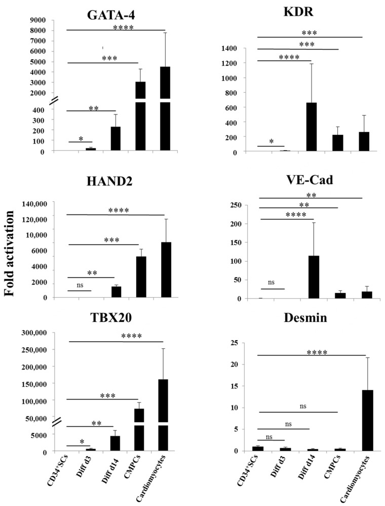
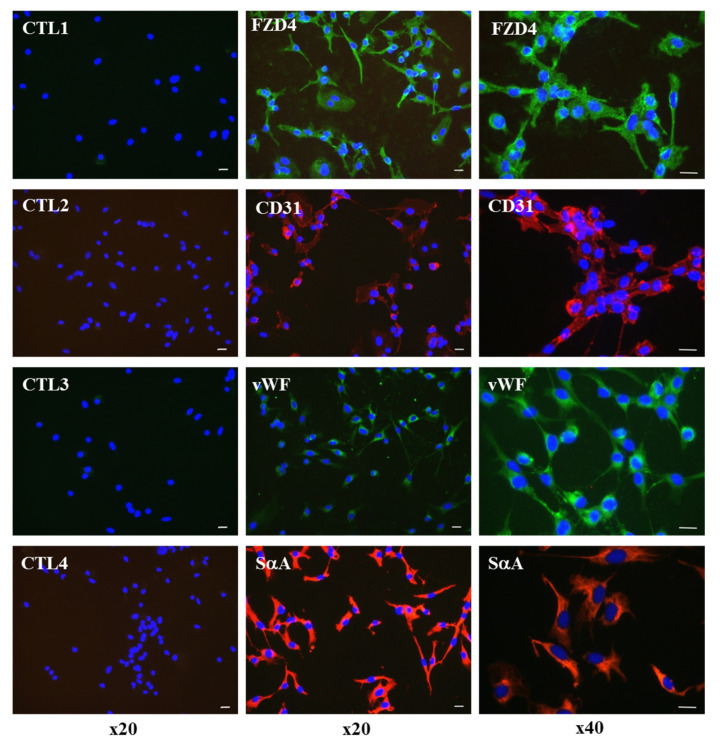
Ex vivo monitored human CD34+ stem cells (SCs) injected into myocardium scar tissue have shown real benefits for the recovery of patients with myocardial infarctions. They have been used previously in clinical trials with hopeful results and are expected to be promising for cardiac regenerative medicine following severe acute myocardial infarctions. However, some debates on their potential efficacy in cardiac regenerative therapies remain to be clarified. To elucidate the levels of CD34+ SC implication and contribution in cardiac regeneration, better identification of the main regulators, pathways, and genes involved in their potential cardiovascular differentiation and paracrine secretion needs to be determined. We first developed a protocol thought to commit human CD34+ SCs purified from cord blood toward an early cardiovascular lineage. Then, by using a microarray-based approach, we followed their gene expression during differentiation. We compared the transcriptome of undifferentiated CD34+ cells to those induced at two stages of differentiation (i.e., day three and day fourteen), with human cardiomyocyte progenitor cells (CMPCs), as well as cardiomyocytes as controls. Interestingly, in the treated cells, we observed an increase in the expressions of the main regulators usually present in cardiovascular cells. We identified cell surface markers of the cardiac mesoderm, such as kinase insert domain receptor (KDR) and the cardiogenic surface receptor Frizzled 4 (FZD4), induced in the differentiated cells in comparison to undifferentiated CD34+ cells. The Wnt and TGF-β pathways appeared to be involved in this activation. This study underlined the real capacity of effectively stimulated CD34+ SCs to express cardiac markers and, once induced, allowed the identification of markers that are known to be involved in vascular and early cardiogenesis, demonstrating their potential priming towards cardiovascular cells. These findings could complement their paracrine positive effects known in cell therapy for heart disease and may help improve the efficacy and safety of using ex vivo expanded CD34+ SCs.
Keywords: CD34+ cells; cardiovascular differentiation; cell therapy; gene expression profiling; umbilical cord blood.
Conflict of interest statement
P.H. is an employee and stockholder of CellProthera. The other authors declare no competing interests.
Figures


Similar articles
-
Generation of Cardiomyocytes From Vascular Adventitia-Resident Stem Cells.Circ Res. 2018 Aug 31;123(6):686-699. doi: 10.1161/CIRCRESAHA.117.312526. Circ Res. 2018. PMID: 30355234
-
FZD4 Marks Lateral Plate Mesoderm and Signals with NORRIN to Increase Cardiomyocyte Induction from Pluripotent Stem Cell-Derived Cardiac Progenitors.Stem Cell Reports. 2018 Jan 9;10(1):87-100. doi: 10.1016/j.stemcr.2017.11.008. Epub 2017 Dec 14. Stem Cell Reports. 2018. PMID: 29249665 Free PMC article.
-
Human cord blood CD34+ progenitor cells acquire functional cardiac properties through a cell fusion process.Am J Physiol Heart Circ Physiol. 2011 May;300(5):H1875-84. doi: 10.1152/ajpheart.00523.2010. Epub 2011 Feb 25. Am J Physiol Heart Circ Physiol. 2011. PMID: 21357510
-
Embryonic template-based generation and purification of pluripotent stem cell-derived cardiomyocytes for heart repair.J Cardiovasc Transl Res. 2012 Oct;5(5):566-80. doi: 10.1007/s12265-012-9391-6. Epub 2012 Jul 18. J Cardiovasc Transl Res. 2012. PMID: 22806916 Review.
-
CD34-positive stem cells: in the treatment of heart and vascular disease in human beings.Tex Heart Inst J. 2011;38(5):474-85. Tex Heart Inst J. 2011. PMID: 22163120 Free PMC article. Review.
Cited by
-
Circulating CD34+ Cells: A New Biomarker of Residual Pulmonary Vascular Obstruction after Pulmonary Embolism.Stem Cell Rev Rep. 2025 Jun;21(5):1501-1511. doi: 10.1007/s12015-025-10865-0. Epub 2025 Mar 14. Stem Cell Rev Rep. 2025. PMID: 40085375
-
Development of a potency assay for CD34+ cell-based therapy.Sci Rep. 2023 Nov 11;13(1):19665. doi: 10.1038/s41598-023-47079-8. Sci Rep. 2023. PMID: 37952030 Free PMC article. Clinical Trial.
-
Transendocardial injection of expanded autologous CD34+ cells after myocardial infarction: Design of the EXCELLENT trial.ESC Heart Fail. 2025 Apr;12(2):1455-1463. doi: 10.1002/ehf2.15124. Epub 2024 Dec 15. ESC Heart Fail. 2025. PMID: 39676512 Free PMC article.
-
APEX1 Polymorphisms Affect Acute Myeloid Leukemia Risk, and Its Expression Is Involved in Cell Proliferation and Differentiation.Int J Lab Hematol. 2025 Apr;47(2):276-287. doi: 10.1111/ijlh.14401. Epub 2024 Nov 13. Int J Lab Hematol. 2025. PMID: 39536468 Free PMC article.
-
Altered lipid metabolism promoting cardiac fibrosis is mediated by CD34+ cell-derived FABP4+ fibroblasts.Exp Mol Med. 2024 Aug;56(8):1869-1886. doi: 10.1038/s12276-024-01309-9. Epub 2024 Aug 29. Exp Mol Med. 2024. PMID: 39198543 Free PMC article.
References
-
- Quyyumi A.A., Vasquez A., Kereiakes D.J., Klapholz M., Schaer G.L., Abdel-Latif A., Frohwein S., Henry T.D., Schatz R.A., Dib N., et al. PreSERVE-AMI: A Randomized, Double-Blind, Placebo-Controlled Clinical Trial of Intracoronary Administration of Autologous CD34+ Cells in Patients With Left Ventricular Dysfunction Post STEMI. Circ. Res. 2017;120:324–331. doi: 10.1161/CIRCRESAHA.115.308165. - DOI - PMC - PubMed
-
- Templin C., Volkmann J., Emmert M.Y., Mocharla P., Müller M., Kraenkel N., Ghadri J.-R., Meyer M., Styp-Rekowska B., Briand S., et al. Increased Proangiogenic Activity of Mobilized CD34+ Progenitor Cells of Patients With Acute ST-Segment–Elevation Myocardial Infarction: Role of Differential MicroRNA-378 Expression. Arterioscler. Thromb. Vasc. Biol. 2017;37:341–349. doi: 10.1161/ATVBAHA.116.308695. - DOI - PubMed
-
- Pasquet S., Sovalat H., Hénon P., Bischoff N., Arkam Y., Ojeda-Uribe M., Bouar R.L., Rimelen V., Brink I., Dallemand R., et al. Long-Term Benefit of Intracardiac Delivery of Autologous Granulocyte–Colony-Stimulating Factor-Mobilized Blood CD34+ Cells Containing Cardiac Progenitors on Regional Heart Structure and Function after Myocardial Infarct. Cytotherapy. 2009;11:1002–1015. doi: 10.3109/14653240903164963. - DOI - PubMed
MeSH terms
Substances
LinkOut - more resources
Full Text Sources
Medical
Molecular Biology Databases
