. 2023 May 31;28(11):4460.
doi: 10.3390/molecules28114460.

Group I Intron as a Potential Target for Antifungal Compounds: Development of a Trans-Splicing High-Throughput Screening Strategy

Affiliations

Group I Intron as a Potential Target for Antifungal Compounds: Development of a Trans-Splicing High-Throughput Screening Strategy

Bastien Malbert et al. Molecules. .

Abstract

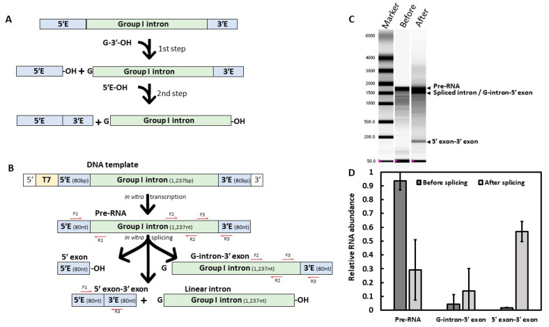
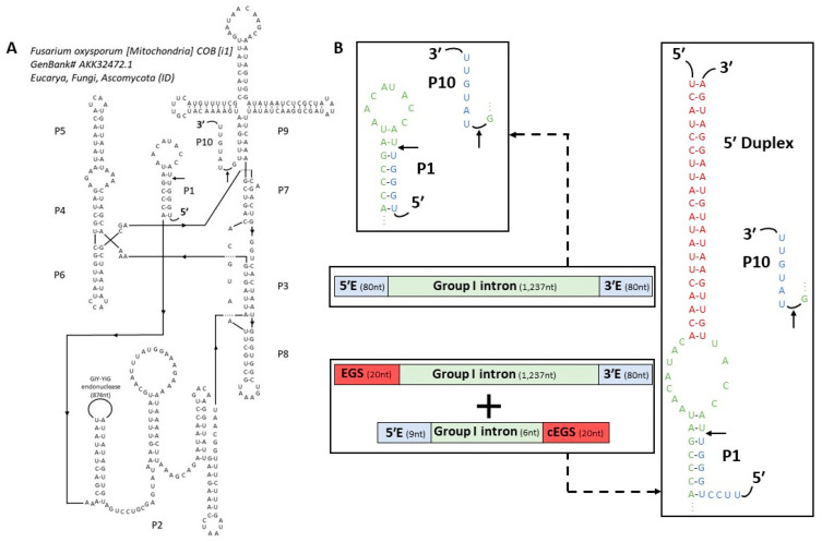
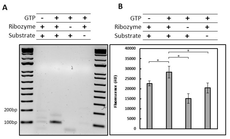
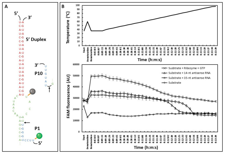
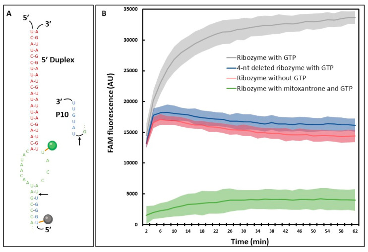
The search for safe and efficient new antifungal compounds for agriculture has led to more efforts in finding new modes of action. This involves the discovery of new molecular targets, including coding and non-coding RNA. Rarely found in plants and animals but present in fungi, group I introns are of interest as their complex tertiary structure may allow selective targeting using small molecules. In this work, we demonstrate that group I introns present in phytopathogenic fungi have a self-splicing activity in vitro that can be adapted in a high-throughput screening to find new antifungal compounds. Ten candidate introns from different filamentous fungi were tested and one group ID intron found in F. oxysporum showed high self-splicing efficiency in vitro. We designed the Fusarium intron to act as a trans-acting ribozyme and used a fluorescence-based reporter system to monitor its real time splicing activity. Together, these results are opening the way to study the druggability of such introns in crop pathogen and potentially discover small molecules selectively targeting group I introns in future high-throughput screenings.

Keywords: Fusarium; HTS; RNA; group I; ribozyme; self-splicing.


Conflict of interest statement

The authors are employees of Bayer Crop Science, a leading developer of agricultural seeds and chemistries.

Figures

Figure 1
Figure 1
In vitro self-splicing of the Fusarium oxysporum cob group I intron. (A) Group I intron splicing mechanism. The 2-steps reaction starts with the addition of a guanosine (G-3′-OH) cofactor to the mix. (B) Experimental design. The Fusarium intron sequence was PCR amplified with 80 bp of border exons and a T7 promoter, then in vitro transcribed prior to the self-splicing reaction. Red arrows correspond to primers used for RT-qPCR. (C) RNA migration of reaction products, before (1) or after (2) the self-splicing reaction. Arrows represent the different splicing products obtained. Because of their similar length, spliced introns could not be distinguished from the intermediate product carrying the 3′ exon. RNA marker (M) sizes in nucleotides are indicated. Representative result of three independent experiments. (D) Quantitative Reverse Transcription PCR (RT-qPCR) of reaction products before and after splicing. Representative result of three independent experiments based on three technical replicates.
Figure 2
Figure 2
Secondary structure prediction. (A) Predicted secondary structure of Fusarium oxysporum cob group I intron. Common Group I domains are designated P1 to P10, and 5′-3′ ends are indicated. Arrows correspond to splice sites. The site of GIY-YIG endonuclease insertion is indicated in domain P2. (B) P1 domain comparison between the group I native intron (top) and the ‘classical designed’ trans-acting ribozyme (bottom). Schematic diagram of base-pairing between the ribozyme and the associated RNA target. Exon sequences are in blue, intron sequences in green and additional sequences EGS and complementary EGS (cEGS) are in red.
Figure 3
Figure 3
In vitro trans-splicing activity of Fusarium ribozyme. (A) Amplification of the 5′ exon-3′ exon ligation product by RT-PCR using 3′ exon specific primer for the RT, 5′ exon and 3′ exon specific primers for the PCR. The 5′ exon primer is an 18-bp oligonucleotide with only 9-bp annealed to the 5′ exon. The RT-PCR product corresponds to a 98-bp amplification (9-bp for the 5′ exon primer extension, 9-bp for the 5′ exon and 80-bp for the 3′ exon). Splicing reactions were performed with (+) or without (-) GTP, Fusarium ribozyme or substrate. (B) Quantitation of the 5′ exon-3′ exon ligation product formed during trans-splicing by a molecular beacon. Splicing reactions were performed with (+) or without (-) GTP, Fusarium ribozyme or substrate. Error bars correspond to standard deviation. Error bars correspond to standard deviation. Representative experiment of three independent experiments, the stars correspond to the p-value of a multi-factor ANOVA followed by a Bonferroni correction (* p < 0.01).
Figure 4
Figure 4
Fluorescence-based assay exploring the different signal components. (A) Substrate used in this experiment. The substrate is composed of a FAM fluorophore in green (maximum excitation: 495 nm; maximum emission: 515 nm) and a DABdT quencher in grey (maximum absorbance: 475 nm); 13-nt separate the quencher from the fluorophore. (B) Fluorescence-based assay. The 35-nt substrate was either mixed with the ribozyme and GTP for a splicing reaction (cross), with a 35-nt antisense RNA (circle), a 14-nt antisense RNA (triangle) or alone (diamond). After 2 min at 37 °C to assess initial fluorescence (start), samples were heated up to 60 °C for 2 min (denaturation) and slowly cooled down to 37 °C during 1 h (renaturation). Samples were then kept at 37 °C for 10 min and the temperature was increased by 2 °C every 7 to 8 s with fluorescence measurements. Representative experiment based on three technical replicates. Error bars correspond to standard deviation.
Figure 5
Figure 5
Fluorescence-based HTS-ready assay to screen inhibitors of Fusarium intron. (A) P1 and 5′ Duplex domains of the fluorescently labeled substrate and the associated ribozyme. The quencher (Iowa Black® Quencher FQ, maximum absorbance: 531 nm), and the FAM fluorophore are represented by a black and a green circle, respectively. (B) Fluorescence measurement of trans-splicing activity with (grey) and without (red) GTP, using the 4-nt deleted ribozyme with GTP (blue) or with the addition of 10 µM of mitoxantrone (green). Shaded areas correspond to standard deviations. Representative experiment of three independent experiments.

Similar articles

Cited by

References

    1. Disney M.D., Yildirim I., Childs-Disney J.L. Methods to Enable the Design of Bioactive Small Molecules Targeting RNA. Org. Biomol. Chem. 2014;12:1029–1039. doi: 10.1039/C3OB42023J. - DOI - PMC - PubMed
    1. Childs-Disney J.L., Disney M.D. Approaches to Validate and Manipulate RNA Targets with Small Molecules in Cells. Annu. Rev. Pharmacol. Toxicol. 2016;56:123–140. doi: 10.1146/annurev-pharmtox-010715-103910. - DOI - PMC - PubMed
    1. Disney M.D. Targeting RNA with Small Molecules To Capture Opportunities at the Intersection of Chemistry, Biology, and Medicine. J. Am. Chem. Soc. 2019;141:6776–6790. doi: 10.1021/jacs.8b13419. - DOI - PMC - PubMed
    1. Mei H.Y., Cui M., Lemrow S.M., Czarnik A.W. Discovery of Selective, Small-Molecule Inhibitors of RNA Complexes—II. Self-Splicing Group I Intron Ribozyme. Bioorganic Med. Chem. 1997;5:1185–1195. doi: 10.1016/S0968-0896(97)00065-5. - DOI - PubMed
    1. Seth P.P., Miyaji A., Jefferson E.A., Sannes-Lowery K.A., Osgood S.A., Propp S.S., Ranken R., Massire C., Sampath R., Ecker D.J., et al. SAR by MS: Discovery of a New Class of RNA-Binding Small Molecules for the Hepatitis C Virus: Internal Ribosome Entry Site IIA Subdomain. J. Med. Chem. 2005;48:7099–7102. doi: 10.1021/jm050815o. - DOI - PubMed

LinkOut - more resources