. 2023 May 31;28(11):4473.
doi: 10.3390/molecules28114473.

Benzoylaconitine Alleviates Progression of Psoriasis via Suppressing STAT3 Phosphorylation in Keratinocytes

Affiliations

Benzoylaconitine Alleviates Progression of Psoriasis via Suppressing STAT3 Phosphorylation in Keratinocytes

Yuanbo Li et al. Molecules. .

Abstract

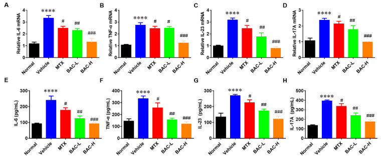
Psoriasis is a chronic and multifactorial skin disease which is caused by inflammatory infiltrates, keratinocyte hyperproliferation, and accumulation of immune cells. As part of the Aconitum species, Benzoylaconitine (BAC) shows potential antiviral, anti-tumor, and anti-inflammatory effects. In this study, we investigated the effects and mechanisms of BAC on tumor necrosis factor-alpha (TNF-α)/LPS-induced HaCaT keratinocytes in a imiquimod(IMQ)-induced mice model. The results showed that BAC could relieve the symptoms of psoriasis by inhibiting cell proliferation, the release of inflammatory factors, and the accumulation of Th17 cells, while no obvious effect on cell viability and safety was observed both in vitro and in vivo. Additionally, BAC can markedly inhibit the protein and mRNA levels of inflammatory cytokines in TNF-α/LPS-induced HaCaT keratinocytes by inhibiting the phosphorylation of STAT3. In brief, our data indicated that BAC could alleviate the progression of psoriasis and may be a potential therapeutic agent for treating psoriasis in clinical practice.

Keywords: Benzoylaconitine; HaCaT keratinocytes; STAT3 signaling; inflammatory cytokine; psoriasis.


Conflict of interest statement

The authors declare no conflict of interest.

Figures

Figure 1
Figure 1
The chemical structure of BAC and its effects on cell proliferation and viability. (A) The chemical structure of BAC. (B,C) Cell viability was measured by Flow cytometry and CCK-8. (D) Detection of cell proliferation after treatment with TNF-α (10 ng/mL) and BAC (0, 10, 20, and 40 μM) for 24 h. (E) Detection of cell proliferation after treatment with LPS (100 ng/mL) and BAC (0, 10, 20, and 40 μM) for 24 h (Mean ± SD; vs. Con, *** p < 0.001; vs. TNF-α, # p < 0.05, ## p < 0.01, ### p < 0.001; NS = not significant).
Figure 2
Figure 2
BAC significantly inhibited the gene and protein expression level of inflammatory cytokines in TNF-α/LPS-induced HaCaT cells. (AH) mRNA level of IL-6, IL-8, TNF-α, and IL-17, and the protein level of IL-6, IL-8, TNF-α, and IL-17 in the treated cells with TNF-α (10 ng/mL) and BAC (0, 10, 20, and 40 μM), respectively. (Mean ± SD; vs. Con, **** p < 0.0001, *** p < 0.001; vs. TNF-α, # p < 0.05, ## p < 0.01, ### p < 0.001).
Figure 3
Figure 3
BAC attenuated IMQ-induced mouse psoriatic lesion. (A) The images of mouse back skin in different groups. (B) PASI scores of skin lesion. (CE) The daily scales, epidermal thickness, and erythema, respectively. (F) H&E staining and statistics of Ki-67, Keratin 1 and Keratin 17-positive cells (Scale bar = 100 μm (200×); Mean ± SD; vs. normal, **** p < 0.0001,*** p < 0.001; vs. vehicle, # p < 0.05, ## p < 0.01, ### p < 0.001).
Figure 4
Figure 4
BAC reduced the accumulation of Th17 cells in IMQ-induced mice. (A,B) The frequency of Th17 cells in the spleen in IMQ-induced mice after analysis of flow cytometry. (C,D) The frequency of Th17 cells in the skin of IMQ-induced mice after analysis of flow cytometry (Mean ± SD; vs. Normal, **** p < 0.0001; vs. Vehicle, ## p < 0.01, ### p < 0.001, #### p < 0.0001).
Figure 5
Figure 5
BAC significantly inhibited the gene and protein expression level of inflammatory cytokines in IMQ-induced mice. (AH) The mRNA level of IL-6, TNF-α, IL-23, and IL-17A, and the protein level of IL-6, TNF-α, IL-23, and IL-17A, respectively (Mean ± SD; vs. Normal, **** p < 0.0001; vs. Vehicle, # p < 0.05, ## p < 0.01, ### p < 0.001).
Figure 6
Figure 6
Safety analysis of BAC for dosing IMQ-induced mice. (A) Body weight of mice in each group. (BD) Serum level of ALP/ALT, Cr, and Bun, respectively.
Figure 7
Figure 7
The suppression of STAT3 phosphorylation by BAC in TNF-α stimulated HaCaT cells. (A) The expression level of p-STAT3Tyr 705, p-STAT3Ser727, STAT3, and Actin with WB analysis in TNF-α- induced HaCaT cells. (B) Histogram of p-STAT3Tyr705, p-STAT3Ser727, and STAT3 expression levels in TNF-α-induced HaCaT cells (Mean ± SD; vs. Con, **** p < 0.0001; vs. TNF-α, ### p < 0.001).

References

    1. Fabian E., Oesch F., Ott K., Landsiedel R., van Ravenzwaay B. A protocol to determine dermal absorption of xenobiotica through human skin in vitro. Arch. Toxicol. 2017;91:1497–1511. doi: 10.1007/s00204-016-1782-z. - DOI - PubMed
    1. Helmick C.G., Lee-Han H., Hirsch S.C., Baird T.L., Bartlett C.L. Prevalence of psoriasis among adults in the US 2003–2006 and 2009–2010 national health and nutrition examination surveys. Am. J. Prev. Med. 2014;4:37–45. doi: 10.1016/j.amepre.2014.02.012. - DOI - PMC - PubMed
    1. Benhadou F., Mintoff D., del Marmol V. Psoriasis: Keratinocytes or immune cells-which is the trigger? Dermatology. 2019;235:91–100. doi: 10.1159/000495291. - DOI - PubMed
    1. Garzorz-Stark N., Eyerich K. Psoriasis pathogenesis: Keratinocytes are back in the spotlight. J. Investig. Dermatol. 2019;139:995–996. doi: 10.1016/j.jid.2019.01.026. - DOI - PubMed
    1. Michalek I.M., Loring B., John S.M. A systematic review of worldwide epidemiology of psoriasis. J. Eur. Acad. Dermatol. 2017;31:205–212. doi: 10.1111/jdv.13854. - DOI - PubMed