. 2023 May 24;12(11):2093.
doi: 10.3390/plants12112093.

Silver Nanoparticles of Artemisia sieberi Extracts: Chemical Composition and Antimicrobial Activities

Affiliations

Silver Nanoparticles of Artemisia sieberi Extracts: Chemical Composition and Antimicrobial Activities

Fatimah Al-Otibi et al. Plants (Basel). .

Abstract

Background: Artemisia sieberi (mugwort) is a member of the daisy family Asteraceae and is widely propagated in Saudi Arabia. A. sieberi has historical medical importance in traditional societies. The current study aimed to assess the antibacterial and antifungal characteristics of the aqueous and ethanolic extracts of A. sieberi. In addition, the study investigated the effect of silver nanoparticles (AgNPs) synthesized from the A. sieberi extract.

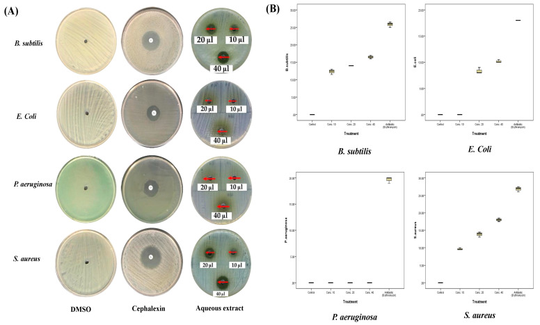
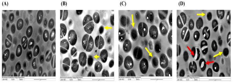
Methods: The ethanolic and aqueous extracts and AgNPs were prepared from the shoots of A. sieberi. The characteristics of AgNPs were assessed by UV-visible spectroscopy, transmission electron microscopy (TEM), Fourier transform infrared spectroscopy (FTIR), and dynamic light scattering (DLS). The antibacterial experiments were performed against Staphylococcus aureus, Bacillus subtilis, Escherichia coli, and Pseudomonas aeruginosa. The fungal species used were Candida parapsilosis, Candida krusei, Candida famata, Candida rhodotorula, and Candida albicans. The antibacterial and antifungal characteristics were evaluated by measuring the diameter of growing organisms in Petri dishes treated with different concentrations of either extracts or AgNPs compared to the untreated controls. Furthermore, TEM imaging was used to investigate any ultrastructure changes in the microbes treated with crude extracts and AgNO3.

Results: The ethanolic and aqueous extracts significantly decreased the growth of E. coli, S. aureus, and B. subtilis (p < 0.001), while P. aeruginosa was not affected. Unlike crude extracts, AgNPs had more substantial antibacterial effects against all species. In addition, the mycelial growth of C. famata was reduced by the treatment of both extracts. C. krusei mycelial growth was decreased by the aqueous extract, while the growth of C. parapsilosis was affected by the ethanolic extract and AgNPs (p < 0.001). None of the treatments affected the growth of C. albicans or C. rhodotorula. TEM analysis showed cellular ultrastructure changes in the treated S. aureus and C. famata compared to the control.

Conclusion: The biosynthesized AgNPs and extracts of A. sieberi have a potential antimicrobial characteristic against pathogenic bacterial and fungal strains and nullified resistance behavior.

Keywords: Artemisia sieberi; TEM analysis; antibacterial; antifungal; pathogenic microbes; silver nanoparticles.


Conflict of interest statement

The authors declare no conflict of interest.

Figures

Figure 1
Figure 1
FTIR results of the aqueous extract of A. sieberi. The results were produced by the Nicolet 6700 FTIR Spectrometer at 500–4000/cm.
Figure 2
Figure 2
FTIR results of AgNPs biosynthesized from the aqueous extract of A. sieberi. The results were produced by the Nicolet 6700 FTIR Spectrometer at 500–4000/cm.
Figure 3
Figure 3
Characterization of the synthesized A. sieberi AgNPs by the JEOL JEM-1400 transmission electron microscope. (A) The synthesis process of AgNPs from the aqueous extract A. sieberi and AgNO3 incubated in sunlight and caused the color change from colorless to brown. (B,C) TEM micrographs of (B) A. sieberi extract and (C) A. sieberi AgNPs, in which the shape and size are shown in µm.
Figure 4
Figure 4
Characterization of the synthesized A. sieberi AgNPs. (A) The UV–visible spectrum of A. sieberi extract and AgNPs; the spectrum was analyzed by the Shimadzu UV–visible spectrophotometer. (B) TEM micrograph of A. sieberi extract in which the shape and size are shown in µm.
Figure 5
Figure 5
Antibacterial activities of the aqueous extract of A. sieberi. The growth inhibition zone was measured by the well diffusion method for the species growing on the Mueller–Hinton agar dishes. (A) Petri dishes of different bacterial strains treated with either DMSO (negative control, <1%), cephalexin disc (positive control, 5 µg/mL), or different amounts of the aqueous extract of A. sieberi (10, 20, and 40%). (B) Box plots showed the antibacterial effect of different concentrations of the aqueous extract of A. sieberi compared to DMSO and cephalexin.
Figure 6
Figure 6
Antibacterial activities of the ethanolic extract of A. sieberi. The growth inhibition zone was measured by the well diffusion method for the species growing on the Mueller–Hinton agar dishes. (A) Petri dishes of different bacterial strains treated with either DMSO (negative control, 1 µL), cephalexin disc (positive control, 5 µg/mL), or different amounts of the ethanolic extract of A. sieberi (1 mg/mL). (B) Box plots showed the antibacterial effect of different concentrations of the ethanolic extract of A. sieberi compared to DMSO and cephalexin.
Figure 7
Figure 7
Antibacterial activities of biosynthesized AgNPs of A. sieberi. The growth inhibition zone was measured by the well diffusion method for the species growing on the Mueller–Hinton agar dishes. (A) Petri dishes of different bacterial strains treated with either DMSO (negative control, 1 µL), cephalexin disc (positive control, 5 µg/mL), AgNO3 (2 mM, 20% v/v), or different amounts of AgNPs. (B) Box plots showed the antibacterial effect of different concentrations of the AgNPs compared to DMSO, cephalexin, and AgNO3 (2 mM).
Figure 8
Figure 8
Antifungal activities of the aqueous extract of A. sieberi. The growth inhibition zone was measured by the well diffusion method for the species growing on the Sabouraud agar dishes. (A) Petri dishes of different fungal species treated with either terbinafine disc (positive control, 5 µg/mL) or different amounts of the aqueous extract of A. sieberi (10, 20, and 40%). (B) Box plots showed the antifungal effect of different concentrations of the aqueous extract of A. sieberi compared to terbinafine.
Figure 9
Figure 9
Antifungal activities of the ethanolic extract of A. sieberi. The growth inhibition zone was measured by the well diffusion method for the species growing on the Sabouraud agar dishes. (A) Petri dishes of different fungal species treated with either terbinafine disc (positive control, 5 µg/mL) or different amounts of the ethanolic extract of A. sieberi (10, 20, and 40%). (B) Box plots showed the antifungal effect of different concentrations of the ethanolic extract of A. sieberi compared to terbinafine.
Figure 10
Figure 10
Antifungal activities of the biosynthesized AgNPs of A. sieberi. The growth inhibition zone was measured by the well diffusion method for the species growing on the Sabouraud agar dishes. (A) Petri dishes of different fungal species treated with either terbinafine disc (positive control, 5 µg/mL), AgNO3 (2 mM, 20% v/v), or different amounts of the ethanolic extract of A. sieberi (10, 20, and 40%). (B) Box plots showed the antifungal effect of different concentrations of the ethanolic extract of A. sieberi compared to terbinafine.
Figure 11
Figure 11
TEM images of S. aureus. The images showed the ultrastructural changes induced by the extracts of A. sieberi at the highest concentration (40 µg/mL) captured by the JEM-1400 transmission electron microscope at the magnification of 3000×. Red arrows indicate abnormal cellular vacuoles, while yellow arrows indicate undivided cells. (A) Control (DMSO), (B) aqueous extract, (C) ethanolic extract, and (D) A. sieberi-biosynthesized AgNPs.
Figure 12
Figure 12
TEM images of C. famata. The images showed the ultrastructural changes induced by extracts of A. sieberi at the highest concentration (40 µg/mL) captured by the JEM-1400 microscope at the magnification of 2000×. Red arrows indicate abnormal cellular vacuoles, while the green arrow indicates raptured cellular membrane. (A) Control (DMSO), (B) aqueous extract, (C) ethanolic extract, and (D) A. sieberi-biosynthesized AgNPs (10000×).

Similar articles

Cited by

References

    1. Batiha G.E.-S., Beshbishy A.M., Tayebwa D.S., Adeyemi O.S., Yokoyama N., Igarashi I. Anti-piroplasmic potential of the methanolic Peganum harmala seeds and ethanolic Artemisia absinthium leaf extracts. J. Protozool. Res. 2019;29:8–25.
    1. Batiha G.E.-S., Olatunde A., El-Mleeh A., Hetta H.F., Al-Rejaie S., Alghamdi S., Zahoor M., Magdy Beshbishy A., Murata T., Zaragoza-Bastida A., et al. Bioactive Compounds, Pharmacological Actions, and Pharmacokinetics of Wormwood (Artemisia absinthium) Antibiotics. 2020;9:353. doi: 10.3390/antibiotics9060353. - DOI - PMC - PubMed
    1. Aati H.Y., Perveen S., Orfali R., Al-Taweel A.M., Aati S., Wanner J., Khan A., Mehmood R. Chemical composition and antimicrobial activity of the essential oils of Artemisia absinthium, Artemisia scoparia, and Artemisia sieberi grown in Saudi Arabia. Arab. J. Chem. 2020;13:8209–8217. doi: 10.1016/j.arabjc.2020.09.055. - DOI
    1. Bunse M., Daniels R., Gründemann C., Heilmann J., Kammerer D.R., Keusgen M., Lindequist U., Melzig M.F., Morlock G.E., Schulz H., et al. Essential oils as multicomponent mixtures and their potential for human health and well-being. Front. Pharmacol. 2022;13:956541. doi: 10.3389/fphar.2022.956541. - DOI - PMC - PubMed
    1. Bisht D., Kumar D., Kumar D., Dua K., Chellappan D.K. Phytochemistry and pharmacological activity of the genus artemisia. Arch. Pharm. Res. 2021;44:439–474. doi: 10.1007/s12272-021-01328-4. - DOI - PMC - PubMed

LinkOut - more resources