. 2023 May 27;12(11):2126.
doi: 10.3390/plants12112126.

Protective Role of an Extract Waste Product from Citrus bergamia in an In Vitro Model of Neurodegeneration

Affiliations

Protective Role of an Extract Waste Product from Citrus bergamia in an In Vitro Model of Neurodegeneration

Jessica Maiuolo et al. Plants (Basel). .

Abstract

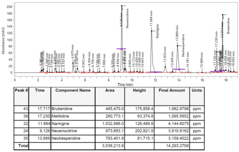
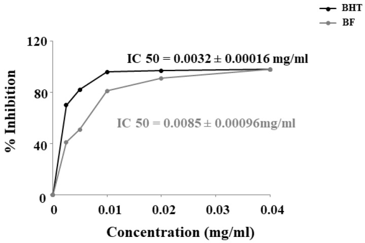
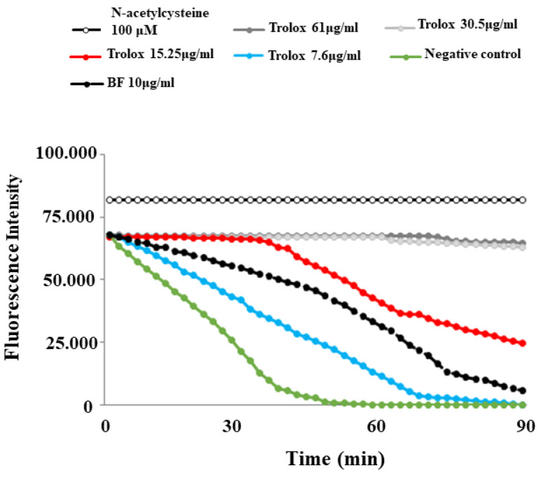
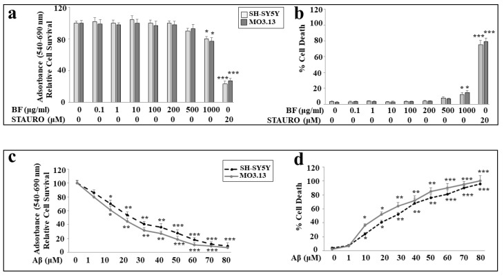
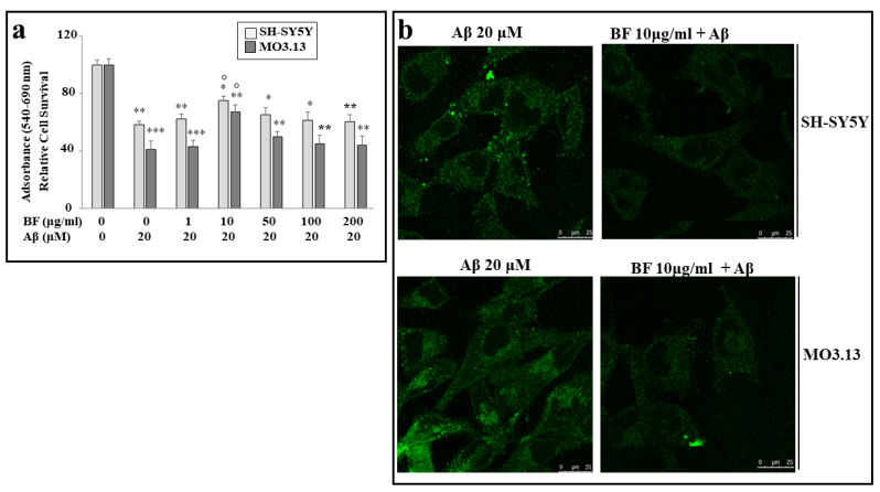
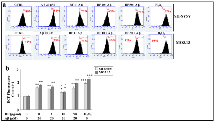
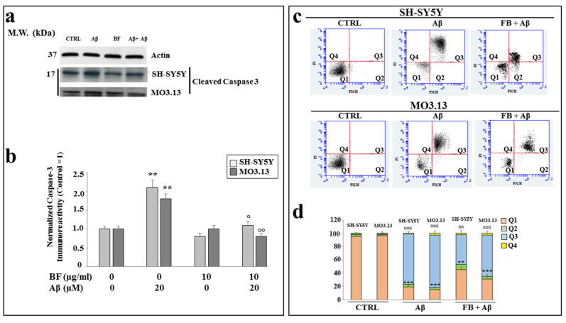
A balanced diet, rich in fruits and vegetables and ensuring the intake of natural products, has been shown to reduce or prevent the occurrence of many chronic diseases. However, the choice to consume large quantities of fruits and vegetables leads to an increase in the amount of waste, which can cause an alteration in environmental sustainability. To date, the concept of a "byproduct" has evolved, now being understood as a waste product from which it is still possible obtain useful compounds. Byproducts in the agricultural sector are a rich source of bioactive compounds, capable of possessing a second life, decreasing the amount of waste products, the disposal costs, and environmental pollution. A promising and well-known citrus of the Mediterranean diet is the bergamot (Citrus bergamia, Risso et Poiteau). The composition of bergamot is known, and the rich presence of phenolic compounds and essential oils has justified the countless beneficial properties found, including anti-inflammatory, antioxidant, anti-cholesterolemic, and protective activity for the immune system, heart failure, and coronary heart diseases. The industrial processing of bergamot fruits leads to the formation of bergamot juice and bergamot oil. The solid residues, referred to as "pastazzo", are normally used as feed for livestock or pectin production. The fiber of bergamot (BF) can be obtained from pastazzo and could exert an interesting effect thanks to its content of polyphenols. The aims of this work were twofold: (a) to have more information (composition, polyphenol and flavonoid content, antioxidant activity, etc.) on BF powder and (b) to verify the effects of BF on an in vitro model of neurotoxicity induced by treatment with amyloid beta protein (Aβ). In particular, a study of cell lines was carried out on both neurons and oligodendrocytes, to measure the involvement of the glia and compare it with that of the neurons. The results obtained showed that BF powder contains polyphenols and flavonoids and that it is able to exercise an antioxidant property. Moreover, BF exerts a protective action on the damage induced by treatment with Aβ, and this defense is found in experiments on the cell viability, on the accumulation of reactive oxygen species, on the involvement of the expression of caspase-3, and on necrotic or apoptotic death. In all these results, oligodendrocytes were always more sensitive and fragile than neurons. Further experiments are needed, and if this trend is confirmed, BF could be used in AD; at the same time, it could help to avoid the accumulation of waste products.

Keywords: Alzheimer’s disease; Citrus bergamia; byproducts; fiber of bergamot; neurons; oligodendrocytes; pastazzo; β-amyloid protein.


Conflict of interest statement

The authors declare no conflict of interest.

Figures

Figure 1
Figure 1
HPLC analysis of BF. Chromatogram of HPLC analysis of bergamot fiber from Citrus bergamia.
Figure 2
Figure 2
DPPH scavenging activity of BHT and BF.
Figure 3
Figure 3
ORAC assay decay curves for Trolox and BF.
Figure 4
Figure 4
Effects of BF and Aβ on vitality of both cell lines. In panel (a), the representation of cell viability following increasing concentrations of BF on neurons and oligodendrocytes is shown. The experiments were conducted using MTT assay. Under the same experimental conditions, cell mortality is shown in panel (b) (Trypan blue exclusion assay). In these experiments, treatment with STAURO, an apoptosis inductor, was used as a positive control. Panels (c,d) show the toxic effects induced by increasing concentrations of Aβ on neurons and oligodendrocytes on cell viability (MTT assay) and cell mortality (Trypan blue exclusion assay), respectively. Three independent experiments were carried out, and the values are expressed as the mean ± standard deviation (sd). * denotes p < 0.05 vs. the control; ** denotes p < 0.01 vs. the control; *** denotes p < 0.001 vs. the control. Analysis of variance (ANOVA) was followed by a Tukey–Kramer comparison test.
Figure 5
Figure 5
Protection by BF against damage induced by Aβ. In panel (a), only pre-treatment with BF 10 μg/mL is able to protect cells from Aβ-induced damage, and this result can be appreciated by observing the significant increase in viability compared with treatment with Aβ alone. All other BF concentrations tested showed no protective effects on neurons or oligodendrocytes. In panel (b), immunofluorescence images of SH-SY5Y and MO3.13 cells are reported. In particular, the cells were incubated with anti-beta-amyloid antibody. Above, each box indicates the specific treatment related to the image. In the panels (a,b), three independent experiments were carried out, and the values were expressed as mean ± sd. * denotes p < 0.05 vs. the control; ** denotes p < 0.01 vs. the control; *** denotes p < 0.001 vs. the control. ° denotes p < 0.05 vs. Aβ. Variance analysis (ANOVA) was followed by a Tukey–Kramer comparison test.
Figure 6
Figure 6
BF 10 μg/mL is able to protect both cell lines from oxidative damage produced by treatment with Aβ. Panel (a) represents the change of cellular fluorescence as a result of the treatment carried out. In particular, every single box is generated following the reading of the fluorescence of the cells: on the x-axis, the fluorescence is represented (among the fluorochromes, we have chosen FITC, fluorescein isothiocyanate, which binds to our fluorescent probe), while the y-axis is relative to the number of cells that we decided to acquire. At the top of each graph, there is a marker (M4), which is arbitrarily drawn in the control and kept the same for all other samples. The part of the peak included in the marker is indicated by a numerical percentage. In panel (b), the quantification is obtained and displayed as representations of the various percentages. The control percentages are arbitrarily made equal to 1, and the other values are related to it. Three independent experiments were performed, and the values were expressed as the mean ± sd. * denotes p < 0.05 vs. the control; ** denotes p < 0.01 vs. the control; *** denotes p < 0.001 vs. the control. ° denotes p < 0.05 vs. Aβ. Variance analysis (ANOVA) was followed by a Tukey–Kramer comparison test.
Figure 7
Figure 7
Results regarding the expression of caspase-3 and annexin V-PI staining. In panel (a), the expression of the cleaved fraction of caspase-3 is shown. The results have been normalized thanks to the housekeeping actin protein. In panel (b), the respective quantification is highlighted. Three independent experiments were carried out, and the values were expressed as the mean ± sd. ** denotes p < 0.01 vs. the control. ° denotes p < 0.05 vs. Aβ. °° denotes p < 0.01 vs. Aβ. Analysis of variance (ANOVA) was followed by a Tukey–Kramer comparison test. In panel (c), annexin V-PI staining is shown. The cytofluorometric analysis, conducted on each sample, generated these plots; in the x-axis, there is the fluorophore FITC that binds to annexin, and in the y-axis, the fluorophore PI that binds to propidium iodide. Each plot is divided into 4 quadrants: Q1, in which the cells are basically viable and are annexin-PI negative; Q2, in which the cells are annexin positive and PI negative, corresponding to cells undergoing an early apoptotic process; Q3, in which there are annexin-PI-positive cells, identifiable with dead cells through a process of late apoptosis; and Q4, which highlights annexin-negative cells and PI-positive cells, represented by dead cells with a necrotic process. In panel (d), relative quantification is highlighted. Three independent experiments were carried out and a representative experiment is displayed. ** denotes p < 0.01 vs. Q1 of the control; *** denotes p < 0.001 vs. Q1 of the control; °° denotes p < 0.01 vs. Q3 of the control; °°° denotes p < 0.001 vs. Q3 of the control. Analysis of variance (ANOVA) was followed by a Tukey–Kramer comparison test.

Similar articles

Cited by

References

    1. Veiga M., Costa E.M., Silva S., Pintado M. Impact of plant extracts upon human health: A review. Crit. Rev. Food Sci. Nutr. 2020;60:873–886. doi: 10.1080/10408398.2018.1540969. - DOI - PubMed
    1. Leitzmann C. Characteristics and Health Benefits of Phytochemicals. Forsch Komplementmed. 2016;23:69–74. doi: 10.1159/000444063. - DOI - PubMed
    1. Zhai K., Siddiqui M., Abdellatif B., Liskova A., Kubatka P., Büsselberg D. Natural Compounds in Glioblastoma Therapy: Preclinical Insights, Mechanistic Pathways, and Outlook. Cancers. 2021;13:2317. doi: 10.3390/cancers13102317. - DOI - PMC - PubMed
    1. López-Gil J.F., Tárraga-López P.J. Research on Diet and Human Health. Int. J. Environ. Res. Public Health. 2022;19:6526. doi: 10.3390/ijerph19116526. - DOI - PMC - PubMed
    1. Leporini M., Loizzo M.R., Sicari V., Pellicanò T.M., Reitano A., Dugay A., Deguin B., Tundis R. Citrus—Clementina Hort. Juice Enriched with Its By-Products (Peels and Leaves): Chemical Composition, In Vitro Bioactivity, and Impact of Processing. Antioxidants. 2020;9:298. doi: 10.3390/antiox9040298. - DOI - PMC - PubMed