

Stimulation of Chondrocyte and Bone Marrow Mesenchymal Stem Cell Chondrogenic Response by Polypyrrole and Polypyrrole/Gold Nanoparticles
- PMID: 37299369
- PMCID: PMC10255679
- DOI: 10.3390/polym15112571
Stimulation of Chondrocyte and Bone Marrow Mesenchymal Stem Cell Chondrogenic Response by Polypyrrole and Polypyrrole/Gold Nanoparticles
Abstract
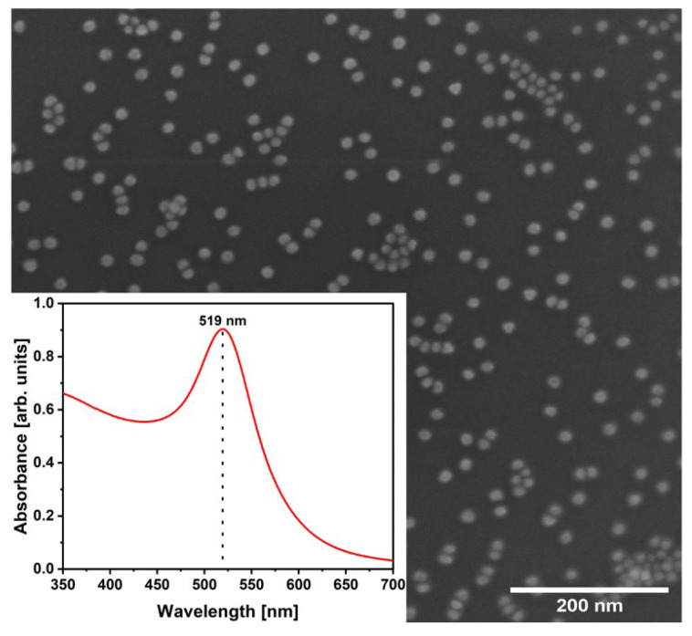
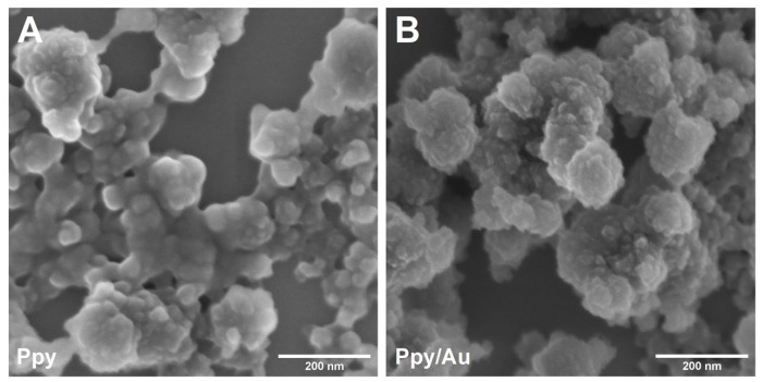
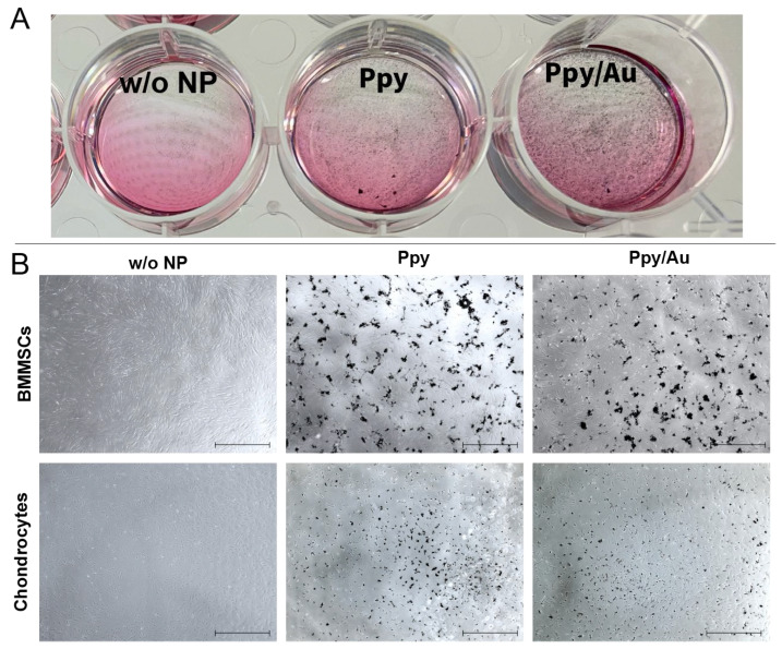
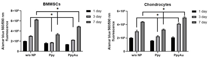
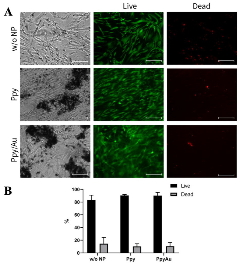
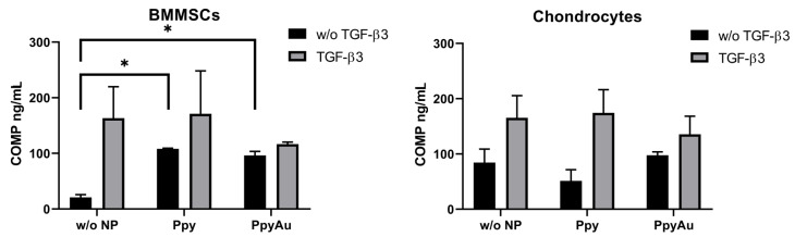
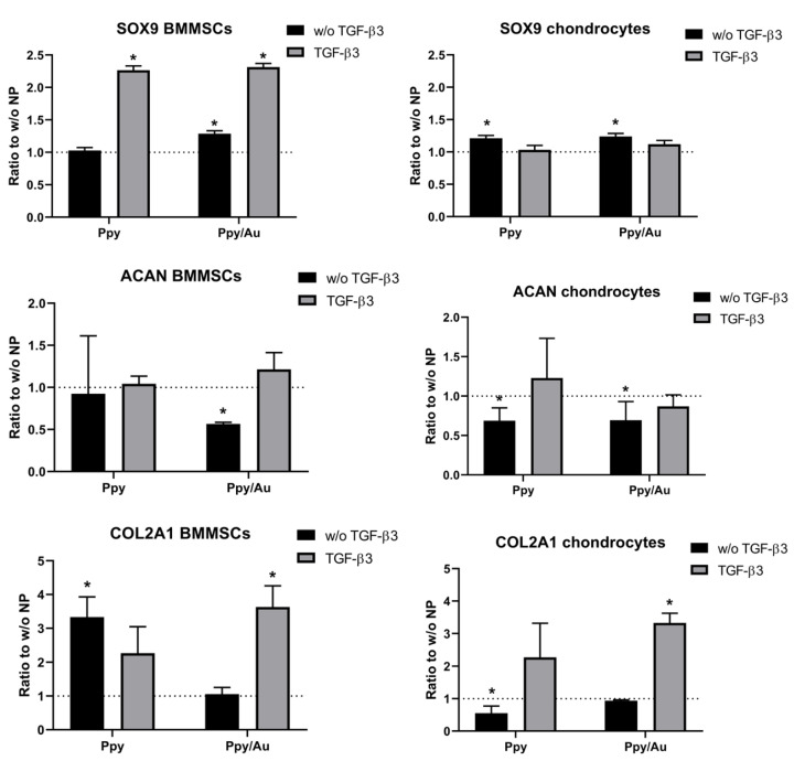
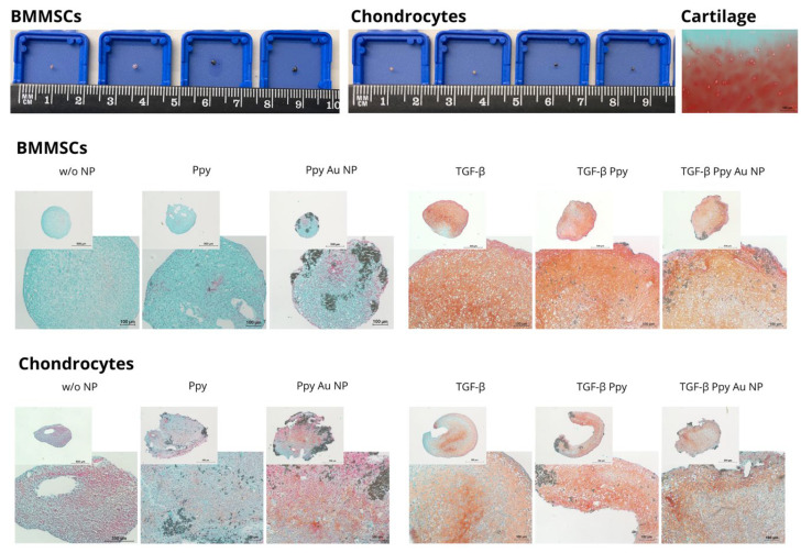
Bone marrow mesenchymal stem cells (BMMSCs) possess a strong ability to differentiate into the chondrogenic lineage, which is important for cartilage regeneration. External stimuli, such as electrical stimulation (ES), are frequently studied for chondrogenic differentiation of BMMSCs; however, the application of conductive polymers such as polypyrrole (Ppy), has never been used for stimulating BMMSCs chondrogenesis in vitro before. Thus, the aim of this study was to evaluate the chondrogenic potential of human BMMSCs after stimulation with Ppy nanoparticles (Ppy NPs) and compare them to cartilage-derived chondrocytes. In this study, we tested Ppy NPs without and with 13 nm gold NPs (Ppy/Au) for BMMSCs and chondrocyte proliferation, viability, and chondrogenic differentiation for 21 days, without the use of ES. The results demonstrated significantly higher amounts of cartilage oligomeric matrix protein (COMP) in BMMSCs stimulated with Ppy and Ppy/Au NPs, as compared to the control. The expression of chondrogenic genes (SOX9, ACAN, COL2A1) in BMMSCs and chondrocytes were upregulated by Ppy and Ppy/Au NPs, as compared to controls. Histological staining with safranin-O indicated higher extracellular matrix production in Ppy and Ppy/Au NPs stimulated samples, as compared to controls. In conclusion, Ppy and Ppy/Au NPs stimulate BMMSC chondrogenic differentiation; however, BMMSCs were more responsive to Ppy, while chondrocytes possessed a stronger chondrogenic response to Ppy/Au NPs.
Keywords: bone marrow mesenchymal stem cells; chondrocytes; chondrogenic differentiation; polypyrrole; polypyrrole/gold nanoparticles.
Conflict of interest statement
Authors declare no conflict of interest.
Figures
Similar articles
-
The Effects of Mechanical Load on Chondrogenic Responses of Bone Marrow Mesenchymal Stem Cells and Chondrocytes Encapsulated in Chondroitin Sulfate-Based Hydrogel.Int J Mol Sci. 2023 Feb 2;24(3):2915. doi: 10.3390/ijms24032915. Int J Mol Sci. 2023. PMID: 36769232 Free PMC article.
-
Different phenotypes and chondrogenic responses of human menstrual blood and bone marrow mesenchymal stem cells to activin A and TGF-β3.Stem Cell Res Ther. 2021 Apr 29;12(1):251. doi: 10.1186/s13287-021-02286-w. Stem Cell Res Ther. 2021. PMID: 33926568 Free PMC article.
-
Centrifugal gravity-induced BMP4 induces chondrogenic differentiation of adipose-derived stem cells via SOX9 upregulation.Stem Cell Res Ther. 2016 Dec 8;7(1):184. doi: 10.1186/s13287-016-0445-6. Stem Cell Res Ther. 2016. PMID: 27931264 Free PMC article.
-
Polypyrrole-based structures for activation of cellular functions under electrical stimulation.Bioelectrochemistry. 2024 Feb;155:108585. doi: 10.1016/j.bioelechem.2023.108585. Epub 2023 Oct 10. Bioelectrochemistry. 2024. PMID: 37847982 Review.
-
Electrical Stimulation in Cartilage Tissue Engineering.Bioengineering (Basel). 2023 Apr 7;10(4):454. doi: 10.3390/bioengineering10040454. Bioengineering (Basel). 2023. PMID: 37106641 Free PMC article. Review.
Cited by
-
Starlike Au nanoparticle unleashing siDDIT3 and photothermal power to combat ferroptosis - driven osteoarthritis.J Nanobiotechnology. 2025 Jul 5;23(1):487. doi: 10.1186/s12951-025-03563-z. J Nanobiotechnology. 2025. PMID: 40615894 Free PMC article.
References
-
- Kaur G., Adhikari R., Cass P., Bown M., Gunatillake P. Electrically conductive polymers and composites for biomedical applications. RSC Adv. 2015;5:37553–37567. doi: 10.1039/C5RA01851J. - DOI
-
- Borges M.H.R., Nagay B.E., Costa R.C., Souza J.G.S., Mathew M.T., Barão V.A.R. Recent advances of polypyrrole conducting polymer film for biomedical application: Toward a viable platform for cell-microbial interactions. Adv. Colloid Interface Sci. 2023;314:102860. doi: 10.1016/j.cis.2023.102860. - DOI - PubMed
Grants and funding
LinkOut - more resources
Full Text Sources
Research Materials
Miscellaneous
