. 2023 Jun 2;15(11):2610.
doi: 10.3390/nu15112610.

Collagen Peptide Exerts an Anti-Obesity Effect by Influencing the Firmicutes/Bacteroidetes Ratio in the Gut

Affiliations

Collagen Peptide Exerts an Anti-Obesity Effect by Influencing the Firmicutes/Bacteroidetes Ratio in the Gut

Ga Hyeon Baek et al. Nutrients. .

Abstract

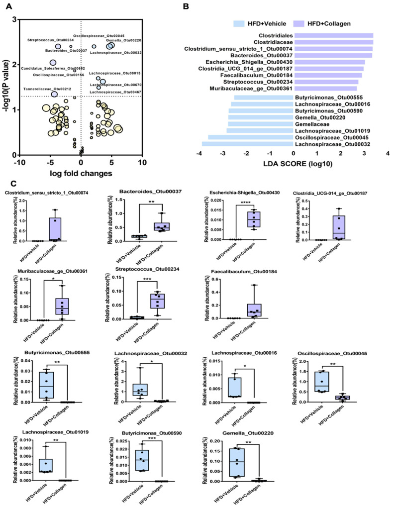
Alterations in the intestinal microbial flora are known to cause various diseases, and many people routinely consume probiotics or prebiotics to balance intestinal microorganisms and the growth of beneficial bacteria. In this study, we selected a peptide from fish (tilapia) skin that induces significant changes in the intestinal microflora of mice and reduces the Firmicutes/Bacteroidetes ratio, which is linked to obesity. We attempted to verify the anti-obesity effect of selected fish collagen peptides in a high-fat-diet-based obese mouse model. As anticipated, the collagen peptide co-administered with a high-fat diet significantly inhibited the increase in the Firmicutes/Bacteroidetes ratio. It increased specific bacterial taxa, including Clostridium_sensu_stricto_1, Faecalibaculum, Bacteroides, and Streptococcus, known for their anti-obesity effects. Consequently, alterations in the gut microbiota resulted in the activation of metabolic pathways, such as polysaccharide degradation and essential amino acid synthesis, which are associated with obesity inhibition. In addition, collagen peptide also effectively reduced all obesity signs caused by a high-fat diet, such as abdominal fat accumulation, high blood glucose levels, and weight gain. Ingestion of collagen peptides derived from fish skin induced significant changes in the intestinal microflora and is a potential auxiliary therapeutic agent to suppress the onset of obesity.

Keywords: Firmicutes/Bacteroidetes ratio; anti-obesity effect; collagen peptide; intestinal microbial flora; prebiotics.


Conflict of interest statement

The authors declare no competing financial interest.

Figures

Figure 1
Figure 1
Screening of peptides with a significant effect on the intestinal microflora. (A) The schematics of the animal study (C57BL/6) design. From days 0–9, the peptide solutions were administered to mice by oral gavage. On day 10, the mice were sacrificed, and the fecal matter was sampled. (B) The relative abundance of the gut microbiota at the phylum levels in six peptide-gavaged mice. The nine most abundant bacteria phyla were obtained from 30 mouse fecal samples from seven groups of mice. (CL; collagen, SA; soybean type A, SB; soybean type B, YA; yeast type A, YB; yeast type B, YC; yeast type C). (C) Firmicutes/Bacteroidetes (F/B) ratio. Unpaired t-tests (two-tailed) were used to analyze variations between the two groups. * p < 0.05. (D) Beta diversity. The PCoA was based on the Jaccard distance, and the principal coordinate with the largest contribution rate was selected for graphical display. The two groups were completely separated. The statistical analyses were performed using AMOVA. (EH) Alpha diversity. Sobs, Shannon, Invsimpson, and Chao indices reflect the differences in richness and evenness among samples.
Figure 2
Figure 2
The anti-obesity effects of collagen peptide screened by gut microbiome analysis. (A) The schematics of the animal study (C57BL/6) design (High-fat diet model). We administered collagen peptide in the HFD mouse model by oral gavage for three weeks and monitored the body weight and food consumption. After three weeks, we sacrificed the mice and sampled the fecal matter. (B) Daily weight of the HFD mouse model in the presence or absence of collagen peptide. The HFD mice administered collagen peptides daily gained less weight than mice fed with HFD alone. Unpaired t-tests (two-tailed) were used to analyze variations between the two groups. ** p < 0.01. (C) The relative abundance of the gut microbiota at the phylum level in RD, HFD + vehicle, and HFD + collagen mice. The 10 most abundant bacterial phyla were obtained from 17 mouse fecal samples from three groups of mice. (D) Firmicutes/Bacteroidetes (F/B) ratio. The one-way ANOVA test was used to analyze variation between the three groups. Different letters indicate statistical significance. (E) Beta diversity. PCoA was based on the Jaccard distance, and the principal coordinate with the largest contribution rate was selected for graphical display. The three groups were completely separated. Statistics analyses were performed using AMOVA. (F) Abdominal body fat comparison in the three groups. The decrease in abdominal body fat of HFD mouse model in the presence of collagen peptide was evident. (GJ) The results of the blood-biochemical analysis. Cholesterol or glucose levels were increased significantly in the mice fed with HFD for three weeks compared with the RD mice. Unpaired t-tests (two-tailed) were used to analyze variations between the two groups. * p < 0.05, ** p < 0.01, *** p < 0.001.
Figure 3
Figure 3
The difference of OTU in two groups (HFD + vehicle and HFD + collagen). (A) Log fold changes. The log2 (vehicle/collagen) is the ratio of the relative abundance of OTUs between the two groups. As the size increases, it means that the relative abundance of the corresponding OTU is high. Fourteen distinguishing taxa (OTUs) were significantly marked among 50 differential OTUs between the two groups. (B) Differentially abundant bacterial taxa in fecal samples from mice models (HFD + vehicle and HFD + collagen). A bar plot showing the LDA score (effect size) of differentially abundant OTUs in the HFD + vehicle (blue, n = 6) and HFD + collagen (purple, n = 6) groups as determined using the Linear Discriminant Effect Size (LEfSe) analysis (α = 0.05, LDA score > 2.0). (C) A box plot comparing the relative abundance of OTUs between the two groups. Unpaired t-tests (two-tailed) were used to analyze variation between the two groups. * p < 0.05, ** p < 0.01, *** p < 0.001, and **** p < 0.0001.
Figure 4
Figure 4
The predicted functional potential changes between HFD + vehicle mice and HFD + collagen mice by using PICRUST2. (A) The boxplots showed the relative abundance in 27 pathways related to obesity. There were 17 significantly increased pathways and 10 decreased pathways in HFD + collagen compared to HFD + vehicle. (B) The PCA analysis of intestinal bacterial metabolic pathway, the orange color indicates HFD + vehicle mice and the green color indicates HFD + collagen mice. (C) The extended error bar chart showed the significant difference in the predicted functional pathways related to obesity between the two groups. The middle value represents the mean differences between the two groups (upper-lower bar value), and the error bar represents the 95% confidence intervals with the effect size of difference in mean proportion. The p-value at the side indicates the significance between the upper and lower bars. (D) Heatmaps were drawn with normalized relative abundances in 27 obesity-related pathways in the two groups. The red colors represent a higher abundance and blue colors a lower abundance. * p < 0.05, ** p < 0.01, *** p < 0.001, and **** p < 0.0001.
Figure 5
Figure 5
The inhibitory effect of collagen peptide on HFD-derived endotoxin excretion in the gut. (A) Endotoxin levels in the sterilized fecal solution were measured by Limulus amebocyte lysate (LAL) assay. (B) Murine macrophage cell line, Raw264.7 cells, were incubated with a sterilized fecal solution for 24 h, and then pro-inflammatory cytokine levels in culture media were measured by ELISA. (A,B) Unpaired t-tests (two-tailed) were used to analyze the variation between the two groups. * p < 0.05, ** p < 0.01, *** p < 0.001.

References

    1. Qin J., Li R., Raes J., Arumugam M., Burgdorf K.S., Manichanh C., Nielsen T., Pons N., Levenez F., Yamada T., et al. A human gut microbial gene catalogue established by metagenomic sequencing. Nature. 2010;464:59–65. doi: 10.1038/nature08821. - DOI - PMC - PubMed
    1. Sokol H., Pigneur B., Watterlot L., Lakhdari O., Bermudez-Humaran L.G., Gratadoux J.J., Blugeon S., Bridonneau C., Furet J.P., Corthier G., et al. Faecalibacterium prausnitzii is an anti-inflammatory commensal bacterium identified by gut microbiota analysis of Crohn disease patients. Proc. Natl. Acad. Sci. USA. 2008;105:16731–16736. doi: 10.1073/pnas.0804812105. - DOI - PMC - PubMed
    1. Schirmer M., Franzosa E.A., Lloyd-Price J., McIver L.J., Schwager R., Poon T.W., Ananthakrishnan A.N., Andrews E., Barron G., Lake K., et al. Dynamics of metatranscription in the inflammatory bowel disease gut microbiome. Nat. Microbiol. 2018;3:337–346. doi: 10.1038/s41564-017-0089-z. - DOI - PMC - PubMed
    1. Mar J.S., LaMere B.J., Lin D.L., Levan S., Nazareth M., Mahadevan U., Lynch S.V. Disease Severity and Immune Activity Relate to Distinct Interkingdom Gut Microbiome States in Ethnically Distinct Ulcerative Colitis Patients. mBio. 2016;7:e01072-16. doi: 10.1128/mBio.01072-16. - DOI - PMC - PubMed
    1. Kudelka M.R., Hinrichs B.H., Darby T., Moreno C.S., Nishio H., Cutler C.E., Wang J., Wu H., Zeng J., Wang Y., et al. Cosmc is an X-linked inflammatory bowel disease risk gene that spatially regulates gut microbiota and contributes to sex-specific risk. Proc. Natl. Acad. Sci. USA. 2016;113:14787–14792. doi: 10.1073/pnas.1612158114. - DOI - PMC - PubMed