. 2023 Dec;52(12):2513-2518.
doi: 10.1007/s00256-023-04383-8. Epub 2023 Jun 10.

(Smoldering) multiple myeloma: mismatch between tumor load estimated from bone marrow biopsy at iliac crest and tumor load shown by MRI

Affiliations

(Smoldering) multiple myeloma: mismatch between tumor load estimated from bone marrow biopsy at iliac crest and tumor load shown by MRI

Fabian Bauer et al. Skeletal Radiol. 2023 Dec.

Abstract

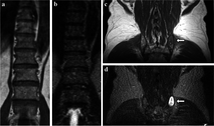
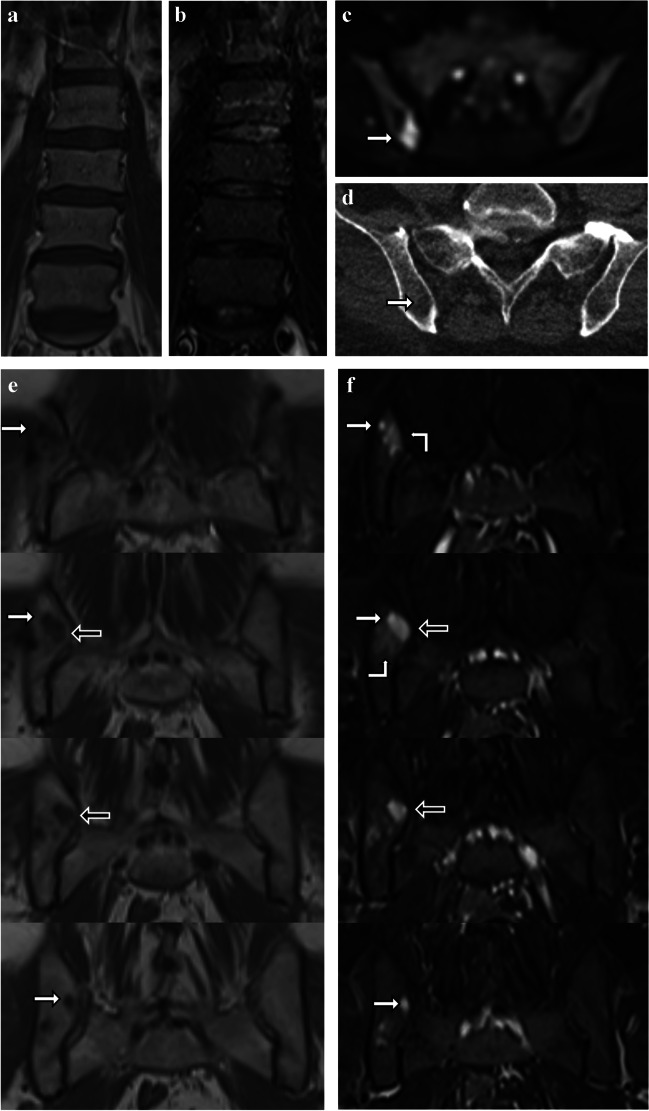
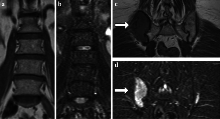
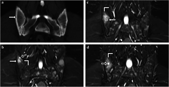
In multiple myeloma and its precursor stages, precise quantification of tumor load is of high importance for diagnosis, risk assessment, and therapy response evaluation. Both whole-body MRI, which allows to investigate the complete bone marrow of a patient, and bone marrow biopsy, which is commonly used to assess the histologic and genetic status, are relevant methods for tumor load assessment in multiple myeloma. We report on a series of striking mismatches between the plasma cell infiltration estimating the tumor load from unguided biopsies of the bone marrow at the posterior iliac crest and the tumor load assessment from whole-body MRI.

Keywords: Bone marrow biopsy; Magnetic resonance imaging; Monoclonal plasma cell disorders; Multiple myeloma; Tumor load.


Conflict of interest statement

Sandra Sauer: travel grants or honoraria for presentations for Celgene, BMS, Janssen, Takeda and Amgen. All other authors declare that they have no conflict of interest.

Figures

Fig. 1
Fig. 1
a–d Case 1. A 62-year-old male with smoldering multiple myeloma after coincidental biopsy of a large focal lesion. Coronal T1tse (a, c) and STIR (b, d) MR images show low to intermediate diffuse infiltration in spine (a, b) and pelvis (c, d). However, a biopsy-based PCI of 90% was reported for the bone marrow. This wb-MRI reveals that a focal lesion at the left posterior iliac bone was coincidentally hit in the biopsy (c, d, white arrows)
Fig. 2
Fig. 2
a–f Case 2. A 62-year-old male with multiple myeloma after coincidental biopsy of a small focal lesion. a, b Coronal T1tse (a) and STIR (b) MR images show physiologic signal of the bone marrow in the spine and pelvis. c, d The focal lesion shows an increased signal in b800 diffusion weighed imaging (c) and an ill-defined, trabecular rarefication in CT imaging (d). e, f Four consecutive coronal T1tse (e) and STIR (f) MR images from dorsal (upper MR images) to ventral show a small circular area (white arrows) with marked T1-hypointense and marked STIR-hyperintense signal corresponding to the biopsy channel. The biopsy channel (black arrows) is passing through a spherical T1-hypointense and STIR-hyperintense focal lesion of 1.2 cm by 0.8 cm. A diffuse, ill-defined, moderate STIR-hyperintensity, which is most likely corresponding to a bone marrow edema caused by biopsy and aspiration, is located next to the focal lesion and the biopsy channel (f, right angle arrows)
Fig. 3
Fig. 3
a–d Case 3. A 60-year-old female with multiple myeloma with 5% PCI after biopsy of mostly unaffected bone marrow in the posterior iliac crest contralateral to a large focal lesion. Coronal T1tse (a, c) and STIR (b, d) MR images show no diffuse infiltration in spine (a, b) or the left pelvis (c, d). Wb-MRI revealed a 6.8 cm by 3.0 cm large focal lesion at the right posterior iliac crest (c, d, white arrows), which would have led to a significantly higher PCI assessment if the right side instead of the left side would have been chosen randomly for biopsy
Fig. 4
Fig. 4
a–d Case 4. A 51-year-old male with multiple myeloma presenting with a marked discrepancy between bone marrow trephine and bone marrow aspirate results. a Coronal CT image of a scan performed one month prior to the wb-MRI scan showing a 1.0 cm by 0.7 cm osteolytic lesion in the right posterior iliac bone, which is the typical location for an unguided bone marrow biopsy (white arrows). b-d Three consecutive coronal STIR MR images from posterior to anterior of the wb-MRI show a small circular area with increased signal in STIR corresponding to the biopsy channel (b, d, black arrows) and a 1.1 cm by 0.9 cm focal lesion with a STIR-hyperintense signal in the corresponding location to the osteolytic lesion (b, c, white arrow). Bone marrow edema with intermediate STIR-hyperintensity surrounding the biopsy channel can be detected (right angle arrow)

References

    1. Rajkumar SV, Dimopoulos MA, Palumbo A, Blade J, Merlini G, Mateos MV, et al. International Myeloma Working Group updated criteria for the diagnosis of multiple myeloma. Lancet Oncol. 2014;15(12):e538–e548. doi: 10.1016/S1470-2045(14)70442-5. - DOI - PubMed
    1. Dimopoulos MA, Hillengass J, Usmani S, Zamagni E, Lentzsch S, Davies FE, et al. Role of magnetic resonance imaging in the management of patients with multiple myeloma: a consensus statement. J Clin Oncol. 2015;33(6):657–664. doi: 10.1200/JCO.2014.57.9961. - DOI - PubMed
    1. Hillengass J, Usmani S, Rajkumar SV, Durie BGM, Mateos MV, Lonial S, et al. International myeloma working group consensus recommendations on imaging in monoclonal plasma cell disorders. Lancet Oncol. 2019;20(6):e302–e312. doi: 10.1016/S1470-2045(19)30309-2. - DOI - PubMed
    1. Greipp PR, San Miguel J, Durie BG, Crowley JJ, Barlogie B, Blade J, et al. International staging system for multiple myeloma. J Clin Oncol. 2005;23(15):3412–3420. doi: 10.1200/JCO.2005.04.242. - DOI - PubMed
    1. Palumbo A, Avet-Loiseau H, Oliva S, Lokhorst HM, Goldschmidt H, Rosinol L, et al. Revised international staging system for multiple myeloma: a report from International Myeloma Working Group. J Clin Oncol. 2015;33(26):2863–2869. doi: 10.1200/JCO.2015.61.2267. - DOI - PMC - PubMed