. 2023:2683:135-151.
doi: 10.1007/978-1-0716-3287-1_11.

Modeling the Blood-Brain Barrier Using Human-Induced Pluripotent Stem Cells

Affiliations

Modeling the Blood-Brain Barrier Using Human-Induced Pluripotent Stem Cells

Louise A Mesentier-Louro et al. Methods Mol Biol. 2023.

Abstract

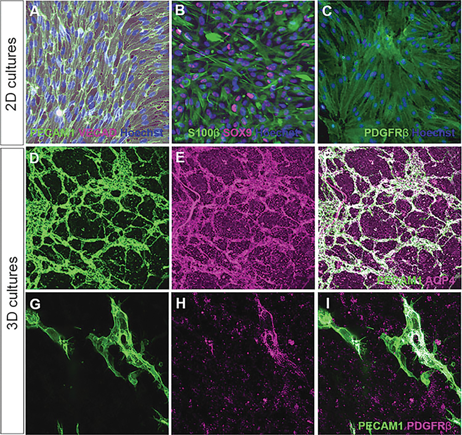
The blood-brain barrier (BBB) is a key physiological component of the brain, protecting the brain from peripheral processes and pathogens. The BBB is a dynamic structure that is heavily involved in cerebral blood flow, angiogenesis, and other neural functions. However, the BBB also creates a challenging barrier for the entry of therapeutics into the brain, blocking more than 98% of drugs from contact with the brain. Neurovascular comorbidities are common in several neurological diseases including Alzheimer's and Parkinson's Disease, suggesting that BBB dysfunction or break down likely has a causal role in neurodegeneration. However, the mechanisms by which the human BBB is formed, maintained, and degenerated in diseases remain largely unknown due to limited access to human BBB tissue. To address these limitations, we have developed an in vitro induced human BBB (iBBB) derived from pluripotent stem cells. The iBBB model can be used for discovery of disease mechanisms, drug targets, drug screening, and medicinal chemistry studies to optimize brain penetration of central nervous system therapeutics. In this chapter, we will explain the steps to differentiate the three cellular components (endothelial cells, pericytes, and astrocytes) from induced pluripotent stem cells, and how to assemble them into the iBBB.

Keywords: Blood-brain barrier; Endothelial cells; Induced pluripotent stem cells; Permeability; Transwell; Vascularization.


Figures

Fig. 1
Fig. 1
Differentiation schemes vascular cell types: Schematic representation of differentiation of induced pluripotent stem cells into astrocytes via induction of (a) neural progenitor cells (NPC), (b) endothelial cells, and (c) pericytes. Details of medium composition can be found in Subheading 2.2. Of note, for endothelial cell differentiation using ETV2 induction, the induction antibiotic (e.g., doxycycline) must be added from differentiation day 0 onwards. Created with BioRender.com
Fig. 2
Fig. 2
In vitro blood-brain barrier models: schematic representations of in vitro blood-brain barrier (BBB) models. (a) After differentiating induced pluripotent stem cells (iPSCs) to endothelial cells, pericytes, and astrocytes, cell types can be combined in two models mimicking the BBB. (b) Endothelial cells can be seeded onto Geltrex-coated transwell membrane cell culture inserts, followed by seeding of astrocytes and pericytes 24 h later to form a barrier. (c) Alternatively, cells can be combined in a 5:1:1 endothelial cell to pericytes to astrocytes ratio in Geltrex to form the iBBB model. (d) Transendothelial electrical resistance (TEER) can be read using chopstick electrodes measuring the current flow impedance over the barrier. (e) Additionally, transwell membrane cell culture inserts allow for the investigation of barrier function through the measurement of FITC-labeled dextran through the barrier. Created with BioRender.com
Fig. 3
Fig. 3
Expression of cell-specific markers in 2D and 3D cultures: Immunofluorescence for cell-specific markers of (a) endothelial cells, (b) astrocytes, (c) and pericytes in 2D cultures. After assembly of 3D cultures, a vascular network composed of (d) PECAM1+ endothelial cells, (e) Aquaporin-4+ astrocyte endfeet, and (h) PDGFRβ+ pericytes is observed

References

    1. Persidsky Y, Ramirez SH, Haorah J et al. (2006) Blood-brain barrier: structural components and function under physiologic and pathologic conditions. J Neuroimmune Pharmacol 1:223–236 - PubMed
    1. Pardridge WM (2012) Drug transport across the blood-brain barrier. J Cereb Blood Flow Metab 32:1959–1972 - PMC - PubMed
    1. Sweeney MD, Kisler K, Montagne A et al. (2018) The role of brain vasculature in neurodegenerative disorders. Nat Neurosci 21:1318–1331 - PMC - PubMed
    1. Kadry H, Noorani B, Cucullo L (2020) A blood-brain barrier overview on structure, function, impairment, and biomarkers of integrity. Fluids Barriers CNS 17:69. - PMC - PubMed
    1. Stamatovic SM, Johnson AM, Keep RF et al. (2016) Junctional proteins of the blood-brain barrier: new insights into function and dysfunction. Tissue Barriers 4:e1154641. - PMC - PubMed

Publication types

LinkOut - more resources