. 2023 Jun 27;42(6):112630.
doi: 10.1016/j.celrep.2023.112630. Epub 2023 May 30.

Extrafollicular IgD-CD27-CXCR5-CD11c- DN3 B cells infiltrate inflamed tissues in autoimmune fibrosis and in severe COVID-19

Affiliations

Extrafollicular IgD-CD27-CXCR5-CD11c- DN3 B cells infiltrate inflamed tissues in autoimmune fibrosis and in severe COVID-19

Hugues Allard-Chamard et al. Cell Rep. .

Abstract

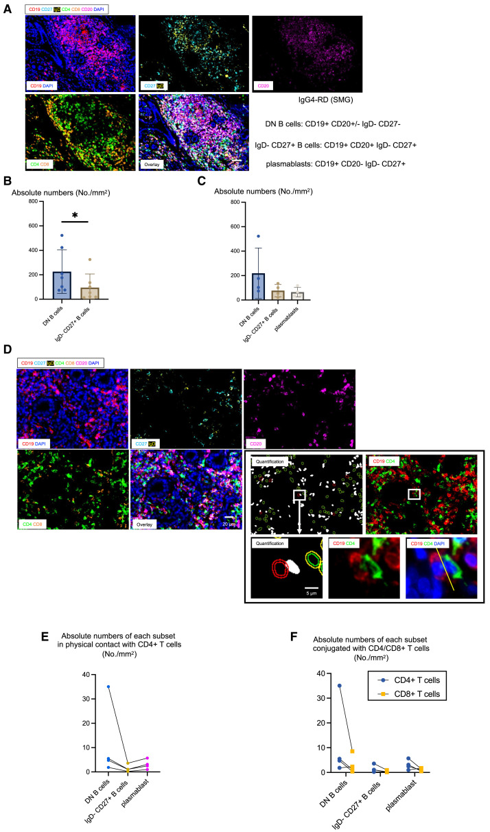
Although therapeutic B cell depletion dramatically resolves inflammation in many diseases in which antibodies appear not to play a central role, distinct extrafollicular pathogenic B cell subsets that accumulate in disease lesions have hitherto not been identified. The circulating immunoglobulin D (IgD)-CD27-CXCR5-CD11c+ DN2 B cell subset has been previously studied in some autoimmune diseases. A distinct IgD-CD27-CXCR5-CD11c- DN3 B cell subset accumulates in the blood both in IgG4-related disease, an autoimmune disease in which inflammation and fibrosis can be reversed by B cell depletion, and in severe COVID-19. These DN3 B cells prominently accumulate in the end organs of IgG4-related disease and in lung lesions in COVID-19, and double-negative B cells prominently cluster with CD4+ T cells in these lesions. Extrafollicular DN3 B cells may participate in tissue inflammation and fibrosis in autoimmune fibrotic diseases, as well as in COVID-19.

Keywords: B cell depletion in disease; COVID-19 pathogenesis; CP: Immunology; DN3 B cells; IgG4-related disease; T-B collaboration in disease; double-negative B cells; extrafollicular B cells; inflammatory fibrosis.


Conflict of interest statement

Declaration of interests S.P. is on the scientific advisory boards of Abpro, Inc., BeBiopharma, Inc., Paratus Biosciences, and Octagon Therapeutics.

Figures

None
Graphical abstract
Figure 1
Figure 1
Several populations of circulating activated B cells distinguish patients with IgG4-RD from healthy controls (A) Multiparametric analysis of circulating B cells using tSNE with overlay of the relative expression of key markers. (B) Gating strategy to examine B cell subsets, segregating plasmablasts and then categorizing IgDCD27 B cells into DN1, DN2, DN3, and DN4 subsets. (C) Patients with IgG4-RD (n = 5) exhibited four distinctive populations of DN (IgDCD27) B cells distinct from those in patients living with HIV, which are FcRL4+ (n = 5). (D) DN B cells from IgG4-RD (n = 38) are different from the DN B cells described in lupus (n = 5). (E) Frequencies of different B cell populations and of SWM B cells, ABC-like B cells, and activated naive B cells in IgG4-RD and controls (n = 38). Multiple comparisons are controlled for by Kruskal-Wallis test. Error bars represent mean ± SEM. p < 0.05; ∗∗p < 0.01; ∗∗∗p < 0.001; ∗∗∗∗p < 0.0001.
Figure 2
Figure 2
Circulating DN B cell populations are enriched in IgG4-RD and COVID-19 (A) Frequencies of circulating B cell subpopulations in patients with IgG4-RD compared with healthy volunteers. (B) DN B cell subsets correlated with plasmablast frequencies. The strongest correlation being with DN3 and DN2 B cells (r = 0.66, r = 0.45) suggesting co-occurrence of these B cell in IgG4-RD. (C) Frequency of DN B cell subsets in controls and moderate, severe, and convalescent (conval) COVID-19 patients from an MGH cohort. The data were extracted from the analyses in Kaneko et al. Two-tailed Mann-Whitney U test was used for pairwise comparisons, and multiple comparisons were controlled for by Kruskal-Wallis test. Error bars represent mean ± SEM. p < 0.05; ∗∗p < 0.01; ∗∗∗p < 0.001; ∗∗∗∗p < 0.0001. (D) Using a logistic regression model and the maximum likelihood method, we confirmed that plasmablasts discriminate between patients with IgG4-RD and healthy controls. We additionally showed that total DN B cells as well as DN2, DN3, and DN4 B cells can independently distinguish IgG4-RD. Significance was tested using the Wald test.
Figure 3
Figure 3
T cell-B cell conjugates in IgG4-RD end organs (A) Immunofluorescence staining of CD19 (red), CD27 (cyan), IgD (yellow), CD20 (purple), CD4 (green), CD8 (orange), and DAPI (blue) in a submandibular gland (SMG) from a patient with IgG4-RD. (B) Absolute numbers and percentages of IgDCD27 DN B cells and IgDCD27+ B cells in IgG4-RD (n = 7). This combines data for which a representative image is shown in Figure S1 and from (A). (C) Absolute numbers of DN B cells (blue), IgDCD27+ B cells (gold), and plasmablasts (gray) in IgG4-RD (n = 4). (D) Immunofluorescence staining of CD19 (red), CD27 (cyan), IgD (yellow), CD20 (purple), CD4 (green), CD8 (orange), and DAPI (blue) and visualization of physically interacting T and B cells in an SMG from a patient with IgG4-RD using the StrataQuest cell-to-cell contact application. Nuclei circled in red depict CD19+ B cell in physical contact with CD4+ T cells. T cells and B cells formed close and extensive intercellular plasma membrane contacts. (E) Absolute numbers of DN B cells (blue), SWM B cells (yellow), and plasmablasts (purple) in physical contact with CD4+ T cells in patients with IgG4-RD (n = 4). (F) Absolute numbers of DN B cells, SWM B cells, and plasmablasts conjugated with CD4+ T cells (blue) and CD8+ T cells (yellow) in patients with IgG4-RD (n = 4).
Figure 4
Figure 4
Comparison of transcriptomes of distinct circulating DN B cell subsets (A) The 4 subsets of DN B cells, isolated from the peripheral blood, of IgG4-RD are transcriptomically distinct and harbor unique signatures that suggest different, non-overlapping biological properties of each population. (B) A list of the representative Gene Ontology annotations that were examined. (C and D) The DN3 subset of DN B cells exhibits signatures of proliferating cells and the unfolded protein response seen in antibody-secreting cells.
Figure 5
Figure 5
DN B cells in draining lymph nodes and in the lungs of COVID-19 patients (A) Representative multicolor immunofluorescence images of CD19 (red), IgD/CD27 (yellow), CXCR5 (green), CD11c (purple), and DAPI (blue) staining in a lymph node from a late COVID-19 patient. Arrows indicate DN B cells. (B) Relative proportions of DN B cells in the pool of CD19+ B cells in lymph nodes from late COVID-19 patients (n = 6). (C) Absolute numbers of each subset of DN B cells in late COVID-19 patients (n = 6) (DN1, blue; DN2, gold; DN3, gray; DN4, cyan). (D) Representative multicolor immunofluorescence images of CD19 (red), IgD/CD27 (yellow), CXCR5 (green), CD11c (purple), and DAPI (blue) staining in a lung from a late COVID-19 patient. Arrows indicate DN B cells. (E) Relative proportions (left) and absolute numbers (right) of DN B cells in the pool of CD19+ B cells in lungs from late COVID-19 (blue) (n = 6) and non-COVID-19 patients (gold) (n = 6). (F) Absolute numbers of each subset of DN B cells in late COVID-19 patients (blue) (n = 6) and non-COVID-19 patients (gold) (n = 6). (G) Relative proportions of each DN B cells in the pool of DN B cells in lungs from late COVID-19 patients (n = 6). Mann-Whitney U test used to calculate p value. Error bars represent mean ± SEM. p < 0.05.
Figure 6
Figure 6
DN B cells infiltrate the SMGs in IgG4-related disease (A) Representative multicolor immunofluorescence images of CD19 (red), IgD (orange), CD27 (green), and DAPI (blue) staining in an SMG from a patient with IgG4-RD. Arrows indicate DN B cells. (B) Relative proportions (left) and absolute numbers (right) of DN B cells in the pool of CD19+ B cells in SMGs from IgG4-RD (blue) (n = 10) and chronic sialadenitis (CS) (gold) (n = 7). (C) Representative multicolor immunofluorescence images of CD19 (red), IgD/CD27 (yellow), CD11c (purple), CXCR5 (green), and DAPI (blue) staining in an SMG from a patient with IgG4-RD (DN1, top left; DN2, top right; DN3, bottom left; DN4, bottom right). (D) Absolute numbers of each subset of DN B cells in IgG4-RD (n = 10) (blue) and CS (gold) (n = 7). (E) Relative proportions of each DN B cell in the pool of DN B cells in SMGs from IgG4-RD (blue) (n = 10) and CS (gold) (n = 7). Mann-Whitney U test used to calculate p value. Error bars represent mean ± SEM. p < 0.05; ∗∗p < 0.01.

References

    1. Jenks S.A., Cashman K.S., Woodruff M.C., Lee F.E.H., Sanz I. Extrafollicular responses in humans and SLE. Immunol. Rev. 2019;288:136–148. doi: 10.1111/imr.12741. - DOI - PMC - PubMed
    1. Woodruff M.C., Ramonell R.P., Nguyen D.C., Cashman K.S., Saini A.S., Haddad N.S., Ley A.M., Kyu S., Howell J.C., Ozturk T., et al. Extrafollicular B cell responses correlate with neutralizing antibodies and morbidity in COVID-19. Nat. Immunol. 2020;21:1506–1516. doi: 10.1038/s41590-020-00814-z. - DOI - PMC - PubMed
    1. Kaneko N., Kuo H.H., Boucau J., Farmer J.R., Allard-Chamard H., Mahajan V.S., Piechocka-Trocha A., Lefteri K., Osborn M., Bals J., et al. Loss of Bcl-6-expressing T follicular helper cells and germinal centers in COVID-19. Cell. 2020;183:143–157.e13. doi: 10.1016/j.cell.2020.08.025. - DOI - PMC - PubMed
    1. Khosroshahi A., Bloch D.B., Deshpande V., Stone J.H. Rituximab therapy leads to rapid decline of serum IgG4 levels and prompt clinical improvement in IgG4-related systemic disease. Arthritis Rheum. 2010;62:1755–1762. doi: 10.1002/art.27435. - DOI - PubMed
    1. Mattoo H., Mahajan V.S., Della-Torre E., Sekigami Y., Carruthers M., Wallace Z.S., Deshpande V., Stone J.H., Pillai S. De novo oligoclonal expansions of circulating plasmablasts in active and relapsing IgG4-related disease. J. Allergy Clin. Immunol. 2014;134:679–687. doi: 10.1016/j.jaci.2014.03.034. - DOI - PMC - PubMed

Publication types