Review
. 2023 Aug:248:108466.
doi: 10.1016/j.pharmthera.2023.108466. Epub 2023 Jun 8.

Noncoding RNA circuitry in melanoma onset, plasticity, and therapeutic response

Affiliations
Review

Noncoding RNA circuitry in melanoma onset, plasticity, and therapeutic response

Katerina Grafanaki et al. Pharmacol Ther. 2023 Aug.

Abstract

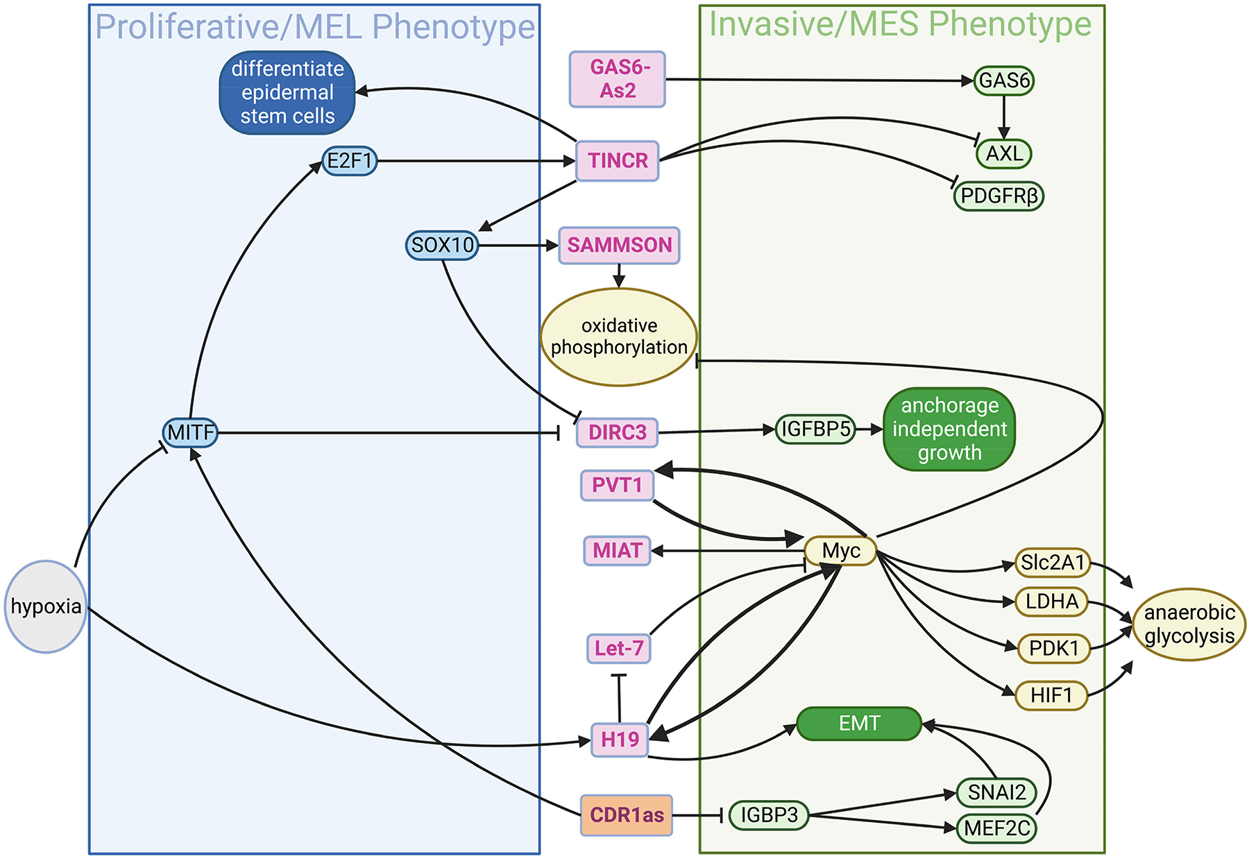
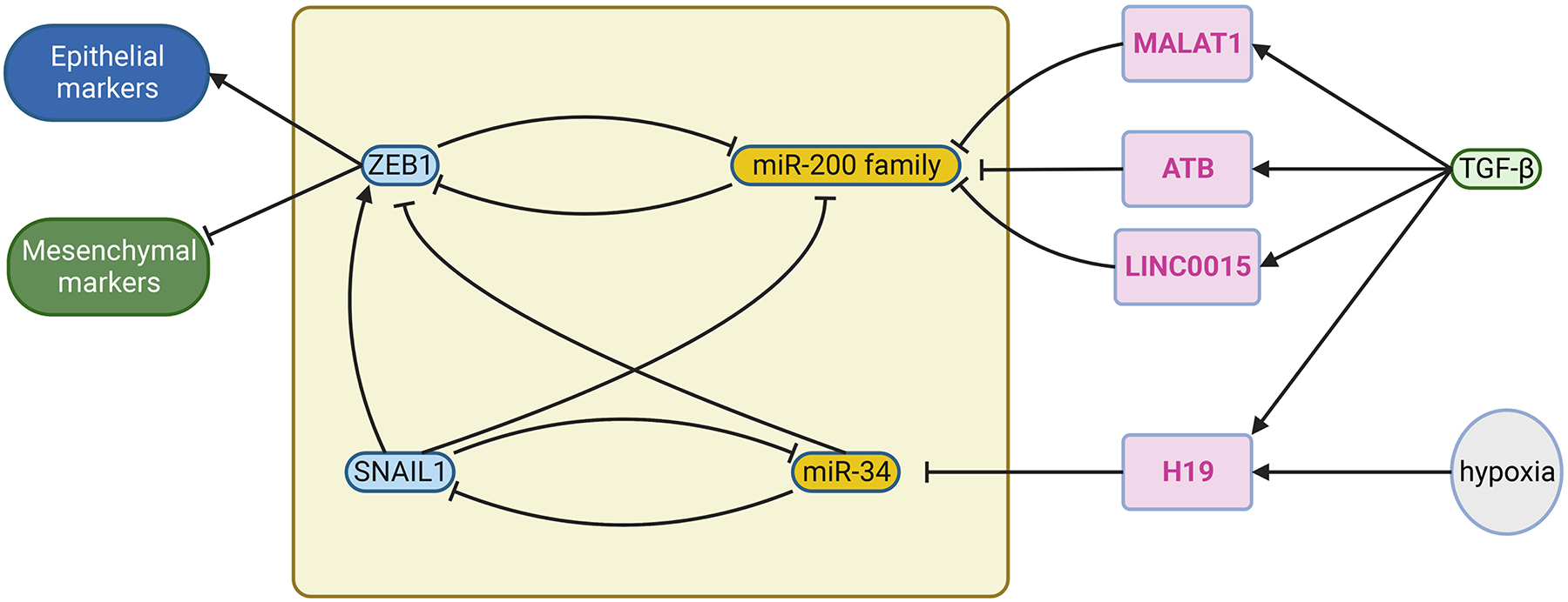
Melanoma, the cancer of the melanocyte, is the deadliest form of skin cancer with an aggressive nature, propensity to metastasize and tendency to resist therapeutic intervention. Studies have identified that the re-emergence of developmental pathways in melanoma contributes to melanoma onset, plasticity, and therapeutic response. Notably, it is well known that noncoding RNAs play a critical role in the development and stress response of tissues. In this review, we focus on the noncoding RNAs, including microRNAs, long non-coding RNAs, circular RNAs, and other small RNAs, for their functions in developmental mechanisms and plasticity, which drive onset, progression, therapeutic response and resistance in melanoma. Going forward, elucidation of noncoding RNA-mediated mechanisms may provide insights that accelerate development of novel melanoma therapies.

Keywords: Circular RNAs (circRNAs); Developmental state; Lineage; Long non-coding RNAs (lncRNAs); Melanoma; Non-coding RNAs (ncRNAs); Plasticity; Translational plasticity; microRNAs (miRNAs).


Conflict of interest statement

Declaration of Competing Interest The authors declare that they have no known competing financial interests or personal relationships that could have appeared to influence the work reported in this paper.

Figures

Figure 1.
Figure 1.
Comparison of NC differentiation and melanoma development on a Waddington-style landscape. Left panel, NC differentiation. NC stem cells (pre-EMT NC) are derived from neural tube. They delaminate, migrate (migrating progenitors), and start specification. Upon reaching the destination, they become committed cell types (autonomic neurons, mesenchymal cells, glial cells, sensory neurons, melanocytes). Each cell type is a stable transcriptional state against perturbation (“valley”) unless receiving specific signals. Right panel, melanoma development. Melanoma is initiated by the stem-like cells that correspond to pre-EMT NC state. As expanding, they differentiate to NC-like cells, mesenchymal-like cells, and melanocytic cells (corresponding to migrating NC progenitors, mesenchymal cells, and melanocytes in the NC differentiation). Like NC development landscape, each differentiated type represents a transcriptional state. However, in contrast to the NC landscape, the plasticity of melanoma cells allows them to switch between transcriptional state (dashed arrows). This can be mediated by genetic circuits that involve noncoding RNAs; see Figure 2 to 5. The left panel is adopted from Marie et al (Marie et al., 2022).
Figure 2.
Figure 2.
Examples of microRNAs and transcription factors forming a dynamic control circuit for switching between transcriptional states. a, ZEB1 activates and suppresses the expression of mesenchymal and epithelial marker genes, respectively. It and miR-200 family reciprocally inhibit each other, constituting a negative feedback loop (upper panel). When the input signal (TGF-β) reaches over a threshold level, the system will switch from epithelial-high to mesenchymal-high state (lower panel; adopted from Stallaert et al (Stallaert et al., 2019)). Solid and dashed curve, stable and unstable state, respectively. This figure is adopted from b, Incorporation of another negative feedback loop (SNAIL1 and miR-34) to ZEB1/miR-200 family circuit (upper panel) results in an additional state (EMT, lower panel; adopted from Zhang et al (Zhang et al., 2014)). As the input signal (TGF-β) keeps increasing, the system will switch from epithelial-high to EMT, and then to mesenchymal-high state (lower panel). c, An example of the collaborative behavior of miRNA. A gene can be targeted by multiple miRNA (e.g. VIM1, MAF, and CXCL12 are targeted by both miR-200c-3p and miR-141–3p), but each miRNA can target multiple genes in which some of them might not be shared with other miRNAs (e.g. miR-200–3p targeting VIM1 and FSTL1) (Guo et al., 2014). Part of the figure is generated using birender.com (2022).
Figure 3.
Figure 3.
The miRNA-protein factor circuit for control the transcriptional states and mediate plasticity in melanoma. In melanoma cells, similar to Fig. 2A and B, miRNAs and protein factors form a negative-feedback circuit for switching between proliferative/MEL and invasive/MES states, putatively in response to input signal such inflammation. miRNAs can be activated from their own promoters by transcriptional factors via input signals (e.g. NFkB and inflammatory signal, respectively), or expressed from the introns of the coding genes (e.g. TRPM1, SLIT1, SLIT3) during their transcription. The function of miRNAs is to suppress expression of coding genes. MEL and MES, melanocytic and mesenchymal phenotype, respectively. Part of the figure is generated using birender.com (2022).
Figure 4.
Figure 4.
Summary of actions of other small RNAs involved in the control of plasticity in melanoma. Part of the figure is generated using birender.com (2022).
Figure 5.
Figure 5.
The lncRNA-protein circuit for control the transcriptional states and metabolic plasticity in melanoma. lncRNAs (marked by the magenta font in the pink circles) can be activated from their own promoters by some transcriptional factors (e.g. MYC) in response to environmental signals (e.g. hypoxia). In contrast, their expression could be suppressed by other transcriptional factors (e.g. MITF). Once expressed, lncRNAs can bind proteins or target genes for their activation or suppression. In melanoma cells, lncRNAs and protein factors form a circuit for the reciprocal inhibition between proliferative/MEL and invasive/MES states, putatively in response to input signal such hypoxia. MEL and MES, melanocytic and mesenchymal phenotype, respectively. Part of the figure is generated using birender.com (2022).
Figure 6.
Figure 6.
Examples of interaction among noncoding RNAs to control the transcriptional state switch. In Fig. 2b, two interacting negative-feedback loops composed of transcriptional factors and miRNA form a genetic circuits for three cell states (epithelial, EMT, and mesenchymal), and SNAIL1 receives the input signal TGFβ. Alternatively, input signal, such as TGFβ and hypoxia, can control this circuit by inducing lncRNAs that suppress miR-200 family (Li et al., 2016; Liu et al., 2018; Raveh et al., 2015; Yuan et al., 2014) or miR-34 by sponging them. This adds another level of regulation of melanoma plasticity. Part of the figure is generated using birender.com (2022).

References

    1. Abdelmohsen K, Panda AC, Munk R, Grammatikakis I, Dudekula DB, De S, Kim J, Noh JH, Kim KM, Martindale JL, et al. (2017). Identification of HuR target circular RNAs uncovers suppression of PABPN1 translation by CircPABPN1. RNA Biol 14, 361–369. - PMC - PubMed
    1. Abildgaard C, and Guldberg P (2015). Molecular drivers of cellular metabolic reprogramming in melanoma. Trends Mol Med 21, 164–171. - PubMed
    1. Adameyko I, and Lallemend F (2010). Glial versus melanocyte cell fate choice: Schwann cell precursors as a cellular origin of melanocytes. Cell Mol Life Sci 67, 3037–3055. - PMC - PubMed
    1. Agarwal V, Bell GW, Nam JW, and Bartel DP (2015). Predicting effective microRNA target sites in mammalian mRNAs. Elife 4. - PMC - PubMed
    1. Alvarez ML, Khosroheidari M, Eddy E, and Kiefer J (2013). Role of microRNA 1207-5P and its host gene, the long non-coding RNA Pvt1, as mediators of extracellular matrix accumulation in the kidney: implications for diabetic nephropathy. PLoS One 8, e77468. - PMC - PubMed

Publication types