. 2023 Oct;23(10):1511-1525.
doi: 10.1016/j.ajt.2023.06.006. Epub 2023 Jun 9.

Memory T follicular helper cells drive donor-specific antibodies independent of memory B cells and primary germinal center and alloantibody formation

Affiliations

Memory T follicular helper cells drive donor-specific antibodies independent of memory B cells and primary germinal center and alloantibody formation

Shan Zeng et al. Am J Transplant. 2023 Oct.

Abstract

Human leukocyte antigen antibodies are important immunologic mediators of renal allograft loss and are difficult to control. The inability to permanently eliminate donor-specific antibodies (DSA) is partly due to an incomplete understanding of the cellular mechanisms driving alloantibody formation, recurrence, and maintenance. Memory T follicular helper (mTfh) cells rapidly interact with memory B cells upon antigen re-exposure for anamnestic humoral responses, but little is known about Tfh memory in transplantation. We hypothesized that alloreactive mTfh cells form after transplantation and play a critical role in DSA formation following alloantigen re-encounter. To test this hypothesis, we utilized murine skin allograft models to identify and characterize Tfh memory and interrogate its ability to mediate alloantibody responses. We identified alloreactive Tfh memory as a mediator of accelerated humoral alloresponses independent of memory B cells and primary germinal center, or DSA, formation. Furthermore, we demonstrate that mTfh-driven alloantibody formation is susceptible to CD28 costimulation blockade. These findings provide novel insight into a pathologic role for memory Tfh in alloantibody responses and strongly support shifting therapeutic focus from the singular targeting of B cell lineage cells and alloantibodies themselves to multimodal strategies that include inhibition of mTfh cells to treat DSA.


Conflict of interest statement

Declaration of competing interest The authors declare the following financial interests/personal relationships which may be considered as potential competing interests:I. Raul Badell reports a relationship with Bristol Myers Squibb Co that includes: funding grants. I. Raul Badell reports a relationship with Veloxis Pharmaceuticals Inc that includes: consulting or advisory.

Figures
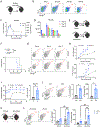
Figure 1.
Figure 1.
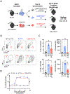
Recall T follicular helper (Tfh) cell response occurs after retransplantation and exhibits characteristics of T cell memory. (A) Naïve B6 mice were transplanted BALB/c skin (mean survival time [MST] 18 days) and euthanized posttransplant for graft-draining lymph node (DLN) and circulating Tfh cell analysis. (B) Representative flow cytometric plots displaying the frequencies of DLN Tfh (CXCR5+PD-1hi, gated on CD4+CD44hiFoxp3(−) T cells) over time. (C) Summary data of the frequencies and numbers of Tfh cells over time (n = 5 per group). (D) Tfh cell phenotype for the indicated markers over time (n = 5 per group). (E) Naïve and sensitized (BALB/c skin-grafted, MST 18 days) B6 mice were transplanted with BALB/c skin (4–6 weeks after primary skin graft rejection) and euthanized 3, 5, 7, and 10 days posttransplant for graft-DLN analysis or serially bled for serum analysis. (F) Kaplan-Meier curves of primary (MST 18 days) and secondary (MST 15 days) BALB/c skin allograft survival. (G) Representative flow cytometric plots displaying the frequencies of DLN Tfh cells in transplanted naïve (primary) and sensitized (secondary) mice. (H) Summary data of the frequencies and numbers of Tfh cells over time (n = 3–5 per group). (I) Summary data of antidonor immunoglobulin G over time and fold change (Δ) on day 10 relative to baseline (day 0) by flow cytometric crossmatch (n = 3–5 per group). (J) BALB/c skin-grafted B6 mice were retransplanted with syngeneic (B6) or allogeneic (BALB/c) skin grafts. Representative flow plots and summary data of the frequencies and numbers of DLN Tfh in syngeneic- or allogeneic-grafted mice 5 days posttransplant (n = 3–5 per group). (K) Naïve B6 mice were unilaterally transplanted with BALB/c skin and then unilaterally retransplanted with BALB/c skin on the dorsum opposite the site of the primary graft. Mice were sacrificed 7 days after the primary or secondary transplant for analysis of the DLNs ipsilateral (Ipsi) and contralateral (Contra) to the respective graft. (L) Representative flow plots and summary data of the frequencies and numbers of ipsilateral and contralateral DLN Tfh in transplanted mice (n = 5 per group). Summary data includes the quantity of Tfh cells 7 days after a unilateral primary or secondary graft in DLNs Ipsi and Contra to the graft (n = 5 per group). Summary data represent mean (±SE) and are representative of 2 to 3 independent experiments. * P <.05, ** P <.01. Abbreviations: Allo, allogeneic; CXC-R5, C-X-C chemokine receptor type 5; ICOS, inducible T cell costimulator; MFI, mean fluorescence intensity; PD1, programmed cell death protein 1; SE, standard error; Syn, syngeneic.
Figure 2.
Figure 2.
Allo-sensitized (Allo-Sen) CD4+ memory T cells rapidly differentiate into T follicular helper (Tfh) cells and mediate enhanced donor-specific antibody (DSA) formation independent of memory B cells upon alloantigen reexposure. (A) CD45.2+ B6 mice were sensitized with BALB/c skin to generate CD4+ memory T cells (mean survival time [MST]: 18 days). Forty-five days posttransplant, splenocytes from these Allo-Sen and nonsensitized (NonSen) control mice were recovered, and 3 × 10⁶ NonSen and Allo-Sen CD4+CD44+ T cells were adoptively transferred into naïve congenic CD45.1+ B6 recipients that were then transplanted with BALB/c grafts. Draining lymph nodes (DLNs) were analyzed 53 and 81 days postpriming (7 and 35 days posttransplant), or mice were serially bled for serum analysis. (B) Kaplan-Meier curves of BALB/c skin allograft survival in mice transferred with NonSen (MST 18 days) and Allo-Sen (MST 18 days) CD4+CD44+ T cells. (C) Representative flow cytometric plots displaying the frequencies of CD45.2(−) and CD45.2+ DLN Tfh (CXCR5+PD-1hi, gated on CD4+CD44hiFoxp3(−) T cells) in mice transferred NonSen or Allo-Sen T cells 53 days postpriming (7 days posttransplant). (D) Summary data of the frequencies and numbers of Tfh cells (n = 5 per group). (E) Representative flow plots displaying the frequencies of CD45.2(−) and CD45.2+ DLN Tfh (CXCR5+PD-1hi, gated on CD4+CD44hiFoxp3(−) T cells) in mice transferred NonSen or Allo-Sen T cells 81 days postpriming (35 days posttransplant). (F) Summary data of the frequencies and numbers of Tfh cells (n = 5 per group). (G) Summary data of the frequencies and numbers of germinal center (GC) B cells (CD95+GL7+, gated on CD19+IgD(−) B cells) 81 days postpriming (35 days posttransplant; n = 5 per group). (H) Summary data of antidonor immunoglobulin G over time using flow cytometric crossmatch (n = 4–5 per group). Summary data represent mean (±SE) and are representative of 2 to 3 independent experiments. * P <.05, ** P <.01. Abbreviations: Allo-S, allogeneic sensitized; CXC-R5, C-X-C chemokine receptor type 5; Non-S, nonsensitized; NS, not significant; PD1, programmed cell death protein 1; SE, standard error.
Figure 3.
Figure 3.
CD4+CXCR5+ T follicular helper (Tfh) cell-derived memory displays enhanced Tfh cells differentiation and expansion and mediates superior donor-specific antibody (DSA) formation after alloantigen rechallenge. (A) CD45.1+ B6 mice were sensitized with BALB/c skin to generate CD45.1+CD44hiCXCR5(−) CD4+ T cells (nonTfh) and CXCR5+PD-1hi Tfh cells. On day 10, 2 × 10⁵ cells were adoptively transferred into naïve CD45.2+ congenic B6 TCRa knockout mice, which were then transplanted with BALB/c skin on day 55 postpriming for alloantigen rechallenges. Draining lymph nodes (DLNs) from BALB/c-grafted mice were analyzed on days 62 and 90 postpriming (7 and 35 days posttransplant) and serially bled for serum analysis. (B) Representative flow cytometric plot (gated on CD19(−)CD4+CD44hiGITR(−) cells) displaying the sorting strategy for CXCR5(−) nonTfh and CXCR5+ Tfh. (C) Summary data of the numbers of transferred CD45.1+ nonTfh and Tfh cells 62 days postpriming (n = 4 per group). (D) Representative flow plots displaying the frequencies of transferred CD45.1+ DLN Tfh (CXCR5+PD-1hi, gated on CD4+CD44hiFoxp3(−) T cells) in mice receiving BALB/c-elicited CXCR5(−) nonTfh cells or CXCR5+ Tfh cells on day 62 (7 days posttransplant). (E) Summary data of the frequencies and numbers of Tfh cells on day 62 (7 days posttransplant; n = 4 per group). (F) Summary data of the numbers of transferred CD45.1+ nonTfh and Tfh cells 90 days postpriming (n = 4 per group). (G) Representative flow plots displaying the frequencies of transferred CD45.1+ DLN Tfh (CXCR5+PD-1hi, gated on CD4+CD44hiFoxp3(−) T cells) in mice receiving BALB/c-elicited CXCR5(−) nonTfh cells or CXCR5+ Tfh cells on day 90 (35 days posttranssplant). Summary data of the frequencies and numbers of (H) Tfh cells and (I) germinal center (GC) B cells (CD95+GL7+ B cells) on day 90 (35 days posttransplant; n = 4 per group). (J) Summary data from BALB/c-grafted mice of DSA (antiBALB/c IgG) over time posttransplant using flow cytometric crossmatch (n = 4 per group). Summary data represent mean (±SE) and are representative of 2 independent experiments. * P <.05. Abbreviations: CXC-R5, C-X-C chemokine receptor type 5; PD1, programmed cell death protein 1; SE, standard error.
Figure 4.
Figure 4.
T follicular helper (Tfh) cells differentiate independent of primary germinal center (GC) formation and the development of de novo donor-specific antibodies (DSAs). Naïve B6 mice were transplanted with BALB/c skin and treated with antiCD154 monoclonal antibody (MR-1) or left untreated and euthanized 10 days posttransplant for draining lymph node (DLN) analysis. (A) Representative flow cytometry plots and summary data of the frequencies and numbers of DLN Tfh (CXCR5+PD-1hi, gated on CD4+CD44hiFoxp3(−) T cells) in treated and untreated mice (n = 4 per group). (B) Representative plots and summary data of the frequencies and numbers of GC B cells (CD95+GL7+, gated on CD19+IgD(−) B cells; n = 4 per group). Next, naïve wild-type (WT) or CD40 knockout (KO) B6 mice were transplanted with BALB/c skin and euthanized posttransplant for DLN analysis or serially bled for serum analysis and DSA assessment. (C) Representative flow plots and (D) summary data of the frequencies and numbers of DLN Tfh and GC B cells in WT and CD40 KO mice (n = 5 per group) 10 days posttransplant. (E) Representative flow plots and (F) summary data of the frequencies and numbers of DLN Tfh cells and GC B cells in WT and CD40 KO mice 35 days posttransplant (n = 4–5 per group). (G) Summary data of antidonor immunoglobulin G (IgG) over time using flow cytometric crossmatch (n = 4 per group). Summary data represent mean (±SE). * P <.05, ** P < .01. Abbreviations: CXC-R5, C-X-C chemokine receptor type 5; MFI, mean fluorescence intensity; PD1, programmed cell death protein 1; Rx, therapy; SE, standard error.
Figure 5.
Figure 5.
Memory T follicular helper (Tfh) cells drive alloantibody formation independent of primary germinal center (GC) and donor-specific antibody (DSA) formation. (A) Wild-type (WT) or CD40 knockout (KO) B6 mice were sensitized with BALB/c skin. WT CD44hiCXCR5(−)PD-1(−) CD4+ T (nonTfh) and CXCR5+PD-1hi Tfh cells, and CD40 KO CXCR5+PD-1hi Tfh cells were isolated, and 5 × 10⁴ cells were sorted and adoptively transferred on day 10 into naïve congenic B6 TCRa KO mice that were then rechallenged with BALB/c skin grafts 55 days postpriming (45 days posttransfer). Mice were serially bled for serum analysis and DLNs were analyzed 90 days postpriming (35 days post alloantigen rechallenge). (B) Representative flow plots of the frequencies of DLN Tfh (CXCR5+PD-1hi, gated on CD4+CD44hiFoxp3(−) T cells) and GC B cells (CD95+GL7+, gated on CD19+IgD(−) B cells) 90 days postpriming (35 days post alloantigen rechallenge). (C) Summary data of the frequencies and number of DLN Tfh and GC B cells in mice transferred WT nonTfh, WT Tfh, or CD40 KO Tfh 90 days postpriming (35 days post alloantigen rechallenge; n = 4–5 per group). (D) Summary data of antidonor immunoglobulin G (IgG) over time by flow cytometric crossmatch (n = 4–5 per group). Summary data represent mean (±SE). * P < .05. Abbreviations: CXC-R5, C-X-C chemokine receptor type 5; MFI, mean fluorescence intensity; PD1, programmed cell death protein 1; SE, standard error.
Figure 6.
Figure 6.
CD28 costimulation blockade inhibits memory T follicular helper (mTfh) cell-mediated donor-specific antibody (DSA) formation. (A) BALB/c-sensitized B6 mice were retransplanted with BALB/c skin and treated with CTLA-4-Ig or left untreated and euthanized 5- and 35-days postretransplant for DLN analysis and serially bled for serum analysis. (B) Representative flow cytometry plots of the frequencies of DLN Tfh (CXCR5+PD-1hi, gated on CD4+CD44hiFoxp3(−) T cells) in treated and untreated mice 5- and 35-days postretransplant. Summary data of the frequencies and numbers of (C) Tfh and (D) GC B (CD95+GL7+, gated on CD19+IgD(−) B cells) cells 5 and 35 days postretransplant (n = 5 per group). (E) Summary data of antidonor IgG MFI and fold change (Δ) over time relative to baseline (day 0) by flow cytometric crossmatch (n = 5 per group). (F) Naïve B6 mice were transplanted with BALB/c skin and 5 × 10⁴ CXCR5+PD-1hi Tfh cells were sorted and transferred on day 10 into naïve B6 TCRa knockout (KO) mice. Mice were then rechallenged with BALB/c skin grafts 55 days postpriming (45 days posttransfer), left untreated, or treated with CTLA-4-Ig and serially bled for serum analysis. (G) Summary data of antidonor IgG over time by flow cytometric crossmatch (n = 4 per group). Summary data represent mean (±SE). * P < .05, ** P <.01. Abbreviations: CTLA-4, cytotoxic T-lymphocyte–associated antigen 4; CXC-R5, C-X-C chemokine receptor type 5; Ig, Immunoglobulin; MFI, mean fluorescence intensity; PD1, programmed cell death protein 1; Rx, therapy; SE, standard error.

Similar articles

References

    1. Loupy A, Hill GS, Jordan SC. The impact of donor-specific anti-HLA antibodies on late kidney allograft failure. Nat Rev Nephrol. 2012;8(6):348–357. 10.1038/nrneph.2012.81. - DOI - PubMed
    1. Loupy A, Lefaucheur C. Antibody-mediated rejection of solid-organ allografts. N Engl J Med. 2018;379(12):1150–1160. 10.1056/NEJMra1802677. - DOI - PubMed
    1. Sapir-Pichhadze R, Tinckam KJ, Laupacis A, Logan AG, Beyene J, Kim SJ. Immune sensitization and mortality in wait-listed kidney transplant candidates. J Am Soc Nephrol. 2016;27(2):570–578. 10.1681/ASN.2014090894. - DOI - PMC - PubMed
    1. Bray RA, Gebel HM. Strategies for human leukocyte antigen antibody detection. Curr Opin Organ Transplant. 2009;14(4):392–397. 10.1097/mot.0b013e32832d31c7. - DOI - PubMed
    1. Hidalgo LG, Campbell PM, Sis B, et al. De novo donor-specific antibody at the time of kidney transplant biopsy associates with microvascular pathology and late graft failure. Am J Transplant. 2009;9(11):2532–2541. 10.1111/j.1600-6143.2009.02800.x. - DOI - PubMed

Publication types

LinkOut - more resources