. 2023 Jun 11;13(1):9493.
doi: 10.1038/s41598-023-36200-6.

Comparison of the different anti-CD16 antibody clones in the activation and expansion of peripheral blood NK cells

Affiliations

Comparison of the different anti-CD16 antibody clones in the activation and expansion of peripheral blood NK cells

Jinho Kim et al. Sci Rep. .

Abstract

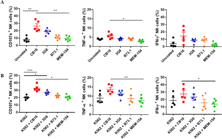
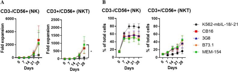
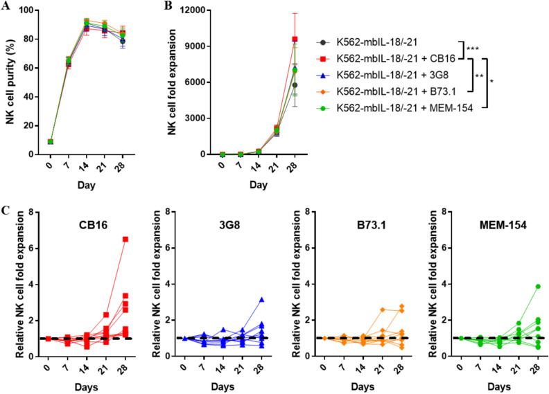
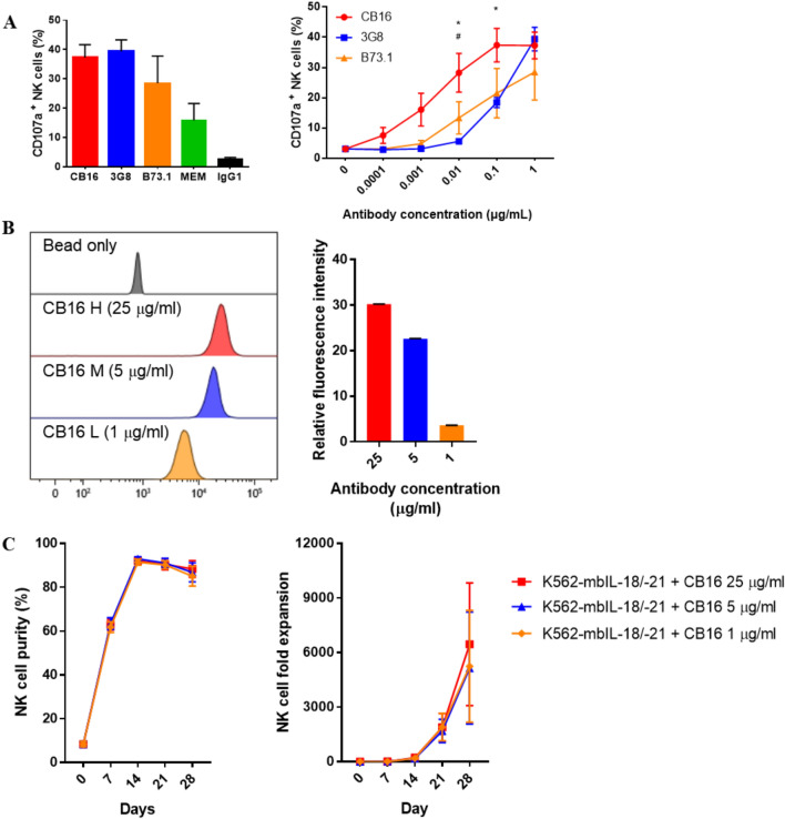
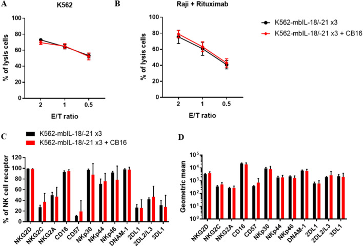
Natural killer (NK) cells are promising tool for cancer treatment. Methods have been developed for large-scale NK cell expansion, including feeder cell-based methods or methods involving stimulation with NK cell activating signals, such as anti-CD16 antibodies. Different clones of anti-CD16 antibodies are available; however, a comprehensive comparison of their differential effects on inducing NK cell activation and expansion has not been conducted among these various clones under the same experimental conditions. Herein, we found that the NK cell expansion rate differed depending on the various anti-CD16 antibodies (CB16, 3G8, B73.1, and MEM-154) coated on microbeads when stimulated with genetically engineered feeder cells, K562‑membrane-bound IL‑18, and mbIL‑21 (K562‑mbIL‑18/-21). Only the CB16 clone combination caused enhanced NK cell expansion over K562‑mbIL‑18/-21 stimulation alone with similar NK cell functionality. Treatment with the CB16 clone once on the initial day of NK cell expansion was sufficient to maximize the combination effect. Overall, we developed a more enhanced NK expansion system by merging a feeder to effectively stimulate CD16 with the CB16 clone.


Conflict of interest statement

The authors declare no competing interests.

Figures

Figure 1
Figure 1
NK cell responses upon stimulation with different anti-CD16 antibody clones. CD107a, TNF-α and IFN-γ of NK cells in PBMCs from healthy donors were measured in the stimulation of (A) antibody alone or (B) costimulation of K562 and antibody. Antibody stimulations were induced by microbeads coated with various clones of CD16 antibodies, CB16, 3G8, B73.1 and MEM-154, respectively, for 6 h. All data are shown as the mean ± SEM (n = 5; *, p < 0.05; **, p < 0.01).
Figure 2
Figure 2
Proliferation capacity of NK and NKT cells upon stimulation with different anti-CD16 antibody clones. (A) Fold expansion of NK cells, CD3-/CD56+ and NKT cells, CD3+/CD56+, in the feeder cell, K562-mbIL-18/-21, or anti-CD16 antibody stimulation. (B) NK and NKT cells purity in the feeder cell, K562-mbIL-18/-21, or anti-CD16 antibody stimulation. Antibody stimulations were induced by microbeads coated with various clones of CD16 antibodies, CB16, 3G8, B73.1 and MEM-154, respectively. All data are shown as the mean ± SEM (n = 6; *, p < 0.05).
Figure 3
Figure 3
Enhanced NK cell expansion via costimulation with feeder cells and anti-CD16 antibodies. (A) NK cell purity and (B) fold expansion of NK cells in PBMCs cultured with feeder cell, K562-mbIL-18/-21, alone or costimulation with various clones of anti-CD16 antibodies, CB16, 3G8, B73.1 and MEM-154, coated on microbeads, respectively. (C) Relative NK cell fold expansion of each PBMC donor in four different costimulation condition. NK cell expansion in K562-mbIL-18/-21 alone was used a standard (dashed line). All data are shown as the mean ± SEM (n = 9; *, p < 0.05; **, p < 0.01; ***, p < 0.001).
Figure 4
Figure 4
Effectiveness of the CB16 clone for NK cell stimulation. (A) CD107a expression on NK cell were measured after four anti-CD16 antibody clones (CB16, 3G8, B73.1, MEM-154) and negative control IgG1, all at a concentration of 1 μg/mL. CB16, 3G8, B73.1 were assessed at various antibody concentration for 6 h. (B) Microbeads coated with various concentration of CB16 clone were quantitatively measured by fluorescence labeled secondary antibodies. (C) NK cell purity and fold expansion of NK cells cultured in feeder cell, K562-mbIL-18/-21 alone or combination of microbeads coated with various concentration, 25, 5, 1 μg/mL, of CB16 clone. All data are shown as the mean ± SEM (n = 6 *#, p < 0.05, *CB16 vs B73.1; #CB16 vs 3G8).
Figure 5
Figure 5
Characteristics of expanded NK cells costimulated with the feeder cell and CB16 clone. Cytotoxic activity of expanded NK against (A) K562 or (B) Raji coated with Rituximab cultured in repeated stimulation, day 0, 7 and 14, of feeder cells, K562-mbIL-18/-21, with or without CB16 coated on microbead for 28 days. (C) Percent expression levels and (D) geometric mean of NK cell surface receptors related to cytotoxic function after culture with indicated stimulation for 28 days. All data are shown as the mean ± SEM (n = 6).

References

    1. Smyth MJ, Hayakawa Y, Takeda K, Yagita H. New aspects of natural-killer-cell surveillance and therapy of cancer. Nat. Rev. Cancer. 2002;2:850–861. doi: 10.1038/nrc928. - DOI - PubMed
    1. French AR, Yokoyama WM. Natural killer cells and viral infections. Curr. Opin. Immunol. 2003;15:45–51. doi: 10.1016/s095279150200002x. - DOI - PubMed
    1. Picano E, et al. Prognostic importance of dipyridamole-echocardiography test in coronary artery disease. Circulation. 1989;80:450–457. doi: 10.1161/01.cir.80.3.450. - DOI - PubMed
    1. Robertson MJ, Ritz J. Biology and clinical relevance of human natural killer cells. Blood. 1990;76:2421–2438. doi: 10.1182/blood.V76.12.2421.2421. - DOI - PubMed
    1. Cooper MA, Fehniger TA, Caligiuri MA. The biology of human natural killer-cell subsets. Trends Immunol. 2001;22:633–640. doi: 10.1016/s1471-4906(01)02060-9. - DOI - PubMed

Publication types