

Precise and comprehensive evaluation of perianal fistulas, classification and related complications using magnetic resonance imaging
- PMID: 37303685
- PMCID: PMC10250967
Precise and comprehensive evaluation of perianal fistulas, classification and related complications using magnetic resonance imaging
Abstract
Objective: This study aimed to comprehensively evaluate perianal fistulas and their related complications using magnetic resonance imaging (MRI).
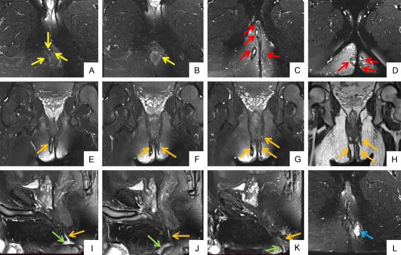
Methods: We enrolled 115 eligible patients who underwent preoperative perianal MRI. Primary fistulas, internal and external openings, and related complications were evaluated using MRI. All fistulas were classified according to Park's classification, Standard Practice Task Force classification, St. James's grade, and the position of the internal opening.
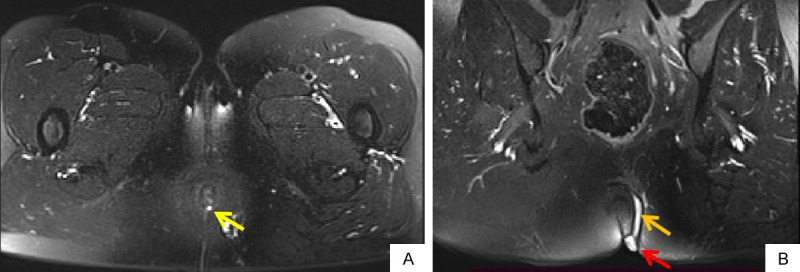
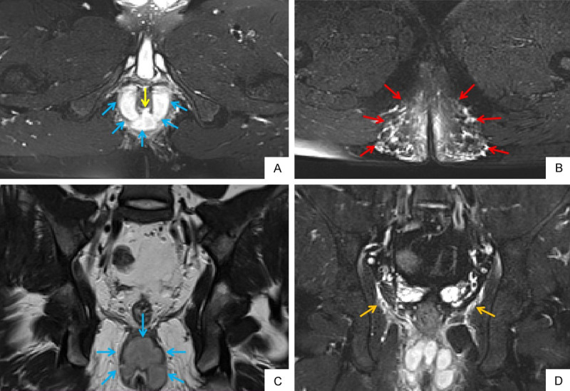
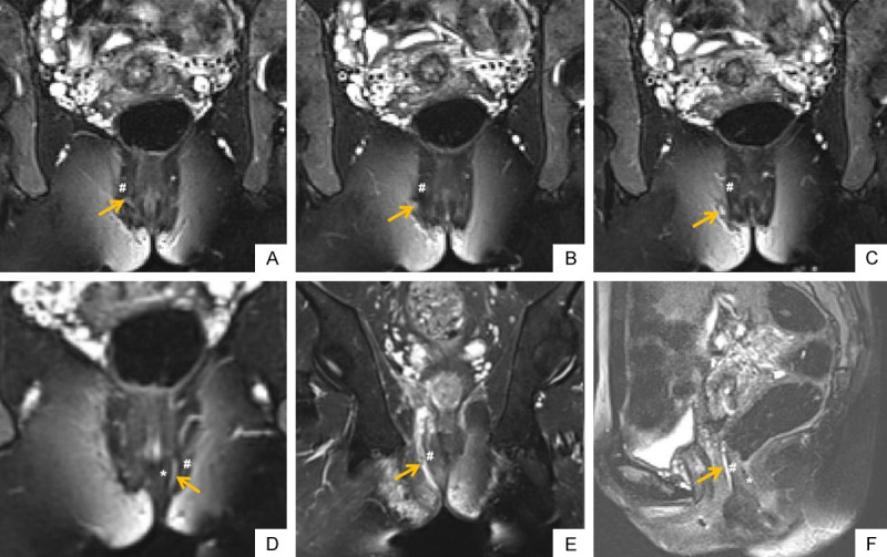
Results: In total, 169 primary fistulas were detected in 115 patients; 73 (63.5%) patients had a single primary tract and 42 (36.5%) patients had multiple primary tracts, and 198 internal and 129 external openings were identified. Based on Park's classification, 150 (88.7%) primary fistulas were classified into the following types: intersphincteric (82, 54.7%), trans-sphincteric (58, 38.6%), suprasphincteric (8, 5.3%), extrasphincteric (1, 0.7%), and diffuse intersphincteric with trans-sphincteric (1, 0.7%) types. Based on St. James's grade, 149 fistulas were classified into grade 1 (52, 34.9%), grade 2 (30, 20.1%), grade 3 (20, 13.4%), grade 4 (38, 25.5%), and grade 5 (9, 6.1%). We detected 92 (54.4%) simple and 77 (45.6%) complex perianal fistulas and 72 (42.6%) high and 97 (57.4%) low perianal fistulas. Furthermore, we detected 32 secondary tracts in 23 (20.0%) patients and 87 abscesses in 60 (52.2%) patients. Levator ani muscle involvement and extensive soft tissue edema were detected in 12 (10.4%) and 24 (20.9%) patients, respectively.
Conclusion: MRI is a valuable and comprehensive tool that can not only be used to determine the general condition of perianal fistulas but also to classify them and identify related complications.
Keywords: Perianal fistula; classification; complications; magnetic resonance imaging.
AJTR Copyright © 2023.
Conflict of interest statement
None.
Figures
References
-
- Jhaveri KS, Thipphavong S, Guo L, Harisinghani MG. MR imaging of perianal fistulas. Radiol Clin North Am. 2018;56:775–789. - PubMed
-
- Iqbal N, Tozer PJ, Fletcher J, Lightner AL, Sackitey C, Corr A, Patel U, Ilangovan R, Lung P. Getting the most out of MRI in perianal fistula: update on surgical techniques and radiological features that define surgical options. Clin Radiol. 2021;76:784.e17–784.e25. - PubMed
-
- Halligan S, Stoker J. Imaging of fistula in ano. Radiology. 2006;239:18–33. - PubMed
LinkOut - more resources
Full Text Sources