. 2023 May 22:14:1183825.
doi: 10.3389/fimmu.2023.1183825. eCollection 2023.

Differential effect of lactate on synovial fibroblast and macrophage effector functions

Affiliations

Differential effect of lactate on synovial fibroblast and macrophage effector functions

Valentina Pucino et al. Front Immunol. .

Abstract

Introduction: The synovial membrane is the main site of inflammation in rheumatoid arthritis (RA). Here several subsets of fibroblasts and macrophages, with distinct effector functions, have been recently identified. The RA synovium is hypoxic and acidic, with increased levels of lactate as a result of inflammation. We investigated how lactate regulates fibroblast and macrophage movement, IL-6 secretion and metabolism via specific lactate transporters.

Methods: Synovial tissues were taken from patients undergoing joint replacement surgery and fulfilling the 2010 ACR/EULAR RA criteria. Patients with no evidence of degenerative or inflammatory disease were used as control. Expression of the lactate transporters SLC16A1 and SLC16A3 on fibroblasts and macrophages was assessed by immunofluorescence staining and confocal microscopy. To test the effect of lactate in vitro we used RA synovial fibroblasts and monocyte-derived macrophages. Migration was assessed via scratch test assays or using trans-well inserts. Metabolic pathways were analysed by Seahorse analyser. IL-6 secretion was determined by ELISA. Bioinformatic analysis was performed on publicly available single cell and bulk RNA sequencing datasets.

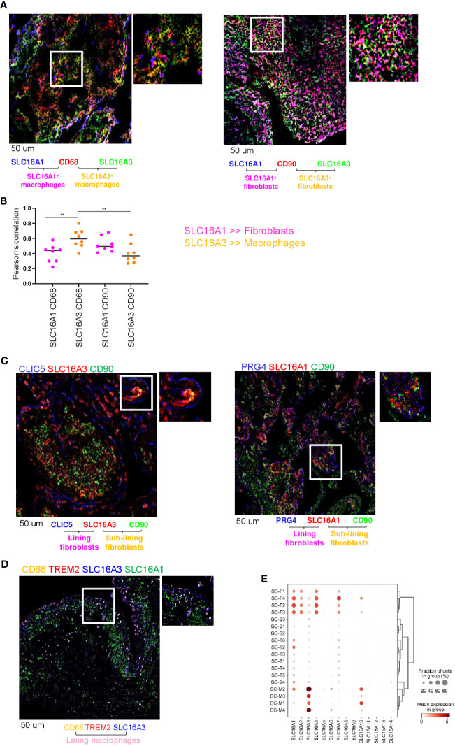
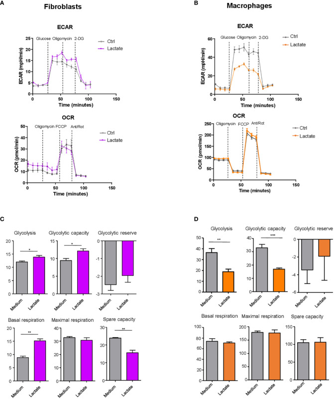
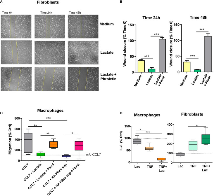
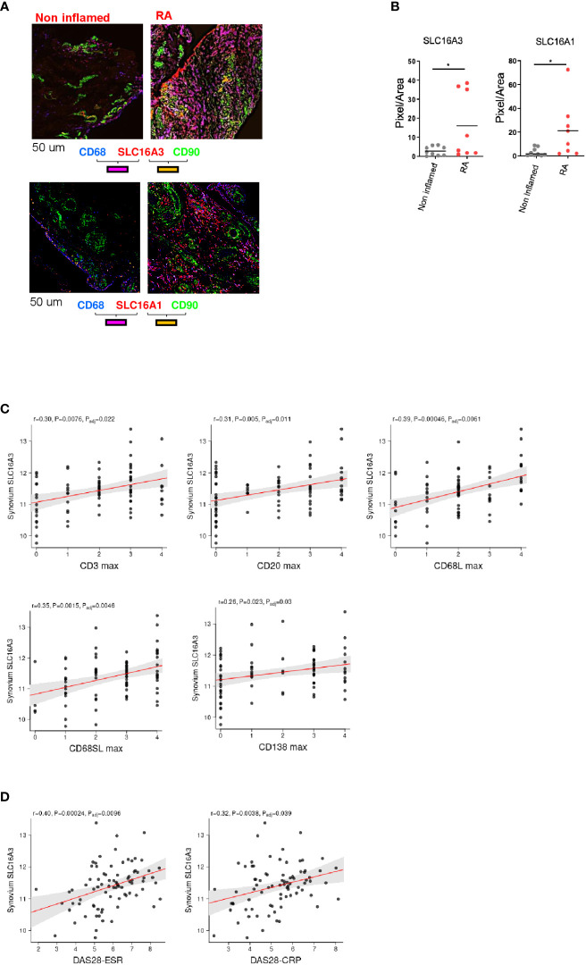
Results: We show that: i) SLC16A1 and SLC16A3 which regulate lactate intake and export respectively, are both expressed in RA synovial tissue and are upregulated upon inflammation. SLC16A3 is more highly expressed by macrophages, while SLC16A1 was expressed by both cell types. ii) This expression is maintained in distinct synovial compartments at mRNA and protein level. iii) Lactate, at the concentration found in RA joints (10 mM), has opposite effects on the effector functions of these two cell types. In fibroblasts, lactate promotes cell migration, IL-6 production and increases glycolysis. In contrast macrophages respond to increases in lactate by reducing glycolysis, migration, and IL-6 secretion.

Discussion: In this study, we provide the first evidence of distinct functions of fibroblasts and macrophages in presence of high lactate levels, opening new insights in understanding the pathogenesis of RA and offering novel potential therapeutic targets.

Keywords: arthritis; cell metabolism; fibroblasts; lactate; macrophages.


Conflict of interest statement

The authors declare that the research was conducted in the absence of any commercial or financial relationships that could be construed as a potential conflict of interest.

Figures

Figure 1
Figure 1
Lactate transporter expression in synovial compartment. (A) Representative IF staining of SLC16A1 (blue) and SLC16A3 (green) expression by synovial fibroblasts (CD90, red) and macrophages (CD68, red) from n=8 RA synovial tissues. (B) Quantification of the colocalization (Pearson’s correlation coefficient, ref 21) between SLC16A1 (blue) and SLC16A3 (green) and CD90 (red) and CD68 (red) within n=8 RA synovial tissues. Data are expressed as mean ± SEM. Anova test **p < 0.05; (C) SLC16A1 and SLC16A3 expression (red) by sub lining (CD90, green) and lining (CLIC5 or PRG4, blue) fibroblasts. (D) SLC16A1 and SLC16A3 expression (blue and green respectively) by sub lining (CD68+TREM2-, orange) and lining (CD68+TREM2+) macrophages. Scale bar 50 um. (E) scRNAseq expression of lactate transporters in synovial cellular subsets: Fibroblast: sub-lining SC-F1 (CD34+CD90+), SC-F2 (HLA-DRAhighCD90+), SC-F3 (DKK+CD90+) and lining SC-F4 (CD55+CD90-PRG4+CLIC5+) subsets (ref 1, 2). Macrophages: sub-lining MerTKnegTREM2neg SC-M1 (IL-1b+CD14+), SC-M4 (IFN-activated), and lining MerTKposTREM2pos SC-M2 (NUPR1+CD14+), SC-M3 (C1QA+CD14+) subsets (ref 2, 5). T cells: SC-T1 (CCR7+), SC-T2 (Treg cells), SC-T3 (Follicular helper T cells), SC-T4 (Granzyme K+), SC-T5 (Granulysin+, Granzyme B+), SC-T6 (Granzyme K+, Granzyme B+). B cells: SC-B1 (Naïve), SC-B2 (Memory), SC-B3 (Autoimmune), SC-B4 (Plasmablasts) (ref 2). Synovial tissues were taken by RA (n=36) and OA (n=15) patients (AMP dataset,ref 1, 2).
Figure 2
Figure 2
Lactate regulation of fibroblast and macrophage metabolism. (A, B) Seahorse analysis of extracellular acidification rate (ECAR, upper) and oxygen consumption rate (OCR, lower) in synovial fibroblasts (left) and monocyte-derived macrophages (right). (C, D) Glycolysis (after glucose injection), glycolytic capacity (after the injection of oligomycin) and glycolytic reserve (difference in ECAR between glucose and oligomycin injections) were calculated. Basal respiration [before oligomycin], maximal respiration [between FCCP and Antimycin+ rotenone injection], spare respiratory capacity [difference between basal and the maximal respiration], were calculated. Data expressed are representative of n=3 biological replicates. Data are expressed as mean ± SEM. Student’s t-test *p < 0.05; **p < 0.03; ***p < 0.01.
Figure 3
Figure 3
Lactate regulation of fibroblast and macrophage motility and IL-6 production. (A) Scratch test assay of synovial fibroblasts from RA patients (n=3 biological replicates). Cells were seeded for 24h and 48h in RPMI supplemented with 1% FBS and treated with or without sodium lactate (10 mM) +/- phroletin. (B) The perimeter (wound gap) of each scratch was measured with ImageJ software. The fold was calculated on Time 0 for each treatment (n=3, each in duplicate). (C) In vitro chemokinesis of monocytes-derived macrophages in response to CCL7 (300 ng/mL; n=6). Cells were cultured in medium containing sodium lactate (10 mM) or fibroblasts conditioned medium (10%) with or without phroletin (41 uM). Untreated macrophages (w/o CCL7, dotted line) were set to 100 (n=3, each in duplicate). (D) IL-6 ELISA from supernatants of fibroblasts and macrophages stimulated with TNFα (10 ng/ml) +/- lactate (n=3, each in duplicate). Untreated cells (dotted line) were set to 1. Data expressed are representative of n=3 biological replicates. Data are expressed as mean ± SEM. Anova test *p < 0.05; **p < 0.03; ***p < 0.01.
Figure 4
Figure 4
Correlation between lactate transporter expression and disease activity. (A) Representative images of SLC16A3 and SLC16A1 expression (red) by fibroblasts (CD90 green) and macrophages (CD68, blue) in RA and non-inflamed normal synovium. Scale bar 50 um. (B) Quantifications (pixel/area) of SLC16A3 and SLC16A1 expression in RA (n=8) and normal synovium (n=9). Data are expressed as mean ± SEM. Student’s t-test *p < 0.05. (C) Synovium SLC16A3 transcript positively correlates with histological score of synovitis (CD3, CD20, CD68 lining and sub lining, CD138) and with (D) the disease activity score (DAS28-ESR, DAS28-CRP) in a cohort of naïve-to treatment RA patients (27).

References

    1. Croft AP, Campos J, Jansen K, Turner JD, Marshall J, Attar M, et al. . Distinct fibroblast subsets drive inflammation and damage in arthritis. Nature (2019) 570(7760):246–51. doi: 10.1038/s41586-019-1263-7 - DOI - PMC - PubMed
    1. Zhang F, Wei K, Slowikowski K, Fonseka CY, Rao DA, Kelly S, et al. . Defining inflammatory cell states in rheumatoid arthritis joint synovial tissues by integrating single-cell transcriptomics and mass cytometry. Nat Immunol (2019) 20(7):928–42. doi: 10.1038/s41590-019-0378-1 - DOI - PMC - PubMed
    1. Wei K, Korsunsky I, Marshall JL, Gao A, Watts GFM, Major T, et al. . Notch signalling drives synovial fibroblast identity and arthritis pathology. Nature (2020) 582(7811):259–64. doi: 10.1038/s41586-020-2222-z - DOI - PMC - PubMed
    1. Culemann S, Grüneboom A, Nicolás-Ávila J, Weidner D, Lämmle KF, Rothe T, et al. . Locally renewing resident synovial macrophages provide a protective barrier for the joint. Nature (2019) 572(7771):670–5. doi: 10.1038/s41586-019-1471-1 - DOI - PMC - PubMed
    1. Alivernini S, MacDonald L, Elmesmari A, Finlay S, Tolusso B, Gigante MR, et al. . Distinct synovial tissue macrophage subsets regulate inflammation and remission in rheumatoid arthritis. Nat Med (2020) 26(8):1295–306. doi: 10.1038/s41591-020-0939-8 - DOI - PubMed

Publication types