Case Reports
. 2023 May 23:47:101204.
doi: 10.1016/j.gore.2023.101204. eCollection 2023 Jun.

A tale of two vulvar angiomyxomas: Two cases and review of literature

Affiliations
Case Reports

A tale of two vulvar angiomyxomas: Two cases and review of literature

Anastasia Navitski et al. Gynecol Oncol Rep. .

Abstract

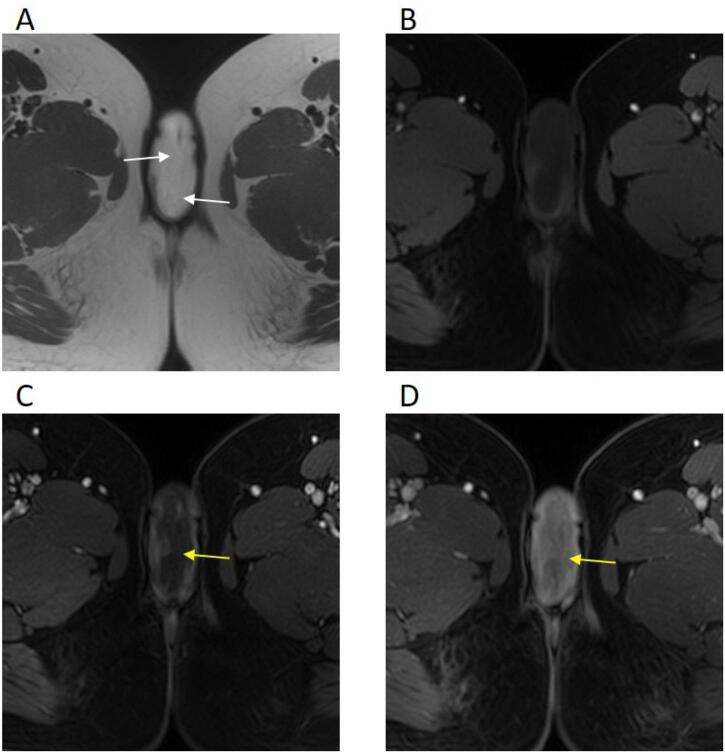
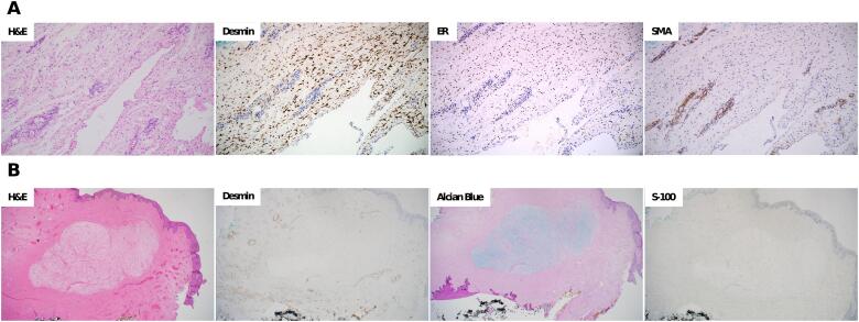
Vulvar angiomyxomas are rare benign mesenchymal neoplasms. Superficial and Aggressive angiomyxomas are two distinct phenotypes that present similarly to other more common vulva-perineal pathologies. Albeit both angiomyxomas carry a risk of recurrence, especially in the setting of incomplete resection, simple excision is insufficient for Aggressive angiomyxoma. It requires wide local excision because of its unique potential for local invasion, infiltration of the paravaginal and pararectal tissue, and more distant metastasis. Here, we present a case of Superficial angiomyxoma and a case of Aggressive angiomyxoma to highlight the diagnostic challenges and management strategies of each tumor. In both cases, angiomyxomas were initially misdiagnosed because of their rarity and nonspecific presentation. Magnetic resonance imaging is the modality of choice for evaluation due to inherent higher spatial resolution of soft tissue anatomical details. Early diagnosis of Aggressive angiomyxoma can prevent incomplete excision and recurrence, spare additional surgery, and offer hormonal therapy options.

Keywords: Aggressive angiomyxoma; Lower genital tract disease; Superficial angiomyxoma; Vulva.


Conflict of interest statement

The authors declare that they have no known competing financial interests or personal relationships that could have appeared to influence the work reported in this paper.

Figures

Fig. 1
Fig. 1
A 28-year-old female with a right labia majora tumor. Magnetic resonance imaging A: Axial T2 weighted image of the vulvar tumor demonstrates a relatively homogenous high signal intensity related to the myxoid matrix with low signal bands (white arrows) giving its characteristic swirling/laminated appearance. B-D: Axial T1 weighted images before (B) and after contrast administration (C and D) showing iso- to hypointense signal relative to skeletal muscles, and progressive enhancement, more intense in late post-contrast images (yellow arrows).
Fig. 2
Fig. 2
Pathology and immunohistochemical staining A. Deep Aggressive angiomyxoma: (H&E) Hematoxylin and eosin stain showing the lesion with dilated lymphatics and entrapped fat; (Desmin) Desmin staining diffusely positive in stromal cells; (ER) Estrogen receptor is diffusely positive in the stromal cells; (SMA) Smooth muscle actin highlights the perivascular smooth muscle while negative in the stromal cells. B. Superficial angiomyxoma: (H&E) Hematoxylin and eosin stain demonstrating a well-circumscribed spindle cell proliferation with a prominent myxoid stroma, numerous small blood vessels, and clear margins of excision; (Desmin) Desmin staining negative in stromal cells; (Alcian Blue) Alcian blue PAS, Periodic Acid Schiff, strongly positive for mucin; (S-100) S-100 staining negative. (For interpretation of the references to colour in this figure legend, the reader is referred to the web version of this article.)

References

    1. Aguilar-Frasco J., Ruben-Castillo C., Rodríguez-Quintero J.H., Medina-Franco H. Aggressive angiomyxoma: giant recurrence successfully treated with wide excision and adjuvant therapy with GnRH analogue. BMJ. Case. Rep. 2018;11 doi: 10.1136/bcr-2018-226973. - DOI - PMC - PubMed
    1. Angelico G., Marletta S., Broggi G., Vigneri P., Vecchio G.M., Salvatorelli L., Magro G. Practical Approach to the Diagnosis of the Vulvo-Vaginal Stromal Tumors: An Overview. Diagnostics. (Basel) 2022;12 doi: 10.3390/diagnostics12020357. - DOI - PMC - PubMed
    1. Baheti A.D., Tirumani S.H., Rosenthal M.H., Howard S.A., Shinagare A.B., Ramaiya N.H., Jagannathan J.P. Myxoid soft-tissue neoplasms: comprehensive update of the taxonomy and MRI features. AJR. Am. J. Roentgenol. 2015;204:374–385. doi: 10.2214/AJR.14.12888. - DOI - PubMed
    1. Fine B.A., Munoz A.K., Litz C.E., Gershenson D.M. Primary medical management of recurrent aggressive angiomyxoma of the vulva with a gonadotropin-releasing hormone agonist. Gynecol. Oncol. 2001;81:120–122. doi: 10.1006/gyno.2000.6119. - DOI - PubMed
    1. Ganeshan D., Amini B., Nikolaidis P., Assing M., Vikram R. Current Update on Desmoid Fibromatosis. J. Comput. Assist. Tomogr. 2019;43:29–38. doi: 10.1097/RCT.0000000000000790. - DOI - PMC - PubMed

Publication types

LinkOut - more resources