. 2023 Jun 2:2023:8150143.
doi: 10.1155/2023/8150143. eCollection 2023.

Optimization of the Imaged cIEF Method for Monitoring the Charge Heterogeneity of Antibody-Maytansine Conjugate

Affiliations

Optimization of the Imaged cIEF Method for Monitoring the Charge Heterogeneity of Antibody-Maytansine Conjugate

Ayat Abbood. J Anal Methods Chem. .

Abstract

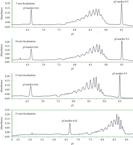
The aim of this study was to develop a whole-column imaging-detection capillary isoelectric focusing (icIEF) method for the analytical characterization of charge heterogeneity of a novel humanized anti-EphA2 antibody conjugated to a maytansine derivative. In addition to focusing time, sample composition was optimized: pH range, percent of carrier ampholytes, conjugated antibody concentration, and urea concentration. A good separation of charge isoforms was obtained with 4% carrier ampholytes of a large (3-10) and narrow pH range (8-10.5) (1 : 1 ratio), conjugated antibody concentration (0.3-1 mg/ml) with a good linearity (R2: 0.9905), 2 M of urea concentration, and 12 minute for focusing. The optimized icIEF method demonstrated a good interday repeatability with RSD values: <1% (pI), <8% (% peak area), and 7% (total peak areas). The optimized icIEF was useful as an analytical characterization tool to assess the charged isoform profile of a discovery batch of the studied maytansinoid-antibody conjugate in comparison to its naked antibody. It exhibited a large pI range (7.5-9.0), while its naked antibody showed a narrow pI range (8.9-9.0). In the discovery batch of maytansinoid-antibody conjugate, 2% of charge isoforms had the same pI as the pI of naked antibody isoforms.


Conflict of interest statement

The author declares that there are no conflicts of interest.

Figures

Figure 1
Figure 1
Analysis of maytansinoid-antibody by icIEF. Experimental conditions: final concentration 1 mg/mL in 0.35% methyl cellulose, 2% pharmalytes 3–10 and 2% pharmalytes 8–10.5 (1 : 1 ratio), 2 M urea, pI markers: 6.61, 9.50. Focusing time: 10 min at 3000 V. λ = 280 nm.
Figure 2
Figure 2
Analysis of different concentrations of maytansinoid-antibody by icIEF (0.3, 0.5, 0.8, 1, and 1.5 mg/ml). Other experimental conditions are as mentioned in Figure 1.
Figure 3
Figure 3
Plot of total peak area against maytansinoid-antibody concentration (0.3, 0.5, 0.8, 1, and 1.5 mg/ml).
Figure 4
Figure 4
Effects of urea concentration on the separation of charge isoforms of maytansinoid-antibody by icIEF (0, 1, 2, and 3 M urea). Other experimental conditions are as mentioned in Figure 1.
Figure 5
Figure 5
Effect of urea concentration on the pI values of main ADC charge variants.
Figure 6
Figure 6
Effects of focusing time on the separation of charge isoforms of maytansinoid-antibody by icIEF. Other experimental conditions are as mentioned in Figure 1.
Figure 7
Figure 7
Effects of % pharmalytes on the separation of charge isoforms of maytansinoid-antibody by icIEF. Other experimental conditions are as mentioned in Figure 1.
Figure 8
Figure 8
icIEF profiles of naked antibody (a) and its corresponding maytansinoid conjugate (b). Final concentration of maytansinoid-antibody in sample matrix is 1 mg/ml diluted in 0.35% methyl cellulose, 4% 3–10 pharmalytes/8–10.5 pharmalytes (1 : 1 ratio), and 2 M urea, pI markers: 8. 18, 9.50 (a) and 6.61, 9.50 (b), and focusing time of 12 min at 3000 V. Other experimental conditions are as mentioned in Figure 1.

References

    1. Kaplon H., Reichert J. M. Antibodies to watch in 2021. mAbs . 2021;13(1) doi: 10.1080/19420862.2020.1860476.1860476 - DOI - PMC - PubMed
    1. Baah S., Laws M., Rahman K. M. Antibody-drug conjugates-A tutorial review. Molecules . 2021;26(10):p. 2943. doi: 10.3390/molecules26102943. - DOI - PMC - PubMed
    1. Khongorzul P., Ling C. J., Khan F. U., Ihsan A. U., Zhang J. Antibody-drug conjugates: a comprehensive review. Molecular Cancer Research . 2020;18(1):3–19. doi: 10.1158/1541-7786.mcr-19-0582. - DOI - PubMed
    1. Sievers E. L., Senter P. D. Antibody-drug conjugates in cancer therapy. Annual Review of Medicine . 2013;64(1):15–29. doi: 10.1146/annurev-med-050311-201823. - DOI - PubMed
    1. Hafeez U., Parakh S., Gan H. K., Scott A. M. Antibody–drug conjugates for cancer therapy. Molecules . 2020;25(20):p. 4764. doi: 10.3390/molecules25204764. - DOI - PMC - PubMed