. 2023 May 26:13:1085908.
doi: 10.3389/fcimb.2023.1085908. eCollection 2023.

Non-typeable Haemophilus influenzae major outer membrane protein P5 contributes to bacterial membrane stability, and affects the membrane protein composition crucial for interactions with the human host

Affiliations

Non-typeable Haemophilus influenzae major outer membrane protein P5 contributes to bacterial membrane stability, and affects the membrane protein composition crucial for interactions with the human host

Yu-Ching Su et al. Front Cell Infect Microbiol. .

Abstract

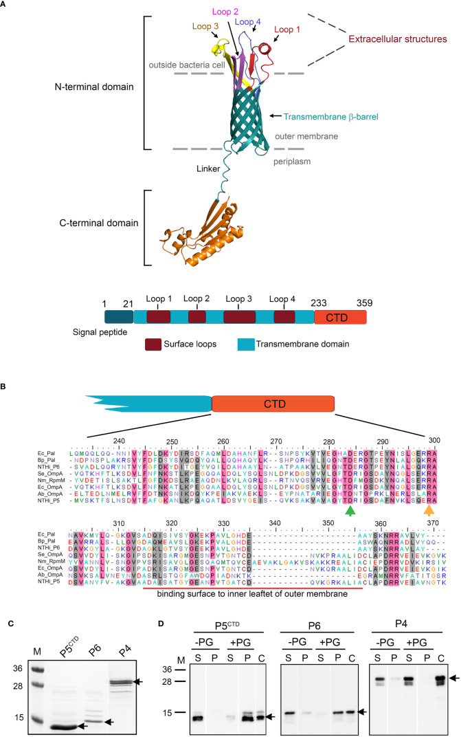
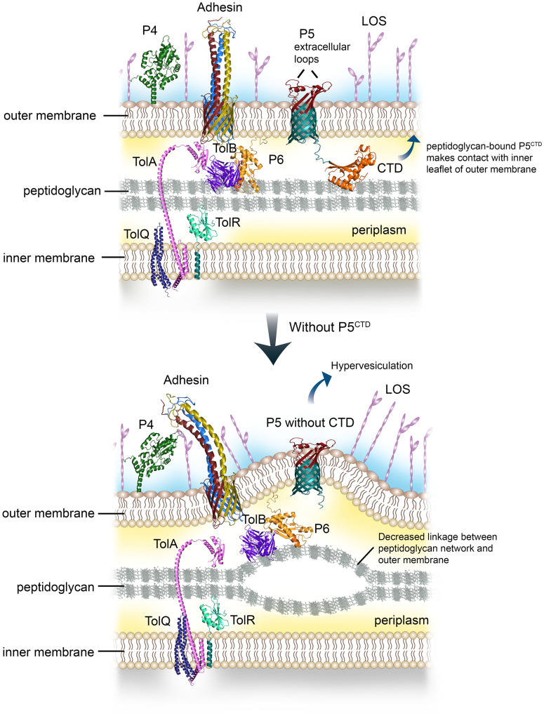
Non-typeable Haemophilus influenzae (NTHi) is a Gram-negative human pathogen that causes a wide range of airway diseases. NTHi has a plethora of mechanisms to colonize while evading the host immune system for the establishment of infection. We previously showed that the outer membrane protein P5 contributes to bacterial serum resistance by the recruitment of complement regulators. Here, we report a novel role of P5 in maintaining bacterial outer membrane (OM) integrity and protein composition important for NTHi-host interactions. In silico analysis revealed a peptidoglycan-binding motif at the periplasmic C-terminal domain (CTD) of P5. In a peptidoglycan-binding assay, the CTD of P5 (P5CTD) formed a complex with peptidoglycan. Protein profiling analysis revealed that deletion of CTD or the entire P5 changed the membrane protein composition of the strains NTHi 3655Δp5CTD and NTHi 3655Δp5, respectively. Relative abundance of several membrane-associated virulence factors that are crucial for adherence to the airway mucosa, and serum resistance were altered. This was also supported by similar attenuated pathogenic phenotypes observed in both NTHi 3655Δp5 CTD and NTHi 3655Δp5. We found (i) a decreased adherence to airway epithelial cells and fibronectin, (ii) increased complement-mediated killing, and (iii) increased sensitivity to the β-lactam antibiotics in both mutants compared to NTHi 3655 wild-type. These mutants were also more sensitive to lysis at hyperosmotic conditions and hypervesiculated compared to the parent wild-type bacteria. In conclusion, our results suggest that P5 is important for bacterial OM stability, which ultimately affects the membrane proteome and NTHi pathogenesis.

Keywords: NTHI; P5; adherence; extracellular matrix; peptidoglycan; serum resistance; virulence.


Conflict of interest statement

The authors declare that the research was conducted in the absence of any commercial or financial relationships that could be construed as a potential conflict of interest.

Figures

Figure 1
Figure 1
In silico analysis and characterization of C-terminal domain (M233-K359) of P5 (P5CTD). (A) 3D-model of P5 from NTHi 3655 based on AlphaFold prediction (Identifier: AF-A0A0H3PCS3-F1) (upper panel). The N-terminal domain of P5 forms a transmembrane β-barrel embedded within the asymmetric lipid bilayer of the outer membrane, and is connected by a linker to a C-terminal domain (CTD) sitting inside the periplasm. Four loops (Loop 1-4) form extracellular structures of P5. For clearer visualization, individual loops are indicated with different colours. The topology of P5 is similar to OmpA proteins from E. coli and A. baumannii (Park et al., 2012; Samsudin et al., 2016). Linear structural features of P5 from NTHi 3655 (lower panel). CTD of P5 is located between residue M233 and K359. (B) Sequence alignment of the P5CTD with OmpA-like domains from other OmpA and Pal family proteins. Clustal Omega (https://www.ebi.ac.uk/Tools/msa/clustalo/ ) was used to perform multiple sequence alignment. Residue D283 (green arrow) and R298 (orange arrow) are key residues in peptidoglycan binding (Park et al., 2012). Conserved residues are in pink shading, and similar residues are in grey shading. Red line indicates the region that mediates contact with the inner leaflet of the OM. Ec_Pal, peptidoglycan-associated lipoprotein (Pal) from Escherichia coli (GenBank accession number: P0A912); Bp_Pal, Pal from Burkholderia pseudomallei (Q63RA7); NTHi_P6, P6 from NTHi 3655 (P10324); Se_OmpA, OmpA from Salmonella typhimurium (ACY87707.1); Nm_RpmM, OmpA from Neisseria meningitidis (P0A0V3); Ec_OmpA, OmpA from E. coli (P0A910); Ab_OmpA, OmpA from Acinetobacter baumannii (Q6RYW5); NTHi_P5, P5 from NTHi 3655 (EDJ92910). (C) Purified His-tagged P5CTD, P6 and P4 of NTHi 3655 on Coomassie blue-stained 12% SDS-PAGE. (D) Peptidoglycan-binding assay of His-tagged P5CTD. Of note, all His-tagged proteins were pre-treated with a peptidoglycan-removal procedure prior to the peptidoglycan-binding assay (Karalus and Murphy, 1999). Purified proteins (3 μM) incubated with 100 µg of peptidoglycan from E. coli K12 (+PG) were pelleted by ultracentrifugation. Supernatant (S) and pellet (P) were analysed by western blotting with HRP-conjugated anti-His pAb. Incubation without peptidoglycan (-PG) was included as a negative control. Lane C was loaded with just the His-tagged protein alone as an anti-His pAb detection control. Black arrows indicate the His-tagged proteins. M, protein marker in kDa (PageRuler™, Prestained Protein Ladder, 10-250 kDa).
Figure 2
Figure 2
Characterization of protein profiles of NTHi 3655 wild-type and isogenic mutants. Analysis of (A) whole cell lysates (1×10⁷ CFU per lane) and (B) membrane fractions (20 µg per lane) of NTHi 3655 wild-type and isogenic mutants on a Coomassie-blue stained 12% SDS-PAGE (left panel) and western blotting (right panel). In western blotting, wild-type P5 (full length) was detected as two signal bands of ~28 kDa and 36 kDa (indicated by black arrows) from the whole cell lysate (panel A), and membrane fraction (panel B) of NTHi 3655 wild-type and NTHi 3655Δp5::p5. Wild-type P5 of NTHi appears as two isoforms (~28 kDa and 36 kDa) on SDS-PAGE due to heat denaturation of protein at 85°C during sample preparation (Roier et al., 2012). In NTHi 3655Δp5CTD , P5 with CTD deletion was detected as a signal band of ~25 kDa (indicated with red arrows) in western blotting (right panels of A, B). We did not identify any western blotting signal of P5 in the NTHi 3655Δp5. Blue arrows in panel (B) indicate protein bands in membrane fractions that have different levels of intensities on SDS-PAGE compared to the NTHi 3655 wild-type. (C) Flow cytometry analysis of detection of P5 extracellular structures on the bacterial surface. Bacteria from mid-log phase (OD600 = 0.5) were washed and resuspended in PBS containing 1% BSA. A bacterial suspension containing 5×10⁷ CFU in 20 µl were incubated with rabbit anti-P5Loop3 pAb for 30 min (Thofte et al., 2021). Samples were thereafter washed followed by incubation with FITC-conjugated goat anti-rabbit IgG, and finally analyzed on a BD FACSVerse flow cytometer. For antibody background control, samples were stained with the FITC-conjugated secondary antibody only. A representative data from three independent experiments is shown. Median values of fluorescence intensity are indicated for each sample. FlowJo v10 software (BD, Williamson Way Ashland, OR) was used for data presentation. Rabbit anti-P5Loop3 pAb targeting extracellular loop 3 of P5 was used in western blot (A, B) and flow cytometry (C) (Thofte et al., 2021). WT, wild-type NTHi 3655; Δp5, p5-knockout mutant (NTHi 3655Δp5); Δp5CTD , mutant expressing P5 without CTD (NTHi 3655Δp5CTD ); Δp5::p5, p5-transcomplemented NTHi (NTHi 3655Δp5::p5). M, protein marker (PageRuler™ Prestained Protein Ladder, 10 to 250 kDa).
Figure 3
Figure 3
Analysis of bacterial pathogenic phenotypes and membrane stability. (A) Bacterial adherence to human type II alveolar epithelial cells (A549) at multiplicity of infection (MOI) of 100 for 30 min. Mean data from three independent experiments (biological replicates) is presented. Bacterial adherence was presented as percentage of CFU recovered per well relative to initial inoculum. (B) Binding of NTHi 3655 wild-type and mutants to human fibronectin. Bacterial (5×10⁷ CFU) binding to human fibronectin (0.8-2.0 µg/ml) in 100 µl reactions was analysed by flow cytometry after incubation for 1 hour at 37°C. Rabbit anti-human fibronectin and FITC-conjugated swine anti-rabbit pAbs were used to detect the bacterial-bound fibronectin. Data represent mean values of three independent experiments. (C) Serum killing of NTHi 3655 wild-type and mutants. Bacterial (1.5×10³ CFU) killing by 5% NHS was analysed by CFU count on chocolate agar. Heat-inactivated serum was included as a negative control and here no bacteria were killed (data not shown). Percentage of bacterial survival was expressed as (Tt CFU/T0 CFU)×100. T0 represents CFU of sample plated at 0 min; and Tt represents CFU of sample plated at indicated time points. Data represent mean values of three independent experiments. (D) Outer membrane vesicles (OMVs) production among NTHi 3655 wild-type and mutants. OMVs from bacterial cultures were sucrose-density gradient purified and subjected to nanoparticle tracking analysis with a NanoSight NS300. OMV samples were diluted in PBS until 20-120 particles per frame were archived. Settings were optimized using 100nm polystyrene beads, and samples were recorded using the same settings (camera level 12, three recordings of 30 sec each). Recordings were thereafter processed using the NanoSight 3.1 software. Data represents mean values from three independent experiments. (E) Spot viability assay of bacterial survival in response to hyperosmotic environment. Bacteria that were serially diluted (109 to 104 CFU/ml) was spotted on chocolate agar without sodium chloride (NaCl) (left panel) or supplemented with 50 mM (middle panel) and 100 mM NaCl (right panel). Images were captured using ProtoCOL 3 HD (Synbiosis, UK). The assay was repeated in three independent experiments, and images from a representative experiment were shown. For panel A-D, error bars indicate standard deviations. Differences between wild-type and mutants were calculated by one-way ANOVA for panel (A, D); and two-way ANOVA for panel (B, C) *, P ≤ 0.05; **, P ≤ 0.01; ***, P ≤ 0.005; ****, P ≤ 0.001. WT, NTHi 3655 wild-type; Δp5, p5-knockout mutant (NTHi 3655Δp5); Δp5CTD , mutant expressing P5 without CTD (NTHi 3655Δp5CTD ); Δp5::p5, p5-transcomplemented NTHi (NTHi 3655Δp5::p5).
Figure 4
Figure 4
Graphical summary of the role of P5 in regulating outer membrane (OM) stability and distribution of surface virulence factors. Peptidoglycan-associated lipoprotein P6 together with P5 bind peptidoglycan via the OmpA-like domain (upper panel). The CTD of P5 will also interact with the inner leaflet of the OM, this will lead to stable contact between the OM and peptidoglycan. Meanwhile, TolAQRB-P6 complex joins the inner membrane with the peptidoglycan. The multiple linkages aid in the stability of NTHi cell envelope, subsequently provide an optimal membrane platform for the assembly and display of various surface virulence factors such as HMW, Hap, P4 and LOS, to name a few. However, interruption or deletion of proteins with OmpA-like domain involved in peptidoglycan binding will decrease the number of linkages or bonds between the OM and the peptidoglycan (lower panel). This will lead to OM protrusion, and increased vesiculation. The OM layer will become more permeable to solutes or antibiotics. The disrupted or collapsed OM will also hamper the translocation and assembly of various membrane proteins at bacterial surface, indirectly reducing bacterial interactions with the human host.

Similar articles

Cited by

References

    1. Atack J. M., Day C. J., Poole J., Brockman K. L., Bakaletz L. O., Barenkamp S. J., et al. . (2018). The HMW2 adhesin of non-typeable Haemophilus influenzae is a human-adapted lectin that mediates high-affinity binding to 2-6 linked n-acetylneuraminic acid glycans. Biochem. Biophys. Res. Commun. 503 (2), 1103–1107. doi: 10.1016/j.bbrc.2018.06.126 - DOI - PMC - PubMed
    1. Avadhanula V., Rodriguez C. A., Ulett G. C., Bakaletz L. O., Adderson E. E. (2006). Nontypeable Haemophilus influenzae adheres to intercellular adhesion molecule 1 (ICAM-1) on respiratory epithelial cells and upregulates ICAM-1 expression. Infect. Immun. 74 (2), 830–838. doi: 10.1128/IAI.74.2.830-838.2006 - DOI - PMC - PubMed
    1. Avila-Calderon E. D., Ruiz-Palma M. D. S., Aguilera-Arreola M. G., Velazquez-Guadarrama N., Ruiz E. A., Gomez-Lunar Z., et al. . (2021). Outer membrane vesicles of gram-negative bacteria: an outlook on biogenesis. Front. Microbiol. 12. doi: 10.3389/fmicb.2021.557902 - DOI - PMC - PubMed
    1. Berenson C. S., Murphy T. F., Wrona C. T., Sethi S. (2005). Outer membrane protein P6 of nontypeable Haemophilus influenzae is a potent and selective inducer of human macrophage proinflammatory cytokines. Infect. Immun. 73 (5), 2728–2735. doi: 10.1128/IAI.73.5.2728-2735.2005 - DOI - PMC - PubMed
    1. Brown M. A., Jabeen M., Bharj G., Hinks T. S. C. (2022). Non-typeable Haemophilus influenzae airways infection: the next treatable trait in asthma? Eur. Respir. Rev. 31 (165), 1–17. doi: 10.1183/16000617.0008-2022 - DOI - PMC - PubMed

Publication types