Review
. 2023 May 18:16:1193590.
doi: 10.3389/fnmol.2023.1193590. eCollection 2023.

Therapeutic effects of phlorotannins in the treatment of neurodegenerative disorders

Affiliations
Review

Therapeutic effects of phlorotannins in the treatment of neurodegenerative disorders

Yoon Ji Kwon et al. Front Mol Neurosci. .

Abstract

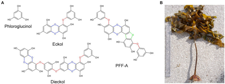
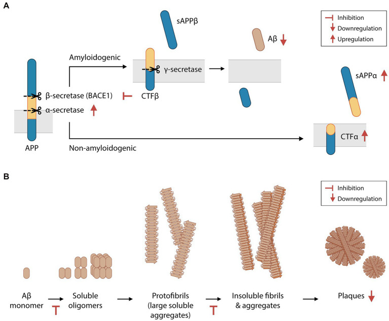
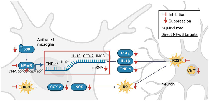
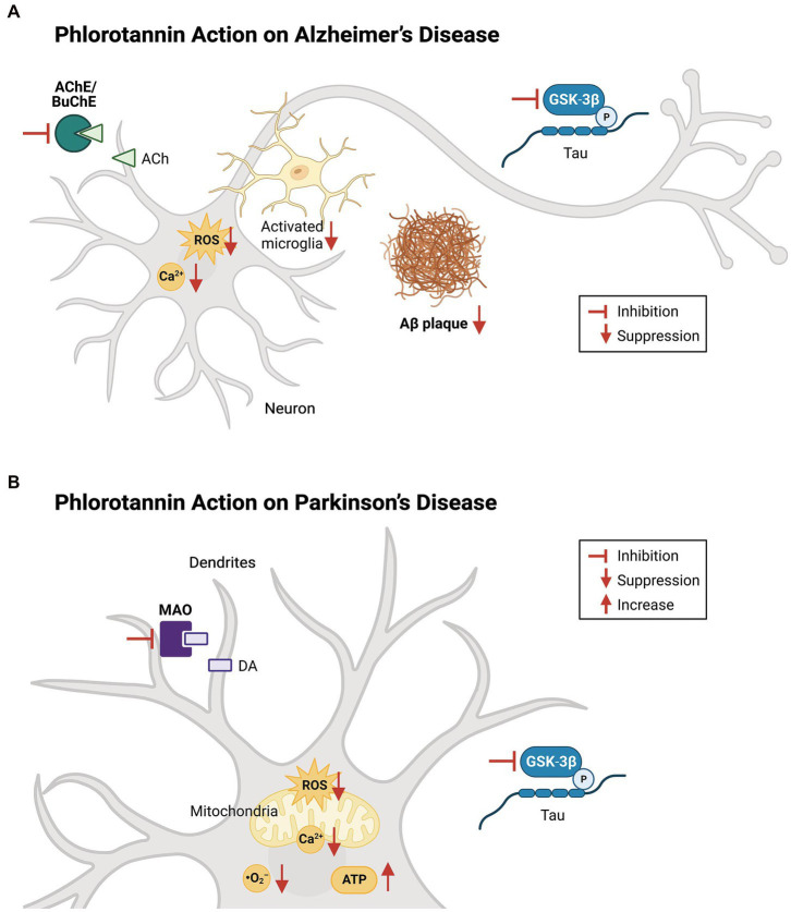
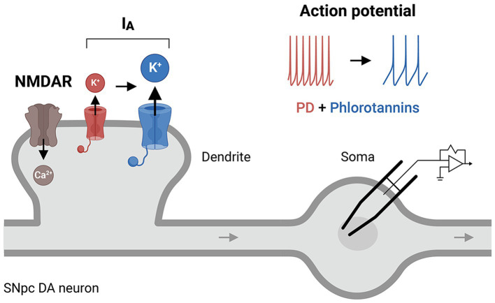
Phlorotannins are natural polyphenolic compounds produced by brown marine algae and are currently found in nutritional supplements. Although they are known to cross the blood-brain barrier, their neuropharmacological actions remain unclear. Here we review the potential therapeutic benefits of phlorotannins in the treatment of neurodegenerative diseases. In mouse models of Alzheimer's disease, ethanol intoxication and fear stress, the phlorotannin monomer phloroglucinol and the compounds eckol, dieckol and phlorofucofuroeckol A have been shown to improve cognitive function. In a mouse model of Parkinson's disease, phloroglucinol treatment led to improved motor performance. Additional neurological benefits associated with phlorotannin intake have been demonstrated in stroke, sleep disorders, and pain response. These effects may stem from the inhibition of disease-inducing plaque synthesis and aggregation, suppression of microglial activation, modulation of pro-inflammatory signaling, reduction of glutamate-induced excitotoxicity, and scavenging of reactive oxygen species. Clinical trials of phlorotannins have not reported significant adverse effects, suggesting these compounds to be promising bioactive agents in the treatment of neurological diseases. We therefore propose a putative biophysical mechanism of phlorotannin action in addition to future directions for phlorotannin research.

Keywords: Alzheimer’s disease; Parkinson’s disease; neurodegenerative disease; phlorotannin; polyphenol.


Conflict of interest statement

YJK was previously employed, and OIK is currently employed by Botamedi Brain Health and Medical Care Company Limited. The remaining authors declare that the research was conducted in the absence of any commercial or financial relationships that could be construed as a potential conflict of interest.

Figures

Figure 1
Figure 1
The chemical structures of phloroglucinol and phlorotannin compounds. (A) Phlorotannins are polyphenolic oligomers of phloroglucinol units that are connected by ether bonds (red), dibenzo-p-dioxin moieties (blue), and dibenzofuran elements (green). (B) Specimen of Ecklonia cava, a species of brown marine algae and common source of phlorotannins. PFF-A, phlorofucofuroeckol A.
Figure 2
Figure 2
Phlorotannins inhibit the biosynthesis and oligomerization of amyloid beta fragments. (A) Aβ is produced via the proteolytic cleavage of APP protein by β-secretase and then γ-secretase. Phlorotannins inhibit β-secretase (BACE1) and γ-secretase activity and promote APP processing by α-secretase, suppressing Aβ production. (B) Aβ monomers aggregate into soluble oligomers, fibrils, and plaques. Phlorotannins inhibit the oligomerization into soluble oligomers as well as fibrilization into fibrils. Ultimately, plaque formation is reduced. Red flat-ended arrows indicate inhibition, red upward arrows indicate upregulation, and red downward arrows indicate downregulation. Aβ, amyloid beta; APP, amyloid precursor protein; BACE1, beta secretase 1; sAPPα, soluble amyloid protein precursor alpha; sAPPβ, soluble amyloid protein precursor beta; CTFα, C-terminal fragment alpha; CTFβ, C-terminal fragment beta. Adapted from Kang et al. (2011) and Yang et al. (2018).
Figure 3
Figure 3
Phlorotannins inhibit the NF-κB pathway of microglial activation and the pro-inflammatory signaling cascade. Phosphorylated p38 activates NF-κB, which in turn promotes transcription of pro-inflammatory signaling molecules. Neuroinflammation generates ROS, NO and Ca2+, induces oxidative damage, and promotes neuron death. Phlorotannins inhibit NF-κB activity and also directly scavenge ROS, thereby reducing microglial recruitment and neuroinflammation, ultimately increasing neuron viability. Red flat-ended arrows indicate direct inhibition; red downward arrows indicate downregulation; asterisks indicate changes observed in Aβ and phlorotannin-treated cells; underlined proteins indicate direct downstream targets of NF-κB. NF-κB, nuclear factor kappa-light-chain-enhancer of activated B cells; TNF-α, tumor necrosis factor alpha; IL-6, interleukin 6; IL-1β, interleukin 1 beta; PGE2, prostaglandin E2; COX-2, cyclooxygenase-2; iNOS, inducible nitric oxide synthase; NO, nitric acid; ROS, reactive oxygen species; Aβ, amyloid beta. Adapted from previous studies in Table 2.
Figure 4
Figure 4
Effects of phlorotannins in Alzheimer’s and Parkinson’s disease. (A) In AD, phlorotannins inhibit AChE and BuChE and combat the loss of cholinergic transmission. Phlorotannins also reduce the formation of Aβ plaques. Aβ-induced spikes in intracellular ROS and Ca2+ levels, as well as microglial activation, recruitment, and pro-inflammatory signaling, are suppressed. Adapted from Lee and Stein (2004) and Choi et al. (2015). (B) In PD, phlorotannins may inhibit MAOs to counteract the loss of dopamine. In mitochondria, phlorotannins reduce mitochondrial loss, glutamate-induced membrane flickering, ROS levels, and Ca2+. Phlorotannins also reduce cytoplasmic ROS and O2 levels, while rescuing ATP levels and the number of TH-positive cells in the substantia nigra. Flat-ended arrows indicate direct inhibition, upward arrows indicate upregulation, and downward arrows indicate downregulation. AChE, acetylcholinesterase; BuChE; butyrylcholinesterase, ACh, acetylcholine; ROS, reactive oxygen species; Aβ, beta amyloid; GSK-3β, glycogen synthase kinase-3 beta; P, phosphorylation; MAO, monoamine oxidase; DA, dopamine; ATP, adenosine triphosphate. Adapted from Choi et al. (2015), Cui et al. (2019), and Seong et al. (2019).
Figure 5
Figure 5
A putative cellular mechanism of phlorotannins to restore IA receptor activity. Phlorotannin treatment may upregulate IA receptors on the presynaptic membrane, thus altering the NMDAR/IA ratio and dendritic excitability, and decreasing the frequency of action potential firing in SNpc DA neurons. These changes may reduce intracellular Ca2+ levels and thus counteract the excitotoxicity and pathogenesis of PD. PD, Parkinson’s disease; NMDAR, N-methyl-D-aspartate receptor; SNpc, substantia nigra pars compacta; DA, dopamine.

References

    1. Aatif M., Muteeb G., Alsultan A., Alshoaibi A., Khelif B. Y. (2021). Dieckol and its derivatives as potential inhibitors of SARS-CoV-2 spike protein (UK strain: VUI 202012/01): a computational study. Mar. Drugs 19:242. doi: 10.3390/md19050242, PMID: - DOI - PMC - PubMed
    1. Ahn G. N., Kim K. N., Cha S. H., Song C.-B., Lee J., Heo M.-S., et al. . (2006). Antioxidant activities of phlorotannins purified from Ecklonia cava on free radical scavenging using ESR and H2O2-mediated DNA damage. Eur. Food Res. Technol. 226, 71–79. doi: 10.1007/s00217-006-0510-y - DOI
    1. Ahn B. R., Moon H. E., Kim H. R., Jung H. A., Choi J. S. (2012). Neuroprotective effect of edible brown alga Eisenia bicyclis on amyloid beta peptide-induced toxicity in PC12 cells. Arch. Pharm. Res. 35, 1989–1998. doi: 10.1007/s12272-012-1116-5, PMID: - DOI - PubMed
    1. Alhola P., Polo-Kantola P. (2007). Sleep deprivation: impact on cognitive performance. Neuropsychiatr. Dis. Treat. 3, 553–567. PMID: - PMC - PubMed
    1. Amendola J., Woodhouse A., Martin-Eauclaire M. F., Goaillard J. M. (2012). Ca2+/cAMP-sensitive Covariation of IA and IH voltage dependences Tunes rebound firing in dopaminergic neurons. J. Neurosci. 32, 2166–2181. doi: 10.1523/JNEUROSCI.5297-11.2012, PMID: - DOI - PMC - PubMed

LinkOut - more resources