Case Reports
. 2023 May 26:13:1164153.
doi: 10.3389/fonc.2023.1164153. eCollection 2023.

Primary cardiac tumor: a case report of right atrial angiosarcoma and review of the literature

Affiliations
Case Reports

Primary cardiac tumor: a case report of right atrial angiosarcoma and review of the literature

Yujian Guo et al. Front Oncol. .

Abstract

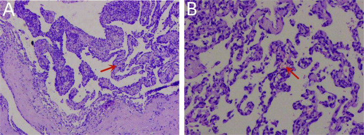
Primary cardiac angiosarcoma is a relatively rare tumor with early metastasis and poor prognosis. Radical resection of the primary tumor remains the primary approach for the optimal survival of patients with early-stage cardiac angiosarcoma without evidence of metastasis. This case involves a 76-year-old man with symptoms of chest tightness, fatigue, pericardial effusion, and arrhythmias who achieved good results after surgery to treat the angiosarcoma in the right atrium. In addition, literature analysis showed that surgery remains an effective way of treating primary early angiosarcoma.

Keywords: angiosarcoma; pericardial effusion; primary cardiac angiosarcoma; primary cardiac tumors; right atrium.


Conflict of interest statement

The authors declare that the research was conducted in the absence of any commercial or financial relationships that could be construed as a potential conflict of interest.

Figures

Figure 1
Figure 1
A chest X-ray showed an enlarged heart shadow and possible left pleural enveloping effusion (A). CT indicated curved fluid density shadows in the bilateral thorax and pericardial cavity and irregular, slightly low-density shadows in the right atrium (B, C).
Figure 2
Figure 2
TTE showed an isoechoic mass of approximately 41 * 32 mm in the right atrium, with a broad base, multiple echo-free zones, and thickened surrounding chamber walls (A–C). A dark area of fluid was visible in the pericardial cavity, and the fluid area was clear (D). TTE, transthoracic echocardiography.
Figure 3
Figure 3
Re-examination of the TTE showed an isoechoic mass of approximately 39 * 37 mm at the top of the right atrium with a wide base and multiple echo-free zones. The surrounding atrial wall was thickened, and the width of the inferior vena cava was approximately 23 mm (A). A dark area of fluid was visible in the pericardial cavity, and the fluid area was not clear (B). TTE, transthoracic echocardiography.
Figure 4
Figure 4
A contrast-enhanced CT of the chest showed arc-shaped fluid density shadows in the bilateral thorax and pericardial cavity, while irregular, slightly low-density shadows with unclear boundaries were evident in the right atrium (red arrow). Most of the lesions were not enhanced, and the edges appear locally enhanced (A–D).
Figure 5
Figure 5
During the operation, a tumor protrusion of approximately 10 * 15 mm was evident on the surface of the right atrium. The serosal surface of the right atrium and superior vena cava showed cowhide changes (A). Although the mass proliferated between the inner and outer membranes of the right atrial wall, it did not breach the atrial cavity (B). The size of the mass was approximately 50 * 40 mm. The tumor morphology was considered for atrial sarcoma diagnosis (D, E). The tumor was completely removed, as well as part of the atrial wall, and the right atrial defect was sutured with a pericardial patch (C).
Figure 6
Figure 6
Histopathology confirmed that the tumor was a primary cardiac angiosarcoma (A). Pathological investigation showed vascular hyperplasia, labyrinth and sinusoid structures, obvious cell atypia, mitosis, and massive necrosis in H&E stains (B).

References

    1. Liu C, Zhao Y, Yin Z, Hu T, Ren J, Wei J, et al. . Right atrial epithelioid angiosarcoma with multiple pulmonary metastasis confirmed by multimodality imaging-guided pulmonary biopsy: a case report and literature review. Med (Baltimore) (2018) 97(30):e11588. doi: 10.1097/MD.0000000000011588 - DOI - PMC - PubMed
    1. Linfeng Q, Xingjie X, Henry D, Zhedong W, Hongfei X, Haige Z. Cardiac angiosarcoma: a case report and review of current treatment. Med (Baltimore) (2019) 98(49):e18193. doi: 10.1097/MD.0000000000018193 - DOI - PMC - PubMed
    1. Ostrowski S, Marcinkiewicz A, Kośmider A, Jaszewski R. Sarcomas of the heart as a difficult interdisciplinary problem. Arch Med Sci (2014) 10(1):135–48. doi: 10.5114/aoms.2014.40741 - DOI - PMC - PubMed
    1. Pacini D, Careddu L, Pantaleo A, Parolari A, Leone O, Daprati A, et al. . Primary malignant tumors of the heart: outcomes of the surgical treatment. Asian Cardiovasc Thorac Ann (2015) 23(6):645–51. doi: 10.1177/0218492315573674 - DOI - PubMed
    1. Brandt RR, Arnold R, Bohle RM, Dill T, Hamm CW. Cardiac angiosarcoma: case report and review of the literature. Z Kardiol (2005) 94(12):824–8. doi: 10.1007/s00392-005-0296-0 - DOI - PubMed

Publication types

LinkOut - more resources