

Network anatomy in logopenic variant of primary progressive aphasia
- PMID: 37306089
- PMCID: PMC10318204
- DOI: 10.1002/hbm.26388
Network anatomy in logopenic variant of primary progressive aphasia
Abstract
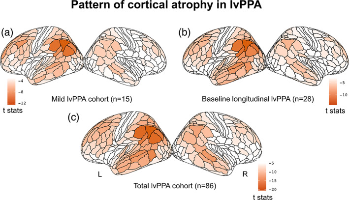
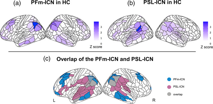
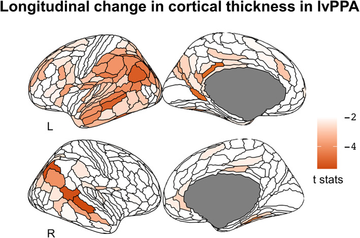
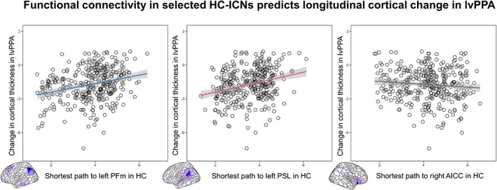
The logopenic variant of primary progressive aphasia (lvPPA) is a neurodegenerative syndrome characterized linguistically by gradual loss of repetition and naming skills resulting from left posterior temporal and inferior parietal atrophy. Here, we sought to identify which specific cortical loci are initially targeted by the disease (epicenters) and investigate whether atrophy spreads through predetermined networks. First, we used cross-sectional structural MRI data from individuals with lvPPA to define putative disease epicenters using a surface-based approach paired with an anatomically fine-grained parcellation of the cortical surface (i.e., HCP-MMP1.0 atlas). Second, we combined cross-sectional functional MRI data from healthy controls and longitudinal structural MRI data from individuals with lvPPA to derive the epicenter-seeded resting-state networks most relevant to lvPPA symptomatology and ascertain whether functional connectivity in these networks predicts longitudinal atrophy spread in lvPPA. Our results show that two partially distinct brain networks anchored to the left anterior angular and posterior superior temporal gyri epicenters were preferentially associated with sentence repetition and naming skills in lvPPA. Critically, the strength of connectivity within these two networks in the neurologically-intact brain significantly predicted longitudinal atrophy progression in lvPPA. Taken together, our findings indicate that atrophy progression in lvPPA, starting from inferior parietal and temporoparietal junction regions, predominantly follows at least two partially nonoverlapping pathways, which may influence the heterogeneity in clinical presentation and prognosis.
Keywords: Alzheimer's disease; cortical atrophy; intrinsic connectivity networks; logopenic variant; longitudinal study; primary progressive aphasia.
© 2023 The Authors. Human Brain Mapping published by Wiley Periodicals LLC.
Figures
Update of
-
Network anatomy in logopenic variant of primary progressive aphasia.medRxiv [Preprint]. 2023 May 16:2023.05.15.23289065. doi: 10.1101/2023.05.15.23289065. medRxiv. 2023. Update in: Hum Brain Mapp. 2023 Aug 1;44(11):4390-4406. doi: 10.1002/hbm.26388. PMID: 37292690 Free PMC article. Updated. Preprint.
References
-
- Ahmed, Z. , Cooper, J. , Murray, T. K. , Garn, K. , McNaughton, E. , Clarke, H. , Parhizkar, S. , Ward, M. A. , Cavallini, A. , Jackson, S. , Bose, S. , Clavaguera, F. , Tolnay, M. , Lavenir, I. , Goedert, M. , Hutton, M. L. , & O'Neill, M. J. (2014). A novel in vivo model of tau propagation with rapid and progressive neurofibrillary tangle pathology: The pattern of spread is determined by connectivity, not proximity. Acta Neuropathologica, 127(5), 667–683. 10.1007/s00401-014-1254-6 - DOI - PMC - PubMed
Publication types
MeSH terms
Grants and funding
LinkOut - more resources
Full Text Sources
Medical
Miscellaneous
