. 2023 Sep;24(9):1093-1106.
doi: 10.1111/mpp.13356. Epub 2023 Jun 12.

A nonclassically secreted effector of Magnaporthe oryzae targets host nuclei and plays important roles in fungal growth and plant infection

Affiliations

A nonclassically secreted effector of Magnaporthe oryzae targets host nuclei and plays important roles in fungal growth and plant infection

Xiaomin Chen et al. Mol Plant Pathol. 2023 Sep.

Abstract

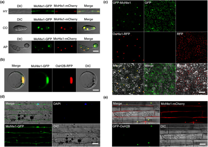
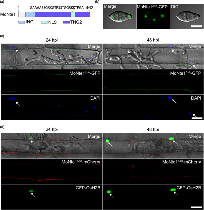
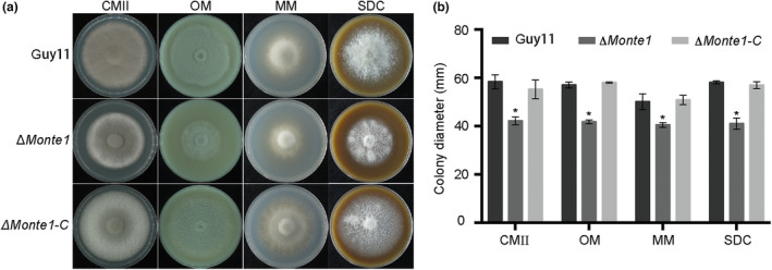
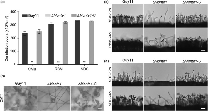
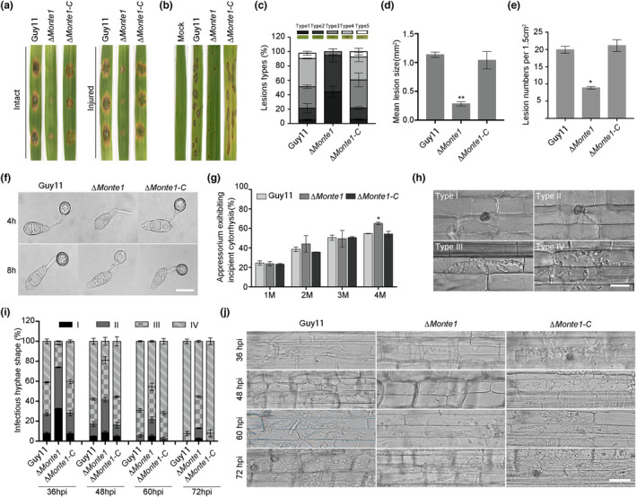
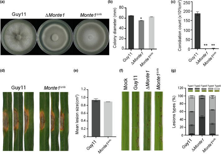
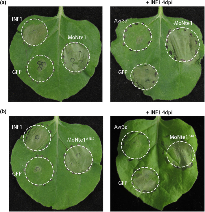
Rice blast caused by Magnaporthe oryzae is one of the most destructive diseases and poses a growing threat to food security worldwide. Like many other filamentous pathogens, rice blast fungus releases multiple types of effector proteins to facilitate fungal infection and modulate host defence responses. However, most of the characterized effectors contain an N-terminal signal peptide. Here, we report the results of the functional characterization of a nonclassically secreted nuclear targeting effector in M. oryzae (MoNte1). MoNte1 has no signal peptide, but can be secreted and translocated into plant nuclei driven by a nuclear targeting peptide. It could also induce hypersensitive cell death when transiently expressed in Nicotiana benthamiana. Deletion of the MoNTE1 gene caused a significant reduction of fungal growth and conidiogenesis, partially impaired appressorium formation and host colonization, and also dramatically attenuated the pathogenicity. Taken together, these findings reveal a novel effector secretion pathway and deepen our understanding of rice-M. oryzae interactions.

Keywords: Magnaporthe oryzae; fungal growth; hypersensitive cell death; nuclear targeting; pathogenicity; rice-Magnaporthe oryzae interactions; signal peptide.


Figures

FIGURE 1
FIGURE 1
Subcellular localization of MoNte1 in Magnaporthe oryzae and plant cells. (a) Localization of MoNte1 at different developmental stages of M. oryzae. MoNte1 was localized to the nuclei at the vegetative hypha (HY), conidium (CO), and appressorium (AP) stages, colocalizing with the nuclear marker (MoHis1‐mCherry). Scale bar = 5 μm. (b) Colocalization assay of MoNte1‐green fluorescent protein (GFP) and OsH2B‐red fluorescent protein (RFP) in a rice protoplast. The MoNte1‐GFP colocalized with the nuclear marker OsH2B‐RFP in the rice protoplast. Scale bar = 10 µm. (c) Colocalization of GFP‐MoNte1 and the nuclear marker OsHis1‐RFP in Nicotiana benthamiana epidermal cells. The subcellular localization of GFP (pGDG) and RFP (pGDR) alone are presented in the right panel. Scale bar = 50 μm. (d) Colocalization of MoNte1‐GFP with the nuclear dye 4',6‐diamidino‐2‐phenylindole (DAPI). The rice sheath was inoculated with the fungal strain expressing MoNte1‐GFP under the control of its native promoter. The rice nuclei were stained with DAPI. Scale bar = 20 μm. (e) Localization pattern of M. oryzae carrying MoNte1‐mCherry in rice cells with nuclear marker (GFP‐OsH2B) transgenic lines at 48 h postinoculation. Scale bar = 20 μm.
FIGURE 2
FIGURE 2
MoNte1nls motif is essential for the nuclear translocation in planta but not in the fungus. (a) Domain architecture and nuclear localization signal (NLS) motif presentation of MoNte1. (b) Subcellular localization of MoNte1Δnls in the conidium. Scale bar = 5 μm. (c) Colocalization of MoNte1Δnls‐GFP with the nuclear dye 4′,6‐diamidino‐2‐phenylindole (DAPI). The rice sheath was inoculated with Magnaporthe oryzae expressing MoNte1Δnls‐GFP under the control of its native promoter. The rice nuclei were stained with DAPI. The photographs were captured at 24 and 48 h postinoculation (hpi). The rice nuclei stained with DAPI are indicated by arrows. Scale bar = 10 μm. (d) Colocalization of MoNte1Δnls‐mCherry in rice cells with nuclear marker GFP‐OsH2B transgenic lines at 24 and 48 hpi. The rice nuclei are indicated by arrows. Scale bar = 10 μm.
FIGURE 3
FIGURE 3
MoNte1 contributes to the vegetative growth of Magnaporthe oryzae. (a) The vegetative growth and colony morphology of the wild‐type Guy11, ΔMonte1, and ΔMonte1‐C strains. The indicated strains were cultured on complete medium (CMII), oatmeal medium (OM), minimal medium (MM), and straw decoction and corn agar (SDC) medium at 28°C in the dark. Photographs were taken after 10 days of culture. (b) The statistics results show the average colony diameters of the Guy11, ΔMonte1, and ΔMonte1‐C strains cultured on the CMII, OM, MM, and SDC for 10 days (*p < 0.05).
FIGURE 4
FIGURE 4
MoNte1 plays a significant role in the asexual development of Magnaporthe oryzae. (a) Conidiation of the wild‐type Guy11, ΔMonte1, and ΔMonte1‐C strains cultured on complete medium (CMII), rice bran medium (RBM), and straw decoction and corn agar (SDC) for 10 days (**p < 0.01). (b) Conidia observation of the Guy11, ΔMonte1, and ΔMonte1‐C strains cultured on CMII for 10 days. (c) The conidiophore development and asexual spore formation of the Guy11, ΔMonte1, and ΔMonte1‐C strains. The strains were cultured on RBM for 10 days. (d) The conidiophore development and asexual spore production of the Guy11, ΔMonte1, and ΔMonte1‐C strains. The strains were cultured on the SDC for 10 days. Scale bar = 20 μm.
FIGURE 5
FIGURE 5
MoNte1 contributes to the full virulence of Magnaporthe oryzae. (a) Pathogenicity assays of the wild‐type Guy11, ΔMonte1, and ΔMonte1‐C strains on barley leaves. Conidial suspension drop of the Guy11, ΔMonte1, and ΔMonte1‐C strains were cultured on intact (unwounded) or injured barley leaves. Photographs were taken at 5 days postinoculation (dpi). (b) Disease development on 2‐week‐old susceptible CO39 rice after spraying with a conidial suspension of the Guy11, ΔMonte1, and ΔMonte1‐C strains. Photographs were taken at 7 dpi. (c) The bar chart shows the lesion types recorded from rice leaves inoculated with a conidial suspension of the Guy11, ΔMonte1, and ΔMonte1‐C strains. (d, e) The lesion size and number of lesions (per 1.5 cm2) were calculated using ImageJ. (f) Conidial germination and appressorium formation assays of the Guy11, ΔMonte1, and ΔMonte1‐C strains. The strains were incubated on the hydrophobic surfaces for 4 and 8 h. Scale bar = 5 μm. (g) The bar chart shows the proportion of collapsed appressoria recorded for the strains under various concentrations (1, 2, 3, and 4 M) of glycerol treatment. The statistical analysis was performed with data obtained from three independent biological experiments consisting of three technical replicates. For each independent biological experiment, a total of 100 appressoria were counted (n = 100 × 3). *p < 0.05. (h) The invasive hyphae were classified into four types: type I represented no penetration, type II represented primary invasive hyphae, type III represented secondary invasive hyphae remaining in initially penetrated rice cells, and type IV represented invasive hyphae penetrating neighbouring cells. Scale bar = 10 μm. (i) The statistical analysis of infectious hyphae types of respective strains at 36, 48, 60, and 72 h postinoculation (hpi). The data were obtained from three independent biological replicates and 100 infectious hyphae were counted for each replicate. (j) The representative images of infectious hyphae types of respective strains at 36, 48, 60, and 72 hpi. Scale bar = 10 μm.
FIGURE 6
FIGURE 6
MoNtenls motif is independent of the full virulence of Magnaporthe oryzae. (a) Vegetative growth assay of the wild type (Guy11), ΔMonte1, and MoNte1 Δnls strains. The strains were cultured on complete medium (CMII) for 10 days then photographed. (b) Statistical analysis for the average colony diameter of Guy11, ΔMonte1, and MoNte1 Δnls strains on CMII for 10 days. The asterisk indicates a significant difference between Guy11 and ΔMonte1 (*p < 0.05). (c) Conidial production of Guy11, ΔMonte1, and MoNte1 Δnls strains on CMII medium for 10 days (**p < 0.01). (d) Pathogenicity assays of the indicated strains on rice leaves. Conidial drop suspension (5 × 105 conidia/mL) of Guy11 and MoNte1 Δnls strains were inoculated on the rice leaves and photographs were taken after 10 days. (e) The bar chart shows the lesion size from rice leaves inoculated with a conidial drop suspension of Guy11 and MoNte1 Δnls strains. (f) Conidial suspensions (5 × 104 conidia/mL) of the Guy11, ΔMonte1, and MoNte1 Δnls strains were sprayed onto 2‐week‐old susceptible CO39 rice leaves. Photographs were taken after 7 days. (g) The bar chart shows the lesion types from rice leaves inoculated with conidial suspensions of Guy11, ΔMonte1, and MoNte1 Δnls strains.
FIGURE 7
FIGURE 7
MoNte1 can induce hypersensitive cell death in Nicotiana benthamiana. (a) Left panel: symptoms of leaves transiently expressing MoNte1, green fluorescent protein (GFP) (as negative control), and INF1 (Phytophthora infestans elicitor INF1, as positive control). Right panel: MoNte1, GFP (as negative control), and Avr3a (P. infestans host‐translocated RXLR‐WY effector, as positive control) were transiently expressed in Nicotiana leaves 24 h before infiltration with INF1. The photographs were taken 5 days after infiltration. (b) Left panel: symptoms of leaves transiently expressing MoNte1ΔNLS, GFP, and INF. Right panel: expression of MoNte1ΔNLS, GFP, and Avr3a 24 h before infiltrating INF1 in leaves. The photographs were taken 5 days after infiltration.

Similar articles

Cited by

References

    1. Abels, E.R. & Breakefield, X.O. (2016) Introduction to extracellular vesicles: biogenesis, RNA cargo selection, content, release, and uptake. Cellular Molecular Neurobiology, 36, 301–312. - PMC - PubMed
    1. Batool, W. , Shabbir, A. , Lin, L. , Chen, X. , An, Q. , He, X. et al. (2021) Translation initiation factor eIF4E positively modulates conidiogenesis, appressorium formation, host invasion and stress homeostasis in the filamentous fungi Magnaporthe oryzae . Frontiers in Plant Science, 12, 646343. - PMC - PubMed
    1. Bendtsen, J.D. , Kiemer, L. , Fausbøll, A. & Brunak, S. (2005) Non‐classical protein secretion in bacteria. BMC Microbiology, 5, 58. - PMC - PubMed
    1. Boradia, V.M. , Malhotra, H. , Thakkar, J.S. , Tillu, V.A. , Vuppala, B. , Patil, P. et al. (2014) Mycobacterium tuberculosis acquires iron by cell‐surface sequestration and internalization of human holo‐transferrin. Nature Communications, 5, 54730. - PubMed
    1. Branco, P. , Francisco, D. , Chambon, C. , Hébraud, M. , Arneborg, N. , Almeida, M.G. et al. (2014) Identification of novel GAPDH‐derived antimicrobial peptides secreted by Saccharomyces cerevisiae and involved in wine microbial interactions. Applied Microbiology Biotechnology, 98, 843–853. - PubMed

Publication types

Supplementary concepts