. 2023 Jun 29;97(6):e0187422.
doi: 10.1128/jvi.01874-22. Epub 2023 Jun 12.

A Cysteine Residue of Human Cytomegalovirus vMIA Protein Plays a Crucial Role in Viperin Trafficking to Control Viral Infectivity

Affiliations

A Cysteine Residue of Human Cytomegalovirus vMIA Protein Plays a Crucial Role in Viperin Trafficking to Control Viral Infectivity

Jeong Jin Kim et al. J Virol. .

Abstract

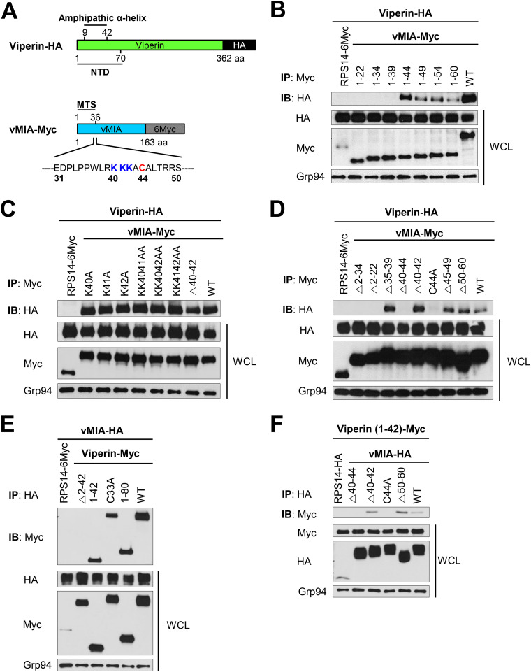
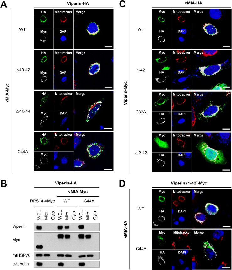
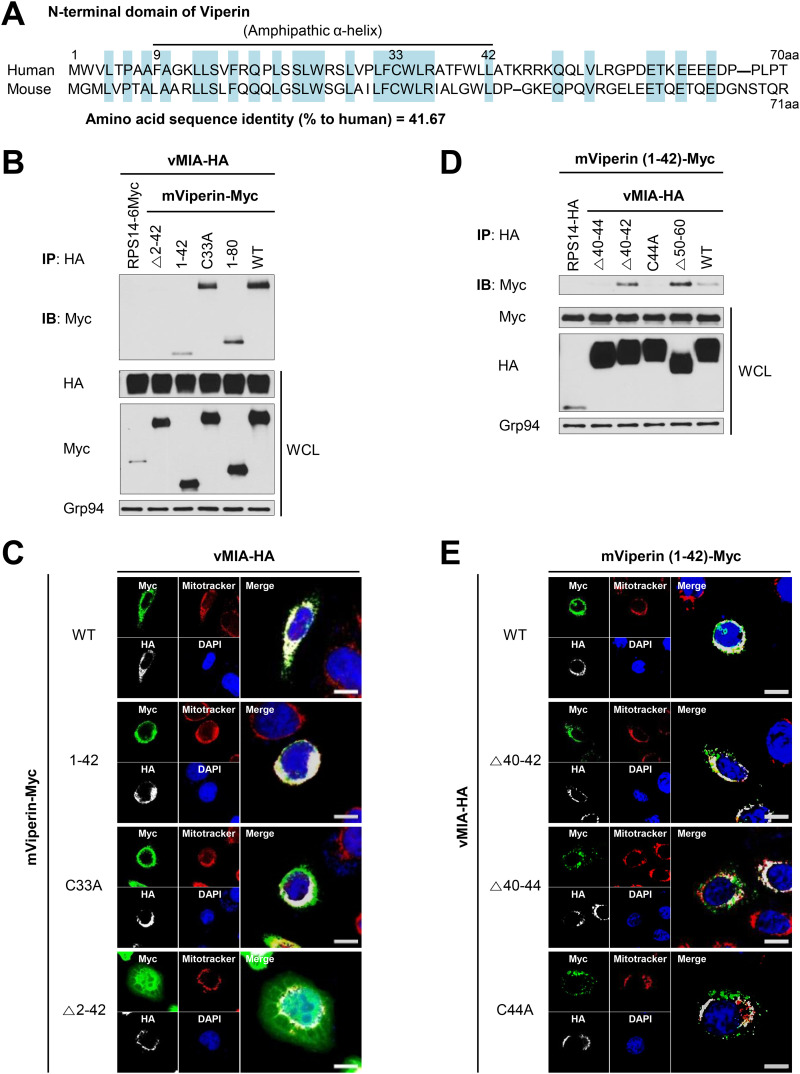
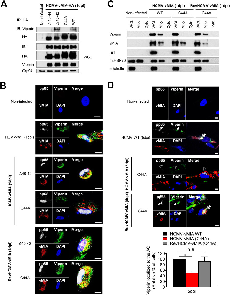
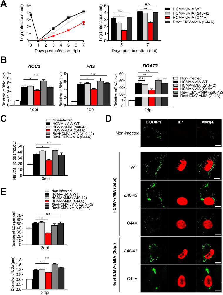
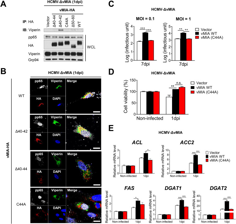
Viperin is a multifunctional interferon-inducible protein that is directly induced in cells by human cytomegalovirus (HCMV) infection. The viral mitochondrion-localized inhibitor of apoptosis (vMIA) interacts with viperin at the early stages of infection and translocates it from the endoplasmic reticulum to the mitochondria, where viperin modulates the cellular metabolism to increase viral infectivity. Viperin finally relocalizes to the viral assembly compartment (AC) at late stages of infection. Despite the importance of vMIA interactions with viperin during viral infection, their interacting residues are unknown. In the present study, we showed that cysteine residue 44 (Cys44) of vMIA and the N-terminal domain (amino acids [aa] 1 to 42) of viperin are necessary for their interaction and for the mitochondrial localization of viperin. In addition, the N-terminal domain of mouse viperin, which is structurally similar to that of human viperin, interacted with vMIA. This indicates that the structure, rather than the sequence composition, of the N-terminal domain of viperin, is required for the interaction with vMIA. Recombinant HCMV, in which Cys44 of vMIA was replaced by an alanine residue, failed to translocate viperin to the mitochondria at the early stages of infection and inefficiently relocalized it to the AC at late stages of infection, resulting in the impairment of viperin-mediated lipid synthesis and a reduction in viral replication. These data indicate that Cys44 of vMIA is therefore essential for the intracellular trafficking and function of viperin to increase viral replication. Our findings also suggest that the interacting residues of these two proteins are potential therapeutic targets for HCMV-associated diseases. IMPORTANCE Viperin traffics to the endoplasmic reticulum (ER), mitochondria, and viral assembly compartment (AC) during human cytomegalovirus (HCMV) infection. Viperin has antiviral activity at the ER and regulates cellular metabolism at the mitochondria. Here, we show that Cys44 of HCMV vMIA protein and the N-terminal domain (aa 1 to 42) of viperin are necessary for their interaction. Cys44 of vMIA also has a critical role for viperin trafficking from the ER to the AC via the mitochondria during viral infection. Recombinant HCMV expressing a mutant vMIA Cys44 has impaired lipid synthesis and viral infectivity, which are attributed to mislocalization of viperin. Cys44 of vMIA is essential for the trafficking and function of viperin and may be a therapeutic target for HCMV-associated diseases.

Keywords: HCMV; interacting residues; interferon-inducible protein; intracellular trafficking; vMIA; viperin.


Conflict of interest statement

The authors declare no conflict of interest.

Figures

FIG 1
FIG 1
Cys44 of vMIA and the N-terminal domain of viperin are essential for their interaction. (A) Constructs of HA-tagged viperin and Myc-tagged vMIA. The N-terminal domain (NTD, residues 1 to 70) and an amphipathic α-helix (residues 9 to 42) of viperin and the N-terminal mitochondrial targeting sequence (MTS, residues 2 to 36) of vMIA are shown. The amino acid sequence of vMIA is listed from aa 31 to 50, with Lys40-42 and Cys44 shown in blue and red, respectively. (B to F) HEK-293T cells were transiently cotransfected with the indicated constructs for 24 h: (B) viperin wild type (WT) and vMIA truncation mutants, (C) viperin WT and vMIA substitution mutants, (D) viperin WT and vMIA deletion mutants, (E) vMIA WT and viperin mutants, and (F) the N-terminal domain of viperin and vMIA mutants. Coimmunoprecipitation was performed, and each protein was detected by immunoblotting using specific monoclonal antibodies. Ribosomal protein subunit 14 (RPS14) was used as a negative control, and Grp94 was used as the loading control. WCL, whole-cell lysate.
FIG 2
FIG 2
Cys44 of vMIA is required for the mitochondrial localization of viperin. (A) HeLa cells were transiently cotransfected with viperin WT and vMIA mutants. Cells were stained with anti-HA and anti-myc antibodies, with MitoTracker Red as the mitochondrial indicator. DAPI (4′,6-diamidino-2-phenylindole) was used to stain the nuclei. A representative image from two independent experiments is shown. Scale bar = 10 μm. (B) HEK-293T cells were transiently cotransfected with viperin and vMIA WT or mutant (C44A) for 24 h. Cells were lysed and subjected to subcellular fractionation. Each protein in mitochondrial (Mito) and cytosolic (Cyto) fractions was detected by immunoblot using specific monoclonal antibodies. Ribosomal protein subunit 14 (RPS14) was used as a negative control. mtHSP70 and α-tubulin were used as mitochondrial and cytoplasmic markers, respectively. WCL, whole-cell lysate. (C and D) HeLa cells were transiently cotransfected with the indicated constructs: (C) vMIA WT and viperin mutants and (D) the N-terminal domain of viperin and vMIA WT or mutant (C44A). Cells were stained with anti-HA and anti-myc antibodies, with MitoTracker Red as the mitochondrial indicator. DAPI was used to stain the nuclei. A representative image from two independent experiments is shown. Scale bar = 10 μm.
FIG 3
FIG 3
The N-terminal domain of mouse viperin interacts with Cys44 of vMIA. (A) The amino acid alignment of human and mouse viperin. Shared amino acid residues are shaded in blue. The amphipathic α-helix, which is shared among mammals, extends from residues 9 to 42. (B) HEK-293T cells were transiently cotransfected with vMIA WT and mouse viperin mutants for 24 h. (B) coimmunoprecipitation was performed, and each protein was detected by immunoblotting using specific monoclonal antibodies. Ribosome protein subunit 14 (RPS14) was used as a negative control, and Grp94 was used as the loading control. WCL, whole-cell lysate. (C) Cells were stained with anti-HA and anti-myc antibodies, with MitoTracker Red as the mitochondrial indicator. DAPI was used to stain the nuclei. A representative image from two independent experiments is shown. Scale bar = 10 μm. HEK-293T cells were transiently cotransfected with the N-terminal domain of mouse viperin and vMIA mutants for 24 h. (D) coimmunoprecipitation and immunoblotting were performed with the indicated antibodies. (E) Cells were stained with anti-HA and anti-myc antibodies, with MitoTracker Red as the mitochondrial indicator. A representative image from two independent experiments is shown. Scale bar = 10 μm.
FIG 4
FIG 4
Recombinant HCMV expressing the Cys44 vMIA mutant has impaired viperin trafficking during viral infection. (A) The interaction between the vMIA mutant of the recombinant virus and endogenous viperin. HFF cells were infected with the recombinant HCMV expressing the HA-tagged vMIA mutant at an MOI of 1. At 1 day postinfection (dpi), coimmunoprecipitation was performed, and each protein was detected by immunoblotting using specific monoclonal antibodies. HCMV protein IE1 was used as the control for viral protein expression, and Grp94 was used as the loading control. WCL, whole cell-lysate. (B and C) Intracellular localization of the vMIA mutant of the recombinant virus and endogenous viperin at the early stages of infection. HFF cells were infected with the recombinant HCMV at an MOI of 1 for 1 day. (B) Cells were stained with antibodies specific to viperin and HCMV proteins vMIA and pp65. pp65 represents a stage of infection that is localized to the nucleus at early stages of infection and to the assembly compartment (AC) at late stages of infection. DAPI was used to stain the nuclei. A representative image from two independent experiments is shown. Scale bar = 10 μm. (C) Cells were lysed and subjected to subcellular fractionation. Each protein in mitochondrial (Mito) and cytosolic (Cyto) fractions was detected by immunoblotting using specific monoclonal antibodies. Noninfected cells were used as a negative control. HCMV protein IE1 was used as the control for viral protein expression. mtHSP70 and α-tubulin were used as mitochondrial and cytoplasmic markers, respectively. WCL, whole-cell lysate. (D) Intracellular localization of the vMIA mutant of the recombinant virus and endogenous viperin at the late stages of infection. HFF cells were infected with the recombinant HCMV at an MOI of 1 for 5 days. Cells were stained with antibodies specific to viperin and HCMV proteins vMIA and pp65. DAPI was used to stain the nuclei. Closed arrows indicate ACs in which viperin and pp65 are colocalized, and open arrows indicate ACs in which pp65 but not viperin is localized. A representative image from two independent experiments is shown. Scale bar = 10 μm. The efficiency of viperin localization to the AC in cells infected with the recombinant virus was quantified. Multiple frames from each coverslip were observed and imaged. A total of 50 to 100 cells displaying pp65-localized AC were counted in each frame (>20 frames/coverslip) and calculated as the ratio of cells with viperin-localized AC to cells with pp65-localized AC. As a control, the ratio in cells infected with HCMV WT was set to 100%. Data are presented as the mean ± SEM of duplicate samples and are representative of three independent experiments. Statistical analysis was performed by one-way ANOVA with Dunnett’s multiple-comparison test. *, P < 0.05.
FIG 5
FIG 5
Cys44 of vMIA is required for viperin-dependent lipid synthesis and virion production. (A) HFF cells were infected with the recombinant HCMV at an MOI of 0.05 for the indicated duration. Virus yield was quantified by a fluorescence-based viral infectivity assay. (B) Relative mRNA levels of lipogenic enzymes in HFF cells infected with the recombinant HCMV at an MOI of 1 for 1 day. ACC2, acetyl-coenzyme A (CoA) carboxylase 2; FAS, fatty acid synthase; DGAT2, diacylglycerol acyltransferases 2. HFF cells were infected with the recombinant virus at an MOI of 1 for 3 days: (C) quantification of total neutral lipids. Total neutral lipids from 3 × 106 cells were extracted and quantified using a fluorescence-based lipid assay kit. (D) Accumulation of lipid droplets (LDs). Cells were stained with BODIPY 493/503 neutral lipid dye, a marker for LDs, and an antibody specific to HCMV protein IE1 to identify infected cells. A representative image from three independent experiments is shown. Scale bar = 20 μm. (E) Quantification of LDs. The LD number is the mean of 20 cells ± SEM; the LD diameter is the mean of 150 LDs ± SEM. Data are presented as the mean ± SEM of triplicate samples and are representative of three independent experiments. Statistical analysis was performed by one-way ANOVA with Dunnett’s multiple-comparison test. *, P < 0.05; **, P < 0.01; ***, P < 0.0001.
FIG 6
FIG 6
Viperin interaction and trafficking in cells stably expressing vMIA mutants during vMIA-deficient HCMV infection. (A and B) HFtelo cells stably expressing HA-tagged vMIA WT and mutants were infected with an HCMV mutant lacking vMIA (HCMV-ΔvMIA) at an MOI of 1 for 1 day. (A) Viperin interaction with vMIA mutants. Coimmunoprecipitation was performed, and each protein was detected by immunoblotting using specific monoclonal antibodies. HCMV protein pp65 was used as the control for viral protein expression, and Grp94 was used as the loading control. WCL, whole-cell lysate. (B) Intracellular localization of vMIA mutants and endogenous viperin during HCMV-ΔvMIA infection. Cells were stained with antibodies specific to HCMV proteins pp65 and HA-tagged vMIA and viperin. DAPI was used to stain the nuclei. A representative image from two independent experiments is shown. Scale bar = 10 μm. (C and D) HFtelo cells stably expressing vMIA WT and the mutant (C44A) were infected with HCMV-ΔvMIA (C) at an MOI of 0.1 or 1 for 7 days and (D) at an MOI of 1 for 1 day. (C) Viral yield was quantified by a fluorescence-based virus infectivity assay. (D) Cell viability was analyzed by a trypan blue dye-exclusion assay. (E) Relative mRNA levels of lipogenic enzymes in HFtelo cells stably expressing vMIA WT and mutant (C44A) during HCMV-ΔvMIA infection. ACL, ATP-citrate lyase; ACC2, acetyl-coenzyme A (CoA) carboxylase 2; FAS, fatty acid synthase; DGAT1 and DGAT2, diacylglycerol acyltransferases 1 and 2. Data are presented as the mean ± SEM of triplicate samples and are representative of three independent experiments. Statistical analysis was performed by one-way ANOVA with Dunnett’s multiple-comparison test. *, P < 0.05; **, P < 0.01; ***, P < 0.0001.

Similar articles

Cited by

References

    1. Fülöp T, Larbi A, Pawelec G. 2013. Human T cell aging and the impact of persistent viral infections. Front Immunol 4:271–271. doi:10.3389/fimmu.2013.00271. - DOI - PMC - PubMed
    1. Meakins JL. 1983. Clinical approach to infection in the compromised host. Ann Surg 197:491–491.
    1. Landolfo S, Gariglio M, Gribaudo G, Lembo D. 2003. The human cytomegalovirus. Pharmacol Ther 98:269–297. doi:10.1016/s0163-7258(03)00034-2. - DOI - PubMed
    1. Warnatsch A. 2013. Impact of proteasomal immune adaptation on the early immune response to viral infection. PhD thesis. Kings College London, London, UK.
    1. Crough T, Khanna R. 2009. Immunobiology of human cytomegalovirus: from bench to bedside. Clin Microbiol Rev 22:76–98. doi:10.1128/CMR.00034-08. - DOI - PMC - PubMed

Publication types