. 2023 Sep 12;7(17):4936-4949.
doi: 10.1182/bloodadvances.2022009590.

Platelet functional abnormalities in pediatric patients with kaposiform hemangioendothelioma/Kasabach-Merritt phenomenon

Affiliations

Platelet functional abnormalities in pediatric patients with kaposiform hemangioendothelioma/Kasabach-Merritt phenomenon

Alexey A Martyanov et al. Blood Adv. .

Abstract

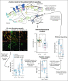
Kaposiform hemangioendothelioma (KHE) is a rare vascular tumor of infancy that is commonly associated with a life-threatening thrombocytopenic condition, Kasabach-Merritt phenomenon (KMP). Platelet CLEC-2, tumor podoplanin interaction is considered the key mechanism of platelet clearance in these patients. Here, we aimed to assess platelet functionality in such patients. Three groups of 6 to 9 children were enrolled: group A with KHE/KMP without hematologic response (HR) to therapy; group B with KHE/KMP with HR; and group C with healthy children. Platelet functionality was assessed by continuous and end point flow cytometry, low-angle light scattering analysis (LaSca), fluorescent microscopy of blood smears, and ex vivo thrombi formation. Platelet integrin activation in response to a combination of CRP (GPVI agonist) and TRAP-6 (PAR1 agonist), as well as calcium mobilization and integrin activation in response to CRP or rhodocytin (CLEC-2 agonist) alone, were significantly diminished in groups A and B. At the same time, platelet responses to ADP with or without TRAP-6 were unaltered. Thrombi formation from collagen in parallel plate flow chambers was also noticeably decreased in groups A and B. In silico analysis of these results predicted diminished amounts of CLEC-2 on the platelet surface of patients, which was further confirmed by immunofluorescence microscopy and flow cytometry. In addition, we also noted a decrease in GPVI levels on platelets from group A. In KHE/KMP, platelet responses induced by CLEC-2 or GPVI activation are impaired because of the diminished number of receptors on the platelet surface. This impairment correlates with the severity of the disease and resolves as the patient recovers.


Conflict of interest statement

Conflict-of-interest disclosure: The authors declare no competing financial interests.

Figures
None
Graphical abstract
Figure 1.
Figure 1.
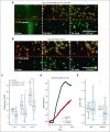
Platelet thrombus formation and aggregation in KHE/KMP. Analysis of platelets from blood samples from healthy children (triangles) and patients with KHE/KMP with (circles) or without (crosses) HR. (A-B) Typical microscopy images of the growing thrombi of patients with KHE/KMP with HR (A) and healthy donors (B) after 5, 10, and 15 minutes of the whole hirudin-anticoagulated blood perfusion through fibrillar collagen-coated flow chamber. Thrombi (highlighted in red) were identified by DiOC-6 fluorescence (green). (C) Scaled thrombi areas at different time points. (D) Typical aggregation curves for the LaSca assays after activation with 800 nM ADP (black curves) or 20 μg/mL collagen (red curve). (E) Initial velocities of platelet aggregation induced by 800 nM ADP or 20 μg/mL collagen. Statistical significance was calculated using Mann-Whitney U test; ∗ P < .05.
Figure 2.
Figure 2.
KHE/KMP affects platelet responsiveness to GPVI-mediated activation. Flow cytometry analysis of platelets and designations of groups of patients are the same as in Figure 1. (A-B) characteristics of platelets in resting state: forward light scattering (FSC) indicating platelet size (A) and cytosolic calcium concentration (B). (C-D) platelet responses to strong activation with a combination of 10 μg/mL CRP and 12.5 μM TRAP-6 (CRP + TRAP-6); or 100 μM of TRAP-6, 100 μM of AYGPKF, and 5 μM of ADP (2TR+ADP): platelet PAC-1 binding (C) and CD62p expression (D). (E-F) platelet responses to mild activation with either 2 μM of ADP (ADP), 2 μg/mL CRP (CRP) or 5 μM of TRAP-6 (TRAP-6): cytosolic calcium concentration (H) and fibrinogen binding (I). Statistical significance was calculated by means of Mann-Whitney U test. ∗P < .05; ∗∗P < .01.
Figure 3.
Figure 3.
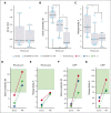
Platelet activation via CLEC-2 and GPVI receptors. Flow cytometry and low-angle light scattering aggregometry analysis and designations of groups of patients are the same as in Figure 1. (A) Initial velocity of platelet aggregation upon activation with 10 nM of rhodocytin. (B-C) Calcium mobilization (B) and fibrinogen binding (C) upon activation with 200 nM of rhodocytin. (D-G) Calcium mobilization (D,F) and fibrinogen binding (E,G) upon platelet activation with 200 nM of rhodocytin (D-E) or 2 μg/mL of CRP (F-G) for patients at the time point of enrollment (without HR, “Enroll.”) and upon HR (“HR”). Red lines correspond to patient 1, blue lines correspond to patient 9, and green lines correspond to patient 11. Green regions correspond to healthy-donor ranges. Statistical significance was calculated using Mann-Whitney U test. ∗P < .05; ∗∗P < .01.
Figure 4.
Figure 4.
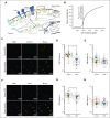
Patients with KHE/KMP have decreased levels of CLEC-2 and GPVI on the platelet surface. (A) Scheme of the computational model of CLEC-2 induced signaling in platelets: CLEC-2 activation results in the receptor clustering and phosphorylation by Syk kinases. In resting platelets, small amount of active Syk is maintained by active SFK kinases, which, in turn, are maintained active by CD148 phosphatases. Nonactive Syk bind to phosphorylated and clustered CLEC-2 receptors and also become active. Active Syk phosphorylate LAT and TULA-2. TULA-2 is the negative regulator of platelet Syk activation. PLCγ2 and PI3K bind to phosphorylated LAT, and PI3K becomes active. PI3K produces PIP3 from PIP2, which is bound by PH-domain of Btk. Hereby, Btk becomes active and activates PLCγ2, which hydrolyzes PIP2 and produces IP3, which initiates cytosolic calcium signaling. (B) Dependance of the maximally achievable cytosolic calcium concentration from CLEC-2 number on the resting platelets, predicted by the computational model. Physiological number of CLEC-2 per platelet is highlighted in red. (C) Typical results of the microscopic immunofluorescence analysis of the CLEC-2 expression on the platelet surface of the patients with KHE/KMP and healthy donors. (D-E) Fluorescence intensities of the anti–CLEC-2 antibodies (D) and anti-NMII antibodies (E) of the patients with KHE/KMP and healthy donors. Blurred corresponds to unique platelet measurements, and bright dots correspond to patients. Each color corresponds to unique patients. Seven healthy donors and 6 patients with KHE/KMP were analyzed. (F) Typical results of the microscopic immunofluorescence analysis of the GPVI expression on the platelet surface (GPVI; NMII) of the patients with KHE/KMP and healthy donors. (G-H) Fluorescence intensities of the anti-GPVI antibodies (G) and anti-NMII antibodies (H) of the KHE/KMP patients and healthy donors. Blurred corresponds to unique platelet measurements, and bright dots correspond to patients. Each color corresponds to unique patients. Seven healthy donors and 6 patients with KHE/KMP were analyzed. Statistical significance was calculated using Mann-Whitney U test. ∗P < .05; ∗∗P < .01. CLEC-2, anti–CLEC-2 antibodies; GPVI, anti-GPVI antibodies; NMII, nonmuscular myozin II (used for platelet identification).
Figure 5.
Figure 5.
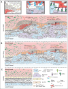
Proposed scheme of the KHE/KMP impact on patient platelets. (A) KHE pathogenesis is mostly based on aberrant VEGFR signaling in LECs. These LECs are constitutively proliferating and subsequently intervene in the blood vessel endothelium, thus causing blood endothelium activation, disruption, and extracellular matrix exposure to the blood flow (Ai)., Exposed collagen causes the initiation of platelet thrombus formation and subsequent thrombocytopenia due to platelet consumption. Thrombi are heterogenous in nature; although platelets in the thrombus core are highly activated by a combination of collagen, thrombin, and platelet granule contents, platelets in the thrombus shell are stimulated mostly by ADP and thromboxane. Here, platelets in the outer layers of the thrombus can leave the thrombus shell and return to the circulation., Meanwhile, even moderate platelet activation can cause platelet GPVI receptor shedding. Thus, these preactivated platelets (orange platelets on the scheme) become less responsive to further GPVI-mediated activation (Ai). In the meantime, pathologically proliferating LECs also intrude the blood flow and, via the podoplanin-CLEC-2 axis, initiate platelet adhesion and activation (Aii-iii)., Active platelets secrete growth factors stored in their granules, which causes additional LEC and EC activation and proliferation, thus acting as a positive feedback loop (Aii)., Finally, platelets are heterogenous because of the mechanisms of their production: platelets are “ripped” from the megakaryocytes, and thus, platelet membrane receptor expression is not uniform. Hereby, it can be expected that some of the platelets in the blood flow have higher CLEC-2 levels (green platelets on the scheme) and some have lesser CLEC-2 levels (white platelets on the scheme). LECs, by exposing podoplanin to the blood flow, capture platelets with higher CLEC-2 levels, acting like filters. Hence, only platelets with lesser CLEC-2 on the surface, those cannot be effectively captured by the LECs, remain in circulation (Aiii). (B) Upon HR, proliferation of the LECs is abrogated because chemotherapy inhibits their VEGF signaling. This allows the blood vessel endothelial cells to cover the exposed extracellular matrix and thus significantly reduce pathological thrombus formation. However, despite being inhibited, LECs still remain in the blood vasculature and still filter platelets with higher CLEC-2 levels (Biii). These platelets secrete growth factors from their granules and thus activate remaining LECs, which maintain LECs’ presence in the blood flow (Bii). (C) Finally, upon recovery, no LECs are exposed to blood flow.

References

    1. Croteau SE, Liang MG, Kozakewich HP, et al. Kaposiform hemangioendothelioma: atypical features and risks of Kasabach-Merritt phenomenon in 107 referrals. J Pediatr. 2013;162(1):142–147. - PMC - PubMed
    1. Mahajan P, Margolin J, Iacobas I. Kasabach-Merritt phenomenon: classic presentation and management options. Clin Med Insights Blood Disord. 2017;10 1179545X17699849. 1-5. - PMC - PubMed
    1. Chinello M, Di Carlo D, Olivieri F, et al. Successful management of kaposiform hemangioendothelioma with long-term sirolimus treatment: a case report and review of the literature. Mediterr J Hematol Infect Dis. 2018;10(1) 1-6. - PMC - PubMed
    1. Enjolras O, Wassef M, Mazoyer E, et al. Infants with Kasabach-Merritt syndrome do not have “true” hemangiomas. J Pediatr. 1997;130(4):631–640. - PubMed
    1. Kasabach H, Merritt K. Capillary hemangioma with extensive purpura. Am J Dis Child. 1940;59(5):1063–1070.

Publication types

MeSH terms

Substances

Supplementary concepts