. 2023 Jun 20;120(25):e2216206120.
doi: 10.1073/pnas.2216206120. Epub 2023 Jun 12.

Trophoblast PR-SET7 dysfunction induces viral mimicry response and necroptosis associated with recurrent miscarriage

Affiliations

Trophoblast PR-SET7 dysfunction induces viral mimicry response and necroptosis associated with recurrent miscarriage

Xiaobo Zhou et al. Proc Natl Acad Sci U S A. .

Abstract

Recurrent miscarriage (RM) is a distressing pregnancy complication. While the etiology of RM remains unclear, growing evidence has indicated the relevance of trophoblast impairment to the pathogenesis of RM. PR-SET7 is the sole enzyme catalyzing monomethylation of H4K20 (H4K20me1) and has been implicated in many pathophysiological processes. However, how PR-SET7 functions in trophoblasts and its relevance to RM remain unknown. Here, we found that trophoblast-specific loss of Pr-set7 in mice led to defective trophoblasts, resulting in early embryonic loss. Mechanistic analysis revealed that PR-SET7 deficiency in trophoblasts derepressed endogenous retroviruses (ERVs), leading to double-stranded RNA stress and subsequent viral mimicry, which drove overwhelming interferon response and necroptosis. Further examination discovered that H4K20me1 and H4K20me3 mediated the inhibition of cell-intrinsic expression of ERVs. Importantly, dysregulation of PR-SET7 expression and the corresponding aberrant epigenetic modifications were observed in the placentas of RM. Collectively, our results demonstrate that PR-SET7 acts as an epigenetic transcriptional modulator essential for repressing ERVs in trophoblasts, ensuring normal pregnancy and fetal survival, which sheds new light on potential epigenetic causes contributing to RM.

Keywords: ERVs; H4K20me1/3; PR-SET7; recurrent miscarriage; viral mimicry.


Conflict of interest statement

The authors declare no competing interest.

Figures

Fig. 1.
Fig. 1.
Trophoblast-specific deletion of Pr-set7 leads to pregnancy loss in mice. (A) Immunohistochemical analysis of PR-SET7 and H4K20me1 expression at E4.5–E8.5 during normal placental development. (B) The strategy for generating mice with trophoblast-specific deletion of Pr-set7 (Pr-set7d/d). (C) H&E staining of implantation sites of Pr-set7f/f and Pr-set7d/d mice at E5.5–E7.5. (D) Representative Immunostaining images of EOMES, AP-2γ, OCT4, and pH3 in Pr-set7f/f and Pr-set7d/d embryos at E5.5. (E) Immunostaining images of H4K20me1 and γH2AX in Pr-set7f/f and Pr-set7f/f embryos at E5.25. Images in A and CE are representative of at least two or three independent experiments, respectively. Em, embryo; ExE, extraembryonic ectoderm; Epi, epiblast; EPC, ectoplacental cone; Ch, chorion; Cp, chorionic plate; S, uterine stromal cells; VE, visceral endoderm.
Fig. 2.
Fig. 2.
PR-Set7 deficiency induces genome instability in TSCs. (A) Immunoblot analysis of PR-SET7, H4K20me1, and H4K20me3 in mTSCs treated with or without Dox for 48 h (Pr-set7d/d and Pr-set7f/f). β-Tubulin and H3 were used as loading controls. n = 3. (B) The MTS assay showed an impaired proliferation of mTSCs upon Pr-set7 knockout. n = 3. (C) FACS analysis revealed significant cell cycle arrest in mTSCs treated with Dox for 24 h (Pr-set7d/d). n = 3. (D) Immunofluorescence analysis of EdU and γH2AX in mTSCs treated with or without Dox for 24 h (Pr-set7d/d and Pr-set7f/f). (E) Immunoblot analysis showed decreased levels of PR-SET7, H4K20me1, and H4K20me3 in the presence of si-PR-SET7 for 48 h. β-Tubulin and H3 were used as loading controls. n = 3. (F) The MTS assay of hTSCs transfected with si-NC or si-PR-SET7 for indicated hours. n = 3. (G) Immunofluorescence analysis of γH2AX and pH3 in hTSCs transfected with si-NC or si-PR-SET7 for 48 h. (H) The quantitative results of G. Images in D and G are representative of at least three independent experiments. In B, C, F, and H, Two-tailed unpaired Student’s t test. *P < 0.05, **P < 0.01, ***P < 0.001.
Fig. 3.
Fig. 3.
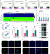
PR-SET7 deficiency results in necroptosis in TSCs. (A and B) GSEA shows the enrichment of genes involved in apoptosis and the p53 pathway in TSCs with PR-SET7 deletion in both mice (A) and humans (B). NES, normalized enrichment score. (C) Heatmap showing upregulation of genes involved in cell death upon Pr-set7 deletion in mTSCs. (D) qRT-PCR confirmed the upregulation of representative genes of cell death. n = 6. (E and F) FACS analysis revealed increased cell death in Pr-set7d/d mTSCs. Two-tailed unpaired Student’s t test, ***P < 0.001. n = 4. (G) qRT-PCR confirmed increased expression of Ripk3 and Mlkl. n = 4. (H) Immunoblots of p-MLKL(S345), MLKL, p-RIPK3, RIPK3, and ZBP1 in Pr-set7d/d versus Pr-set7f/f mTSCs. β-Actin was used as a loading control. n = 3. (I and J) Immunostaining of p-MLKL(S345) in mTSCs treated with or without UNC0379 for 48 h (I) and in Pr-set7f/f and Pr-set7d/d embryos at E5.25 (J). ExE, extraembryonic ectoderm; Epi, epiblast; S, uterine stromal cells. The images are representative of at least three independent experiments. In D and G, the values are normalized to Gapdh and indicated as the mean ± SEM. Two-tailed unpaired Student’s t test, *P < 0.05, **P < 0.01, ***P < 0.001.
Fig. 4.
Fig. 4.
PR-SET7 deletion activates interferon response in TSCs. (A) Gene ontology (GO) analysis of the DEGs in PR-SET7-knockdown versus control hTSCs. BP, Biological Process; CC, Cellular Component; MF, Molecular Function. (B) GSEA reveals the changes in the indicated gene signatures in hTSCs upon PR-SET7 deletion. (C) Heatmap showing the upregulation of genes involved in inflammatory response and interferon response in hTSCs with PR-SET7 knockdown. (D and E) qRT-PCR confirmed the upregulation of representative genes of the inflammatory response and interferon response in hTSCs with PR-SET7 knockdown (D) and treated with UNC0779 (E), respectively. (F) GSEA reveals the changes in the indicated gene signatures in mTSCs upon Pr-set7 knockout. (G) Heatmap showing the upregulation of genes involved in the interferon response in Pr-set7d/d mTSCs. (H) qRT-PCR confirmed the upregulation of representative genes related to interferon response signaling pathway. In D, E, and H, the values are normalized to Gapdh and indicated as the mean ± SEM. Two-tailed unpaired Student’s t test, *P < 0.05, **P < 0.01, ***P < 0.001. n = 4.
Fig. 5.
Fig. 5.
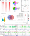
PR-SET7-H4K20me1 represses interferon-related genes indirectly in TSCs. (A) Heatmap representation of H4K20me1, H4K20me3, and H3K4me3 ChIP-seq tag density within 3,000 bp around TSS in hTSCs. (B) Genomic read distributions of H4K20me1 and H4K20me3 binding sites in hTSCs. (C) Average ChIP-seq tag density plots of H3K4me3 (Upper) and H4K20me1 (Lower) on highly expressed or non-expressed genes in the region −3,000 bp upstream of TSS and 3,000 bp downstream of TES. (D) Average ChIP-seq read density of H4K20me1 on DEGs from RNA-seq data of hTSCs. (E and F) Venn diagram showing the overlap of H4K20me1 targets and upregulated genes induced by si-PR-SET7 (E) and GO analysis of the common genes (F) in hTSCs. (G) Genome browser view of normalized RNA-seq tracks and ChIP-seq signals of H4K20me1 at the interferon signaling-associated gene locus. (H) Representative RNA-seq tracks, the ChIP-seq signals of H4K20me1 and H3K4me3 at the ISGs locus. TSS, Transcription Start Site; TES, Transcription End Site.
Fig. 6.
Fig. 6.
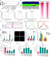
PR-SET7 deficiency induces ERV expression and dsRNA formation in TSCs. (A) KEGG enrichment analysis showed that the upregulated genes were associated with multiple viral infection responses. (B) Heatmap showing the differential expression of ERVs upon PR-SET7 KD in hTSCs. (C) Heatmap representation of ChIP-seq tag density of H4K20me1 and H4K20me3 binding to TEs. (D) The analysis of ChIP-seq data and ERV transcripts expression revealed an inverse correlation between H4K20me1 binding and ERV expression. (E) Average ChIP-seq tag density plots of H4K20me1 and H4K20me3 on ERVs in the region upstream of TSS to downstream of TES. (F and G) Representative UCSC tracks and ChIP-seq analysis of H4K20me3 and H4K20me1 at HERVH-int: ERV1: LTR locus. (H) qRT-PCR analysis of the ERVs captured by J2 antibody in the pull-down assay. (I) Immunostaining of J2 in hTSCs transfected with si-NC or si-PR-SET7 for 48 h. n = 3. (J) qRT-PCR analysis revealed the upregulation of dsRNA sensors in hTSCs treated with si-PR-SET7. n = 4. (K) qRT-PCR analysis revealed the increased expression of interferon response genes in hTSCs treated with si-PR-SET7 was partially reversed by TLR3 inhibitor TLR3-IN-1. n = 3. (L) The analysis of cell viability in hTSCs treated with UNC0379 (2.5 μM), with or without zVAD-fmk (10 mM) or/and TLR3-IN-1(20 μM). (M and N) The analysis of cell death in hTSCs (M) and mTSCs (N) treated with UNC0379 (5 μM in mTSCs) for 48 h with or without zVAD-fmk (10 mM) or/and GSK872 (2 mM). Two-tailed unpaired Student’s t test, NS, no significance, ***P < 0.001. In J and K, the values are normalized to GAPDH and indicated as the mean ± SEM. Two-tailed unpaired Student’s t test, *P < 0.05, **P < 0.01, ***P < 0.001.
Fig. 7.
Fig. 7.
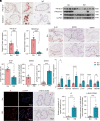
Decreased expression of PR-SET7 is associated with recurrent miscarriage in humans. (A) Immunohistochemical analysis of PR-SET7 and H4K20me1 in the placental villous tissues from normal pregnancies. (B) Immunoblot analysis of PR-SET7 and H4K20me1 in the villi from normal and RM pregnancies. Nor, normal control; RM, recurrent miscarriage. GAPDH and H4 were used as loading controls. (C) Quantitative results of B. (D) Immunostaining of Ki-67, MCM2, and γH2AX in the villi from normal and RM pregnancies. (E) Quantitative results of D. (F) qRT-PCR analysis of ERVs in the villi from normal and RM pregnancies. (G) Immunostaining analysis of J2 and p-MLKL(S358) in the villi from normal and RM pregnancies. (H and I) Quantitative results of G. The data shown in C, E, F, H, and I represent the mean ± SEM. Two-tailed unpaired Student’s t test, ***P < 0.001, ****P < 0.0001. n = 10 for Nor, and n = 14 for RM. CT, cytotrophoblast; ST, syncytiotrophoblast.
Fig. 8.
Fig. 8.
Schematic illustration showing how PR-SET7 is essential for the survival of trophoblasts. PR-SET7 suppresses interferon response and necroptosis via inhibiting the transcription of ERVs and dsRNA formation. Under a PR-SET7 dysfunctional condition, aberrantly upregulated dsRNAs lead to an overwhelming interferon response and necroptosis, potentially contributing to the pathogenesis of RM.

Similar articles

Cited by

References

    1. Dimitriadis E., Menkhorst E., Saito S., Kutteh W. H., Brosens J. J., Recurrent pregnancy loss. Nat. Rev. Dis. Primers 6, 98 (2020). - PubMed
    1. Lv S., et al. , The attenuation of trophoblast invasion caused by the downregulation of EZH2 is involved in the pathogenesis of human recurrent miscarriage. Mol. Ther. Nucleic Acids 14, 377–387 (2019). - PMC - PubMed
    1. Wang X. H., et al. , Low chorionic villous succinate accumulation associates with recurrent spontaneous abortion risk. Nat. Commun. 12, 3428 (2021). - PMC - PubMed
    1. Saha B., et al. , TEAD4 ensures postimplantation development by promoting trophoblast self-renewal: An implication in early human pregnancy loss. Proc. Natl. Acad. Sci. U.S.A. 117, 17864–17875 (2020). - PMC - PubMed
    1. Hemberger M., Hanna C. W., Dean W., Mechanisms of early placental development in mouse and humans. Nat. Rev. Genet. 21, 27–43 (2020). - PubMed

Publication types