. 2023 Jun 20;120(25):e2219431120.
doi: 10.1073/pnas.2219431120. Epub 2023 Jun 12.

Cross talk between Paneth and tuft cells drives dysbiosis and inflammation in the gut mucosa

Affiliations

Cross talk between Paneth and tuft cells drives dysbiosis and inflammation in the gut mucosa

Nathalie Coutry et al. Proc Natl Acad Sci U S A. .

Abstract

Gut microbiota imbalance (dysbiosis) is increasingly associated with pathological conditions, both within and outside the gastrointestinal tract. Intestinal Paneth cells are considered to be guardians of the gut microbiota, but the events linking Paneth cell dysfunction with dysbiosis remain unclear. We report a three-step mechanism for dysbiosis initiation. Initial alterations in Paneth cells, as frequently observed in obese and inflammatorybowel diseases patients, cause a mild remodeling of microbiota, with amplification of succinate-producing species. SucnR1-dependent activation of epithelial tuft cells triggers a type 2 immune response that, in turn, aggravates the Paneth cell defaults, promoting dysbiosis and chronic inflammation. We thus reveal a function of tuft cells in promoting dysbiosis following Paneth cell deficiency and an unappreciated essential role of Paneth cells in maintaining a balanced microbiota to prevent inappropriate activation of tuft cells and deleterious dysbiosis. This succinate-tuft cell inflammation circuit may also contribute to the chronic dysbiosis observed in patients.

Keywords: Paneth cells; antimicrobial peptides; gut inflammation; microbiota dysbiosis; tuft cells.


Conflict of interest statement

The authors declare no competing interest.

Figures

Fig. 1.
Fig. 1.
Maintenance of intestinal Paneth cell terminal differentiation requires the Sox9 transcription factor. (A) Representative immunofluorescence costaining of Lyz and β-catenin showing the presence of Lyz+ Paneth cells 2 wk after tamoxifen-induced Sox9 deletion in adult epithelial cells (Sox9LoxP/LoxP;Villin-CreERT2 mice). Delocalized Paneth cells are indicated with a white arrowhead. Images are representative of 3 mice per genotype, 3 independent experiments. (B) Representative immunofluorescence costaining of Sox9, Lyz and β-catenin showing deletion of Sox9 in stem cells, Paneth cells and progenitors in Sox9LoxP/LoxP;Villin-CreERT2 mice (white arrowhead) 2 wk following tamoxifen treatment. Sox9 expression is maintained in Paneth cells (yellow arrowhead) in Sox9LoxP/LoxP;P450a1-Cre mice (3 mice per genotype), 3 independent experiments. (C) Electron micrographs from small intestine highlighting physiological secretory granules in Paneth cells (arrows) in control Sox9LoxP/LoxP mice (n = 4), whereas secretory granules in Sox9-deficient Paneth cells from Sox9LoxP/LoxP;Villin-CreERT2 mice (n = 5, >30 d post tamoxifen treatment) are less electron dense and exhibit heterogeneity in size, shape, and number. In contrast, secretory granules exhibit normal structure in mice with a Sox9 deletion in all epithelial cells except for Paneth cells (Sox9LoxP/LoxP;P450a1-Cre; n = 2, 1 wk post-βNF treatment). (D) Representative immunofluorescence costaining of Muc2, Lyz, and β-catenin in Sox9LoxP/LoxP and Sox9LoxP/LoxP;Villin-CreERT2 mice (n = 3 per genotype, 2 wk following tamoxifen treatment). Muc2 expression in Paneth cells was only detected following Sox9 deletion in all epithelial cells (white arrowheads). (E) Expression of Paneth cell markers in Sox9LoxP/LoxP and Sox9LoxP/LoxP;Villin-CreERT2 small intestine was evaluated by RT-qPCR analysis in Sox9LoxP/LoxP;Villin-CreERT2 and Sox9LoxP/LoxP mice (n = 4 and n = 5, respectively; 2 wk after tamoxifen treatment). Data are shown as a ratio of expression relative to Sox9LoxP/LoxP mice. [Scale bars: 20 µm (A, B, and D), 2 µm (C).] (E) Mann–Whitney U test; *P < 0.05; **P < 0.01. See also SI Appendix, Fig. S1.
Fig. 2.
Fig. 2.
Alterations of gut microbiota and intestinal barrier are associated with abnormal Paneth cells. (A) β diversity in microbial communities from Sox9LoxP/LoxP and Sox9LoxP/LoxP;Villin-CreERT2 mice (n = 8 mice per genotype). Unweighted principal component analyses (PCA) of UniFrac distance after 16S rRNA sequencing of fecal microbial populations are presented. (B) Intestinal permeability, assessed by oral gavage of FITC-dextran, was evaluated in control mice (Sox9LoxP/LoxP 2 wk post tamoxifen treatment, n = 7), following deletion of Sox9 in all epithelial cells (Sox9LoxP/LoxP;Villin-CreERT2 mice; 2 wk post tamoxifen treatment, n = 7) or under conditions where Sox9 expression was maintained in Paneth cells (Sox9LoxP/LoxP;P450a1-Cre mice; 2 wk post βNF treatment, n = 3). (B) Kruskal–Wallis test with Dunn's post hoc test; NS: not significant; *P < 0.05. The line indicates the median, the box marks the 25th and 75th percentiles, and the whiskers indicate the minimal to maximum values.
Fig. 3.
Fig. 3.
Presence of a type 2 immune response in mice with abnormal Paneth cells. (A) Interleukins and transcription factors associated with type 1, 2, and 17 immunity were evaluated in Sox9LoxP/LoxP (n = 4) and Sox9LoxP/LoxP;Villin-CreERT2 (n = 5) small intestine by RT-qPCR analysis. Data are shown as a ratio relative to expression in Sox9LoxP/LoxP mice. (B) Representative immunostainings of Mbp, a marker of eosinophilia, and Gata3, a marker of Th2 lymphocytes and ILC2, in Sox9-deficient lamina propria in the small intestinal epithelium of Sox9LoxP/LoxP and Sox9LoxP/LoxP;Villin-CreERT2 mice. n = 3 mice per genotype, killed 2 wk after tamoxifen treatment. (C) Quantification of MBP and Gata3+ cells in Sox9LoxP/LoxP and Sox9LoxP/LoxP;Villin-CreERT2 mice. Cells positive for MBP or Gata3 were counted in n = 50 crypt-villus units per mouse (n = 3 mice per genotype). (D) Representative immunostainings illustrating tissue remodeling in the small intestinal epithelium from Sox9LoxP/LoxP;Villin-CreERT2 mice. Dclk1 and PAS stainings, respectively, show tuft cell and goblet hyperplasia in Sox9-deficient small intestine. Retnlβ staining reveals the production of Retnlβ molecule by goblet cells, which are costained with Alcian blue. n = 3 mice per genotype, killed 2 wk after tamoxifen treatment. (E) Quantification of type 2 immune response markers in Sox9LoxP/LoxP and Sox9LoxP/LoxP;Villin-CreERT2 mice. Cells positive for Dclk1, PAS, Retnlβ Gata3, or Alcian blue were counted in n = 50 crypt-villus units per mouse (n = 3 mice per genotype). Mice were killed 2 wk after tamoxifen treatment. [Scale bars: 20 µm (B and D).] (A, C, and E) Mann–Whitney U test. *P < 0.05; **P < 0.01; ***P < 0.001 (A, C, and E) Data are shown as mean ± SEM. See also SI Appendix, Fig. S2.
Fig. 4.
Fig. 4.
The presence of tuft cells is required for the development of type 2 immunity and impaired intestinal permeability caused by altered Paneth cells. (A) Representative immunofluorescence costaining of Muc2, Lyz, and β-catenin in Sox9LoxP/LoxP;Villin-CreERT2;Pou2f3−/− mice. Paneth cells lacking Sox9 in a tuft cell-deficient context display an altered differentiation state since Muc2 is present in Paneth cells in Sox9LoxP/LoxP;Villin-CreERT2 as well as Sox9LoxP/LoxP;Villin-CreERT2;Pou2f3−/− mice (white arrowhead), and Lyz staining is diffuse in both genotypes compared to control mice (yellow arrowhead). n = 3 mice per genotype, representative of 2 independent experiments. Mice were killed 2 wk after tamoxifen treatment. (B) Intestinal permeability, assessed by FITC-dextran gavage, remains intact when Sox9 is deleted in a tuft cell-deficient mouse model. FITC–dextran was measured in serum from Sox9LoxP/LoxP;Pou2f3+/+ mice (n = 4), Sox9LoxP/LoxP;Villin-CreERT2;Pou2f3+/+mice (n = 4), Sox9LoxP/LoxP;Pou2f3−/− mice (n = 6), and Sox9LoxP/LoxP;Villin-CreERT2;Pou2f3−/− mice (n = 7). Mice were killed 2 wk after tamoxifen treatment. (C) The type 2 immune response found in Sox9LoxP/LoxP;Villin-CreERT2 mice is abolished in tuft cell-deficient Sox9LoxP/LoxP;Villin-CreERT2;Pou2f3−/− mice as compared to Sox9LoxP/LoxP;Villin-CreERT2;Pou2f3+/+ mice. Cells positive for Dclk1, Retnlβ or Gata3 were counted in n = 50 crypt-villus units per mouse (n = 5 to 6 mice per genotype). [Scale bars, 20 µm (A).] (B and C) Kruskal–Wallis test with Dunn's post hoc test; NS: not significant; *P < 0.05; ****P < 0.0001. (B) The line indicates the median, the box marks the 25th and 75th percentiles, and the whiskers indicate the minimal to maximum values. (C) Data are shown as mean ± SEM. See also SI Appendix, Fig. S4.
Fig. 5.
Fig. 5.
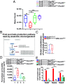
Increased succinate-producers in the remodeled microbiota of mice with altered Paneth cells lead to tuft cell activation via a succinate–SucnR1 pathway. (A) Gene richness in cecum from Sox9LoxP/LoxP;Pou2f3+/+Sox9LoxP/LoxP;Villin-CreERT2;Pou2f3+/+Sox9LoxP/LoxP;Pou2f3−/−, and Sox9LoxP/LoxP;Villin-CreERT2;Pou2f3−/− mice. n = 8 mice per genotype. Mice were killed 4 wk after tamoxifen treatment. (B) Schema of the succinate biosynthetic pathway predominant under anaerobic conditions. The two final steps of the succinate production are shown: i) the conversion of malate to fumarate by the fumarate hydratase encoded by the fumABC genes and ii) the transformation of fumarate to succinate by the fumarate reductase encoded by the frdABCD genes. For each enzymatic reaction, all KO (KEGG Ortholog) alternatives (v1 to v3) found in the KEGG database are shown. Color codes refer to their presence (black) or absence (gray) in the mouse BGI 2.6 million genes catalog. (C) The global completeness of the succinate production pathway is significantly increased in Sox9LoxP/LoxP;Villin-CreERT2 mice compared to Sox9LoxP/LoxP mice. The global completeness for succinate production, i.e., the proportion of KOs detected necessary to produce succinate in each microbial species, was computed for each differentially abundant MSP species found in Sox9LoxP/LoxP (blue) and Sox9LoxP/LoxP;Villin-CreERT2 mice (red). (D) Intestinal permeability, assessed by FITC–dextran gavage, remains intact when Sox9 is deleted in SucnR1-deficient mice. FITC-dextran was measured in serum from Sox9LoxP/LoxP;SucnR1+/+ mice (n = 5), Sox9LoxP/LoxP;Villin-CreERT2;SucnR1+/+mice (n = 5), Sox9LoxP/LoxP;SucnR1−/− mice (n = 5), and Sox9LoxP/LoxP;Villin-CreERT2;SucnR1−/− mice (n = 6). Mice were killed 2 wk after tamoxifen treatment. (E) Type 2 immune response is abolished in the absence of SucnR1. Cells positive for Dclk1, Retnlβ or Gata3 were counted in n = 50 crypt-villus units per mouse (n = 5 to 6 mice per genotype). (A) Unpaired Wilcoxon rank-sum test. (C) Unpaired Wilcoxon signed-rank test, P = 0.017. (D and E) Kruskal–Wallis test with Dunn's post hoc test; NS: not significant; *P < 0.05; **P < 0.01; ***P < 0.001; ****P < 0.0001. (A, C, and D) The line indicates the median, the box marks the 25th and 75th percentiles, and the whiskers indicate the minimal to maximum values. (E) Data are shown as mean ± SEM. See also SI Appendix, Figs. S5 and S6.
Fig. 6.
Fig. 6.
Dysbiosis is dependent on a cross talk between tuft cells and Paneth cells via the downregulation of the antimicrobial RegIII. (A) RT-qPCR analysis of antimicrobial peptides Lyz1, Mmp7, Defa29, and RegIII in organoids derived from Sox9LoxP/LoxP and Sox9LoxP/LoxP;Villin-CreERT2 small intestine. Organoids were analyzed 5 d after 4-OH-tamoxifen treatment (n = 2 independent organoid cultures, replicated 3 times). (B) RT-qPCR analysis of antimicrobial peptides Lyz1, Mmp7, Defa29, and RegIII in Sox9LoxP/LoxP;Villin-CreERT2 and Sox9LoxP/LoxP;Villin-CreERT2;Pou2f3−/− small intestine. n = 6 to 8 mice per genotype. Mice were killed 2 wk after tamoxifen treatment. (A) Mann–Whitney U test. (B) Kruskal–Wallis test with Dunn's post hoc test; NS: not significant; *P < 0.05; **P < 0.01; ***P < 0.001. The line indicates the median, the box marks the 25th and 75th percentiles, and the whiskers indicate the minimal to maximum values. See also SI Appendix, Fig. S7.
Fig. 7.
Fig. 7.
A three-step mechanism for establishment of dysbiosis and inflammation due to the cross Talk between altered Paneth cells and tuft cells through succinate–SucnR1 signaling and type 2 cytokines. In homeostatic conditions, Paneth cells regulate microbiota composition, and the presence of tuft cells is rare. Step 1: In the absence of Sox9, Paneth cells are altered and express normal levels of RegIII but lower levels of Lyz1, leading to mild alterations in the microbiota and increased succinate production potential. Step 2: Sensing of increased succinate levels by tuft cells, via the SucnR1, results in the initiation of a type 2 immune response. Step 3: Type 2 cytokines cause IL4rα-dependent remodeling of epithelial cells, including ectopic Retnlβ expression in goblet cells, tuft cell lineage amplification, and reduction of RegIII levels. This remodeling accentuates the deficiency in microbiota regulation by epithelial cells, eventually causing dysbiosis. As tuft cells cooperate with altered Paneth cells to drive dysbiosis, pharmacological inhibition of tuft cell activity may represent a strategy for controlling inflammation and dysbiosis in predisposed patients with altered Paneth cells. See also SI Appendix, Figs. S8 and S9.

References

    1. Kundu P., Blacher E., Elinav E., Pettersson S., Our gut microbiome: The evolving inner self. Cell 171, 1481–1493 (2017). - PubMed
    1. Turnbaugh P. J., et al. , An obesity-associated gut microbiome with increased capacity for energy harvest. Nature 444, 1027–1031 (2006). - PubMed
    1. Ni J., Wu G. D., Albenberg L., Tomov V. T., Gut microbiota and IBD: Causation or correlation? Nat. Rev. Gastroenterol. Hepatol. 14, 573–584 (2017). - PMC - PubMed
    1. Järbrink-Sehgal E., Andreasson A., The gut microbiota and mental health in adults. Curr. Opin. Neurobiol. 62, 102–114 (2020). - PubMed
    1. Settanni C. R., Ianiro G., Bibbò S., Cammarota G., Gasbarrini A., Gut microbiota alteration and modulation in psychiatric disorders: Current evidence on fecal microbiota transplantation. Progress in Neuro-Psychopharmacol. Biol. Psychiatry 109, 110258 (2021). - PubMed

Publication types