. 2023 Jun 20;120(25):e2302254120.
doi: 10.1073/pnas.2302254120. Epub 2023 Jun 12.

MBD2a-NuRD binds to the methylated γ-globin gene promoter and uniquely forms a complex required for silencing of HbF expression

Affiliations

MBD2a-NuRD binds to the methylated γ-globin gene promoter and uniquely forms a complex required for silencing of HbF expression

Shengzhe Shang et al. Proc Natl Acad Sci U S A. .

Abstract

During human development, there is a switch in the erythroid compartment at birth that results in silencing of expression of fetal hemoglobin (HbF). Reversal of this silencing has been shown to be effective in overcoming the pathophysiologic defect in sickle cell anemia. Among the many transcription factors and epigenetic effectors that are known to mediate HbF silencing, two of the most potent are BCL11A and MBD2-NuRD. In this report, we present direct evidence that MBD2-NuRD occupies the γ-globin gene promoter in adult erythroid cells and positions a nucleosome there that results in a closed chromatin conformation that prevents binding of the transcriptional activator, NF-Y. We show that the specific isoform, MBD2a, is required for the formation and stable occupancy of this repressor complex that includes BCL11A, MBD2a-NuRD, and the arginine methyltransferase, PRMT5. The methyl cytosine binding preference and the arginine-rich (GR) domain of MBD2a are required for high affinity binding to methylated γ-globin gene proximal promoter DNA sequences. Mutation of the methyl cytosine-binding domain (MBD) of MBD2 results in a variable but consistent loss of γ-globin gene silencing, in support of the importance of promoter methylation. The GR domain of MBD2a is also required for recruitment of PRMT5, which in turn results in placement of the repressive chromatin mark H3K8me2s at the promoter. These findings support a unified model that integrates the respective roles of BCL11A, MBD2a-NuRD, PRMT5, and DNA methylation in HbF silencing.

Keywords: BCL11A; DNA methylation; MBD2a–NuRD; PRMT5; fetal hemoglobin.


Conflict of interest statement

The authors declare no competing interest.

Figures
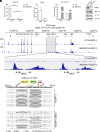
Fig. 1.
Fig. 1.
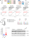
MBD2–NuRD occupies the HBG gene promoter in HUDEP-2 cells and CD34+ cells and positions a nucleosome that enforces a closed chromatin configuration to block NF-Y binding at the HBG promoters. (A) ChIP-qPCR assay shows that MBD2 occupies the HBG promoter but not β-globin (HBB) gene promoter in HUDEP-2 cells (mean ± SD, = 2) and CD34+ cells. The CD34+ ChIP result is shown as the mean ± SD of three technical repeats. *< 0.05, ** < 0.01. (B) ChIP-qPCR assay shows that NF-YA binds at the HBG gene promoter in differentiated parental and M2KO HUDEP-2 cells. The result is shown as the mean ± SD of three technical repeats and is representative of two biological repeats. (C) Protein level of NF-YA and BCL11A in parental and M2KO HUDEP-2 cells. (D) ATAC-seq showing open chromatin at the HBG1 and HBG2 genes in MBD2KO HUDEP-2 cells. (E) NOMe-seq showing a fixed nucleosome at position −110 to +36 bp relative to the transcription initiation site (TSS) in parental HUDEP-2 cells that is evicted in MBD2KO cells.
Fig. 2.
Fig. 2.
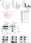
DNA methylation and the GR domain enhance the binding of the MBD2 methylcytosine–binding domain to the γ-globin promoter DNA sequences. (A) Schematic representation of the protein structure of MBD2a vs. MBD2b and MBD3. (B) Schematic illustration of the structure of the peptides used in binding affinity studies: MBD2-MBD, GRMBD, and GRYMBD(Y178H). (C) Binding affinity plots for MBD2-MBD, MBD2-GRMBD, and MBD2- GRMBD(Y178H) binding to methylated DNA encompassing the proximal [2xmCpG (−53/−50)] and distal [1xmCpG (−162)] CpG sites of the HBG promoters were determined by isothermal titration calorimetry. The KD and N values represent the mean ± SD for three to four replicates. (D) A bar graph depicts the KD for each of the four complexes, compared using Welch’s unpaired t test. (E) Sanger sequencing result showing the specific adenine base editing to introduce the Y178H mutation in endogenous MBD2 genomic DNA. In wild-type clones, the amino acid sequence is 177VYY179. In edited clones, the Y178 is changed to H, the amino acid sequence is 177VHY199. The edited base is indicated by red arrow. (F) Q-PCR results showing the γ/(γ+β) mRNA ratio in ABE8e-MBD2 Y178–edited HUDEP-2 cell single colonies. P value was calculated by Welch’s unpaired t test. Blue dots represent the scramble guide clones without editing at MBD2Y178 (= 26), red dots represent the clones with editing at MBD2Y178H amino acid position (= 47). (G) Immunoblotting panel showing equivalent levels of total MBD2 protein in edited single clones and scramble guide control clones. *< 0.05, **< 0.01, ***< 0.001.
Fig. 3.
Fig. 3.
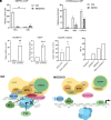
MBD2a–NuRD and not MBD2b–NuRD specifically associates with both BCL11A and PRMT5. (A) Q-PCR in differentiated parental or MBD2KO HUDEP-2 cells with MBD2a versus MBD2b-TAPtag addback shows that MBD2a but not MBD2b enforces γ-globin gene repression. P value is with respect to control (mean ± SD, = 2, the P value was calculated by the Student’s t test. *< 0.05, **< 0.01, ***< 0.001). (B) Venn diagram of unbiased subtraction proteomic analysis of nuclear extract proteins immunoprecipitated with MBD2a versus MBD2b TAP-tagged bait. (C) Peptide enumeration showing the presence of core NuRD components in both MBD2a and MBD2b immunoprecipitated protein samples and differential association of PRMT5/MEP50 peptides only in the MBD2a immunoprecipitation. The same result was obtained with the corresponding whole-cell extract. EV represents empty vector control. (D) Strep-Tactin immunoprecipitation western blot assay showing only MBD2a-TAPtag associated with both BCL11A and PRMT5. (E) Reciprocal immunoprecipitation of endogenous MBD2, BCL11A, and PRMT5 proteins in parental HUDEP-2 cells showing mutual association of all the three proteins. The immunoprecipitation results shown are representative of three independent biological repeats.
Fig. 4.
Fig. 4.
MBD2a recruits PRMT5/MEP50 to the promoter which contributes to γ-globin silencing. (A) MEP50 ChIP-qPCR results showing MEP50/PRMT5 enrichment at the γ-globin promoter in parental HUDEP-2 cells but not in MBD2KO cells (mean ± SD, = 2). *< 0.05, **P<0.01. (B) ChIP-qPCR assay of histone H3R8me2s occupancy showing that the repressive marker is more enriched at the HBG promoter in parental vs MBD2KO HUDEP-2 cells. The result is shown as the mean ± SD of three technical repeats and is representative of two biological repeats. (C) Q-PCR assay of γ/(γ+β) mRNA ratio in PRMT5KD of both HUDEP-2 cells (mean ± SD, = 3) and CD34+ progenitor cells (mean ± SD, = 3) and MBD2KO of HUDEP-2 cells (mean ± SD, = 5). *< 0.05, **< 0.01, ***< 0.001. (D) LC–MS analysis of globin chains showing the relative Hb percent of total hemoglobin in parental, PRMT5-depleted, MBD2KO, and M2KO plus PRMT5-depleted HUDEP-2 cells. (E) Schematic illustration of the HBG gene corepressor complex model showing the requirement for MBD2a–NuRD for maintenance of a positioned nucleosome at the HBG promoter to prevent binding of the transcriptional activator, NF-Y, and the MBD2a-dependent cooccupancy of PRMT5 in the complex in wild-type HUDEP-2 cells. In MBD2a knockout cells, the MBD2a–NuRD complex along with PRMT5 is dissociated from the promoter, the nucleosome is evicted, and the NF-Y complex binds at the CCAAT box at position −89 relative to the TSS to activate transcription.

References

    1. Sankaran V. G., et al. , Human fetal hemoglobin expression is regulated by the developmental stage-specific repressor BCL11A. Science 322, 1839–1842 (2008). - PubMed
    1. Borg J., et al. , Haploinsufficiency for the erythroid transcription factor KLF1 causes hereditary persistence of fetal hemoglobin. Nat. Genet. 42, 801–805 (2010). - PMC - PubMed
    1. Masuda T., et al. , Transcription factors LRF and BCL11A independently repress expression of fetal hemoglobin. Science 351, 285–289 (2016). - PMC - PubMed
    1. Vinjamur D. S., et al. , ZNF410 represses fetal globin by singular control of CHD4. Nat. Genet. 53, 719–728 (2021). - PMC - PubMed
    1. Lan X., et al. , ZNF410 uniquely activates the NuRD component CHD4 to silence fetal hemoglobin expression. Mol. Cell 81, 239–254.e8 (2021). - PMC - PubMed

Publication types