Review
. 2023 Aug 31;193(1):195-216.
doi: 10.1093/plphys/kiad337.

Monitoring nutrients in plants with genetically encoded sensors: achievements and perspectives

Affiliations
Review

Monitoring nutrients in plants with genetically encoded sensors: achievements and perspectives

Mayuri Sadoine et al. Plant Physiol. .

Abstract

Understanding mechanisms of nutrient allocation in organisms requires precise knowledge of the spatiotemporal dynamics of small molecules in vivo. Genetically encoded sensors are powerful tools for studying nutrient distribution and dynamics, as they enable minimally invasive monitoring of nutrient steady-state levels in situ. Numerous types of genetically encoded sensors for nutrients have been designed and applied in mammalian cells and fungi. However, to date, their application for visualizing changing nutrient levels in planta remains limited. Systematic sensor-based approaches could provide the quantitative, kinetic information on tissue-specific, cellular, and subcellular distributions and dynamics of nutrients in situ that is needed for the development of theoretical nutrient flux models that form the basis for future crop engineering. Here, we review various approaches that can be used to measure nutrients in planta with an overview over conventional techniques, as well as genetically encoded sensors currently available for nutrient monitoring, and discuss their strengths and limitations. We provide a list of currently available sensors and summarize approaches for their application at the level of cellular compartments and organelles. When used in combination with bioassays on intact organisms and precise, yet destructive analytical methods, the spatiotemporal resolution of sensors offers the prospect of a holistic understanding of nutrient flux in plants.


Conflict of interest statement

Conflict of interest statement. None declared.

Figures
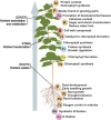
Figure 1.
Figure 1.
Nutrient distribution in plants. Schematic representation of nutrient distribution within a plant. Macro- and micronutrients are involved in multiple processes that occur in different plant areas. For example, P, K, Fe, and Ca are involved in the aerial and ground parts and act as key factors during such processes as growth and development, sugar and protein translocation, chlorophyll formation, and N fixation, as well as cellulose content. N, B, Mn, Zn, and Cu are mostly found in growing parts and are important to chlorophyll, protein, and sugar synthesis, as well as plant growth regulation. Mg and Mo are both primarily found in the roots, where they are involved in root formation and symbiotic N fixation. Additionally, Mg is required to synthesize chlorophyll in photosynthetic tissues.
Figure 2.
Figure 2.
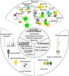
Holistic nutrient analysis approaches in plants. A) In vivo nutrient imaging including sensor imaging with indirect sensors, i.e. transcriptional fluorescent reporters, translational fluorescent reporters, Degron-FP fusion, and direct sensors, i.e. intrinsic sensors using FP as recognition element, extrinsic sensors including FRET-based sensors and intensiometric cpFP-based sensor; and ratiometric Matryoshka design with nested reference FP. B) Bioassays, i.e. organ architecture, leaf color, growth assays, and biotic assays. C) Chemical approaches, i.e. spectrometric analysis (e.g. ICP-OES, ICP-MS, and AAS), ()electrode, spectrophotometry, dye/tracer (e.g. radiotracer and fluorescent/chemical dyes), and X-ray analysis (e.g. XRMA). ICP-OES, Inductively Coupled Plasma Optical Emission Spectrometry; ICP-MS, inductively coupled plasma mass spectrometry; AAS, atomic absorption spectrometry; XRMA, X-ray microanalysis; TF, transcription factor.
Figure 3.
Figure 3.
Subcellular targeting of sensors in plants. Subcellular compartmentalization of nutrients that have been investigated using targeting of sensors to specific organelles in plant cells. RER, rough endoplasmic reticulum; SER, smooth endoplasmic reticulum. Distribution of sensors targeting the plastids (amyloplast and chloroplast), plasma membrane PM, or no target (cytosol) are represented.

Similar articles

Cited by

References

    1. Akerboom J, Chen T-W, Wardill TJ, Tian L, Marvin JS, Mutlu S, Calderón NC, Esposti F, Borghuis BG, Sun XR, et al. . Optimization of a GCaMP calcium indicator for neural activity imaging. J Neurosci. 2012:32(40):13819–13840. 10.1523/JNEUROSCI.2601-12.2012 - DOI - PMC - PubMed
    1. Akerboom J, Rivera JDV, Guilbe MMR, Malavé ECA, Hernandez HH, Tian L, Hires SA, Marvin JS, Looger LL, Schreiter ER, et al. . Crystal structures of the GCaMP calcium sensor reveal the mechanism of fluorescence signal change and aid rational design. J Biol Chem. 2009:284(10):6455–6464. 10.1074/jbc.M807657200 - DOI - PMC - PubMed
    1. Alejandro S, Höller S, Meier B, Peiter E. Manganese in plants: from acquisition to subcellular allocation. Front Plant Sci. 2020:11:300. 10.3389/fpls.2020.00300 - DOI - PMC - PubMed
    1. Allen GJ, Kwak JM, Chu SP, Llopis J, Tsien RY, Harper JF, Schroeder JI.. Cameleon calcium indicator reports cytoplasmic calcium dynamics in Arabidopsis guard cells. Plant J. 1999b:19(6):735–747. 10.1046/j.1365-313x.1999.00574.x - DOI - PubMed
    1. Anderson LA, Palmer T, Price NC, Bornemann S, Boxer DH, Pau RN. Characterisation of the molybdenum-responsive ModE regulatory protein and its binding to the promoter region of the modABCD (molybdenum transport) operon of Escherichia coli. Eur J Biochem. 1997:246(1):119–126. 10.1111/j.1432-1033.1997.00119.x - DOI - PubMed

Publication types