

Management and Outcome of Intracranial Dural Arteriovenous Fistulas That Have Caused a Hemorrhage in the Posterior Fossa : A Clinical Study
- PMID: 37308452
- PMCID: PMC10641416
- DOI: 10.3340/jkns.2023.0065
Management and Outcome of Intracranial Dural Arteriovenous Fistulas That Have Caused a Hemorrhage in the Posterior Fossa : A Clinical Study
Abstract
Objective: We evaluated the diagnosis, treatment, and long-term results of patients with dural arteriovenous fistula (dAVF), which is a very rare cause of posterior fossa hemorrhage.
Methods: This study included 15 patients who underwent endovascular, surgical, combined, or Gamma Knife treatments between 2012 and 2020. Demographics and clinical features, angiographic features, treatment modalities, and outcomes were analyzed.
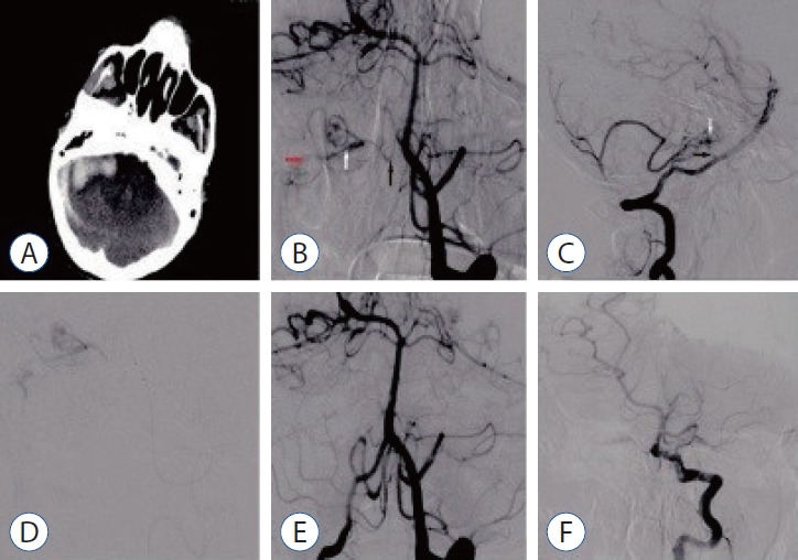
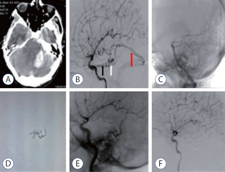
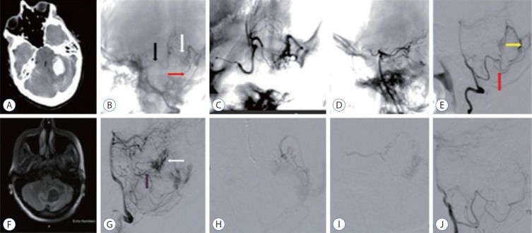
Results: The mean age of the patients was 40±17 years (range, 17-68), and 68% were men (11/15). Seven of the patients (46.6%) were in the age group of 50 years and older. While the mean Glasgow coma scale was 11.5±3.9 (range, 4-15), 46.3% presented with headache and 53.7% had stupor/coma. Four patients (26.6%) had only cerebellar hematoma and headache. All dAVFs had cortical venous drainage. In 11 patients (73.3%), the fistula was located in the tentorium and was the most common localization. Three patients (20%) had transverse and sigmoid sinus localizations, while one patient (6.7%) had dAVF located in the foramen magnum. Eighteen sessions were performed on the patients during endovascular treatment. Sixteen sessions (88.8%) were performed with the transarterial (TA) route, one session (5.5%) with the transvenous (TV) route, and one session (5.5%) with the TA+TV route. Surgery was performed in two patients (14.2%). One patient (7.1%) passed away. While there were nine patients (64.2%) with a Rankin score between 0 and 2, the total closure rate was 69.2% in the first year of control angiograms.
Conclusion: In the differential diagnosis of posterior fossa hemorrhages, the differential diagnosis of dAVFs, which is a very rare entity, should be considered, even in the middle and elderly age groups, in patients presenting with good clinical status and pure hematoma. The treatment of such patients can be done safely and effectively in a multidisciplinary manner with a good understanding of pathological vascular anatomy and appropriate endovascular treatment approaches.
Keywords: Arteriovenous fistula; Endovascular procedure; Hemorrhage; Posterior fossa hemorrhages; Therapeutic embolization.
Conflict of interest statement
No potential conflict of interest relevant to this article was reported.
Figures
References
-
- Brinjikji W, Cloft HJ, Lanzino G. Clinical, angiographic, and treatment characteristics of cranial dural arteriovenous fistulas with pial arterial supply. J Neurointerv Surg. 2021;13:331–335. - PubMed
-
- Chung SJ, Kim JS, Kim JC, Lee SK, Kwon SU, Lee MC, et al. Intracranial dural arteriovenous fistulas: analysis of 60 patients. Cerebrovasc Dis. 2002;13:79–88. - PubMed
-
- Cognard C, Gobin YP, Pierot L, Bailly AL, Houdart E, Casasco A, et al. Cerebral dural arteriovenous fistulas: clinical and angiographic correlation with a revised classification of venous drainage. Radiology. 1995;194:671–680. - PubMed
-
- Daniels DJ, Vellimana AK, Zipfel GJ, Lanzino G. Intracranial hemorrhage from dural arteriovenous fistulas: clinical features and outcome. Neurosurg Focus. 2013;34:E15. - PubMed
-
- Datar S, Rabinstein AA. Cerebellar hemorrhage. Neurol Clin. 2014;32:993–1007. - PubMed
LinkOut - more resources
Full Text Sources
