. 2023 Jun 12;14(1):3463.
doi: 10.1038/s41467-023-38976-7.

Mechanism of small molecule inhibition of Plasmodium falciparum myosin A informs antimalarial drug design

Affiliations

Mechanism of small molecule inhibition of Plasmodium falciparum myosin A informs antimalarial drug design

Dihia Moussaoui et al. Nat Commun. .

Abstract

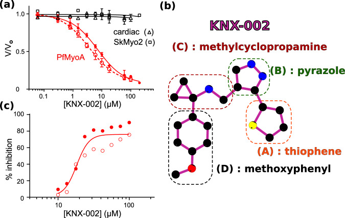
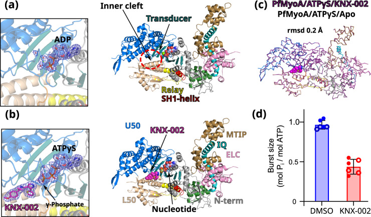
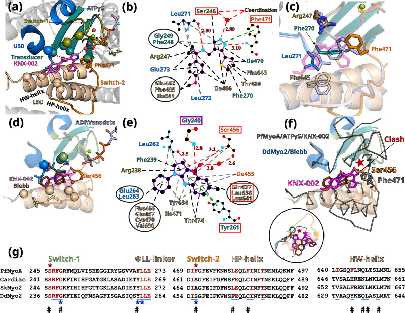
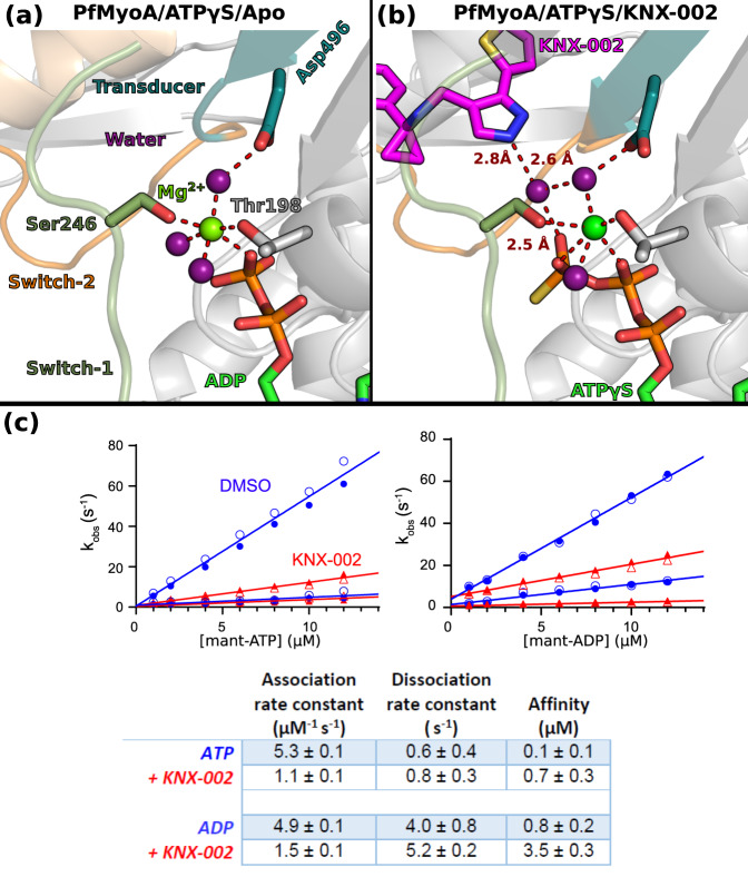
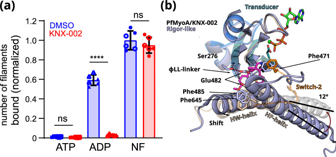
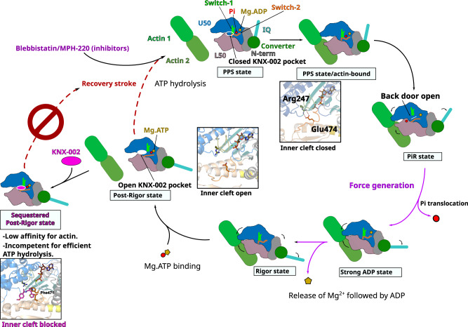
Malaria results in more than 500,000 deaths per year and the causative Plasmodium parasites continue to develop resistance to all known agents, including different antimalarial combinations. The class XIV myosin motor PfMyoA is part of a core macromolecular complex called the glideosome, essential for Plasmodium parasite mobility and therefore an attractive drug target. Here, we characterize the interaction of a small molecule (KNX-002) with PfMyoA. KNX-002 inhibits PfMyoA ATPase activity in vitro and blocks asexual blood stage growth of merozoites, one of three motile Plasmodium life-cycle stages. Combining biochemical assays and X-ray crystallography, we demonstrate that KNX-002 inhibits PfMyoA using a previously undescribed binding mode, sequestering it in a post-rigor state detached from actin. KNX-002 binding prevents efficient ATP hydrolysis and priming of the lever arm, thus inhibiting motor activity. This small-molecule inhibitor of PfMyoA paves the way for the development of alternative antimalarial treatments.


Conflict of interest statement

A.H. receives research funding from Cytokinetics and consults for Kainomyx. All other authors have no competing interests.

Figures

Fig. 1
Fig. 1. KNX-002 inhibits PfMyoA.
a KNX-002 inhibits actin-activated (filled red circles, IC50 = 7.2 µM, (95% CI, 5.8–9.0 µM), n = 3) and basal ATPase activity (open red circles, IC50 = 3.6 µM (95% CI, 3.0–4.5 µM), n = 3). KNX-002 has little effect on the actin-activated ATPase of skeletal myosin (SkMyo2, black squares, IC50 > 200 µM, n = 2) or cardiac myosin (black triangles, IC50 > 100 µM, n = 2). PfMyoA data are represented as mean values ± SD. b The constituent groups of the KNX-002 structure are indicated. c The inhibition of asexual parasite blood stage growth by KNX-002 was quantified (IC50 = 18.2 µM, n = 2). Source data are provided as a Source Data file.
Fig. 2
Fig. 2. KNX-002 targets the post-rigor (PR) state.
a Structure of PfMyoA in the apo condition (PfMyoA/ATPγS/Apo). Only ADP is found in the 2Fo-Fc electron density map contoured at 1.0 σ (on the left). ATPyS was thus hydrolyzed by myosin, in contrast to the same experiment performed in the presence of KNX-002. b Structure of PfMyoA complexed with KNX-002 and MgATPyS (PfMyoA/ATPγS/KNX-002). The compound, MgATPyS, the water molecules and the Mg2+ ion can be clearly identified in the 2Fo-Fc electron density map contoured at 1.0 σ (on the left). The gamma-phosphate of ATPyS is present in the density. U50, Upper 50 kDa subdomain; L50, Lower 50 kDa subdomain. c The compound does not induce major structural rearrangements upon binding. PfMyoA/ATPγS/KNX-002 and PfMyoA/ATPγS/Apo are both in a PR state and superimpose quite well with a rmsd of 0.2 Å using the Cα atoms. Zoom on the regions with maximum differences between the two structures show local displacements of side chains (see Supplementary Fig. 3a). d Manual quenching experiments show a decreased phosphate burst from 0.97 ± 0.05 mol Pi/mol ATP in the absence of compound to 0.44 ± 0.08 mol Pi/mol ATP in the presence of 100 µM KNX-002 (p < 0.0001, two-tailed t-test with Welch’s correction). Data represent two experiments each performed in triplicate with independent protein preparations (open and filled circles). Data bars are mean ± SD. Source data are provided as a Source Data file.
Fig. 3
Fig. 3. The inner pocket in which KNX-002 binds greatly differs from that of Blebbistatin (Blebb).
a KNX-002 binding pocket. Regions involved in binding are: Switch-2 (orange); Transducer (deep teal cyan); U50 (marine blue); L50 (wheat). Residues are displayed as spheres when involved in apolar interactions; as sticks when involved in electrostatic or π-stacking bonds. b Schematic representation of the binding pocket of KNX-002. Each type of interaction is represented differently (Polar interactions, red dashed lines; π-stacking, green; apolar, black). Squares indicate residues involved in different types of bonds for KNX-002 and Blebbistin (shown in e). c Superimposition of PfMyoA/ATPγS/KNX-002 (colored by subdomains) and PfMyoA/ATPγS/Apo (gray-blue), both in the post-rigor state. Residues with different conformation (sticks), indicate how adjustments are required to bind KNX-002. d Blebb binding pocket in Dictyostelium discoideum myosin 2 (DdMyo2, PDB code 1YV3) with the U50 subdomain orientation as in 3a for PfMyoA. KNX-002 (pale purple) does not fit in this pocket as the pre-powerstroke conformation of Switch-2 clashes with the KNX-002 position. Supplementary Table 2 and Supplementary Movie 3 further illustrate how the KNX-002 and Blebb binding sites differ. e Schematic representation of interactions around Blebb. Residues that differ from PfMyoA but bind Blebb are in a red box; conserved residue involved in different bond types (purple box). f KNX-002 and Blebb target different pockets. DdMyo2/Blebb (cartoon, colored by subdomain) and PfMyoA/ATPγS/KNX-002 (ribbon, colored in black) are superimposed on the U50 subdomain (residues 182-463 and 604-631). KNX-002 and Blebb binding pockets strongly differ in the conformation of Switch-2 and in the orientation of HP- and HW-helices. Another orientation is represented as a zoom in a circle. g Sequence alignment of PfMyoA, β-cardiac MYH7 (cardiac), skeletal muscle myosin 2 (SkMyo2) and DdMyo2 for analysis of the conservation of residues involved in KNX-002 and Blebb binding. Residues involved in KNX-002 binding are colored red in PfMyoA and in other myosins when conserved. Residues involved in Blebb binding are underlined (blue). Residues involved in electrostatic or stacking interactions with KNX-002 (red star), and those involved with Blebb (blue star) are indicated. Remarkable positions that distinguish KNX-002 and Blebb binding modes are marked with a #.
Fig. 4
Fig. 4. Effects of KNX-002 on the active site.
The Mg2+ ion is hexa-coordinated in both the Apo (a) and in the KNX-002 bound (b) structures. a Although ATPγS was used for the crystallization in both structures, the nucleotide is hydrolyzed in the absence of KNX-002 and ADP is found in the active site (a). b When KNX-002 occupies its pocket, the compound stabilizes a water molecule that also binds the γ-Pi of ATP and a water molecule that coordinates the Mg2+ ion. Supplementary Fig. 7 indicates that this additional interaction does not change the hexa-coordination of the Mg2+ ion. c KNX-002 slows both mant-ATP and mant-ADP binding to PfMyoA. Rates in the absence (blue circles) or presence of 100 µM KNX-002 (red triangles) of the fast and slow phases from the biphasic transients are plotted. The association (slope) and dissociation (y-intercept) rate constants and resulting affinity are given in the Table. Data represent two experiments with independent protein preparations. Source data are provided as a Source Data file.
Fig. 5
Fig. 5. PfMyoA.ADP binds actin weakly in the presence of KNX-002.
a KNX-002 weakens the affinity of actin for M.ADP but has no effect on binding in the presence of ATP or with nucleotide-free (NF)-PfMyoA. Number of actin filaments bound ± SD to surface immobilized PfMyoA (see Methods). See Supplementary Fig. 8 for examples of the raw data and Supplementary Fig. 9 for data with an expanded y-axis scale. The difference between ADP ± KNX-002 is significant (p < 0.0001) (one-sided ANOVA followed by Tukey’s post-hoc test), but there were no significant differences between ATP ± KNX-002 (p > 0.999), nor between NF ± KNX-002 (p = 0.64). Data represent two experiments each performed in triplicate with independent protein preparations (open and filled circles). Data bars are mean ± SD. Source data are provided as a Source Data file. b The KNX-002 binding pocket does not exist in the Rigor state. PfMyoA/ATPγS/KNX-002 (colored by subdomains as in Fig. 2b) and PfMyoA in the Rigor-like state (light blue) (PDB code 6I7D) are superimposed on the U50 subdomain and show how a change in the conformation and the orientation of the L50 subdomain would close the inner cleft and would thus not be compatible with KNX-002 binding. Indeed, the L50HP-helix rotates by 12° and the L50HW-helix shifts, changing the position of F485 and F645, two residues involved in KNX-002 binding. The reorientation of Switch-2 also leads to a new position for the key F471 residue that is incompatible with KNX-002 docking into this site in the Rigor state.
Fig. 6
Fig. 6. Mechanism of the inhibition by compound KNX-002.
Schematic representation of the motor cycle of PfMyoA, where the state of nucleotide bound governs (i) the conformation of the motor and (ii) the affinity for the actin filament. In the post-rigor (PR) state, PfMyoA binds Mg.ATP and has low affinity for actin since the actin-binding cleft between U50 and L50 subdomains is open. After the recovery stroke that primes the lever arm, PfMyoA adopts the pre-powerstroke (PPS) state in which ATP is hydrolyzed. The weak association of the PPS to actin initiates a transition towards the Pi release state (PiR), the first force-producing state. This initiation of the powerstroke allows the opening of the Pi release tunnel allowing Pi translocation. After Pi release, a large swing of the lever arm (powerstroke) leads to a Strong ADP state in which the actin-binding cleft is closed, allowing stronger association with actin. Mg2+ and ADP are finally released from the Rigor state after a small swing of the lever arm and reorientation of the N-terminal subdomain. An ATP molecule can then bind, which leads to a fast transition towards the post-rigor state that detaches from actin. In contrast to the previously described Myo2 inhibitors, Blebbistatin and MPH-220 which target PPS, KNX-002 targets PR, which prevents the recovery stroke and ATP hydrolysis. When KNX-002 intercalates between the U50 and the L50, it stabilizes a state of poor affinity for F-actin that is not compatible with efficient ATP hydrolysis.

References

    1. Talman AM, Domarle O, McKenzie FE, Ariey F, Robert V. Gametocytogenesis: the puberty of Plasmodium falciparum. Malar. J. 2004;3:24. doi: 10.1186/1475-2875-3-24. - DOI - PMC - PubMed
    1. Phillips MA, et al. Malaria. Nat. Rev. Dis. Prim. 2017;3:17050. doi: 10.1038/nrdp.2017.50. - DOI - PubMed
    1. Organization, W. H. World malaria report 2021 (World Health Organization, 2021).
    1. Haldar K, Bhattacharjee S, Safeukui I. Drug resistance in Plasmodium. Nat. Rev. Microbiol. 2018;16:156–170. doi: 10.1038/nrmicro.2017.161. - DOI - PMC - PubMed
    1. Balikagala B, et al. Evidence of artemisinin-resistant malaria in Africa. N. Engl. J. Med. 2021;385:1163–1171. doi: 10.1056/NEJMoa2101746. - DOI - PubMed

Publication types