. 2023 Jun 13;14(1):3475.
doi: 10.1038/s41467-023-39274-y.

Ceramide sensing by human SPT-ORMDL complex for establishing sphingolipid homeostasis

Affiliations

Ceramide sensing by human SPT-ORMDL complex for establishing sphingolipid homeostasis

Tian Xie et al. Nat Commun. .

Abstract

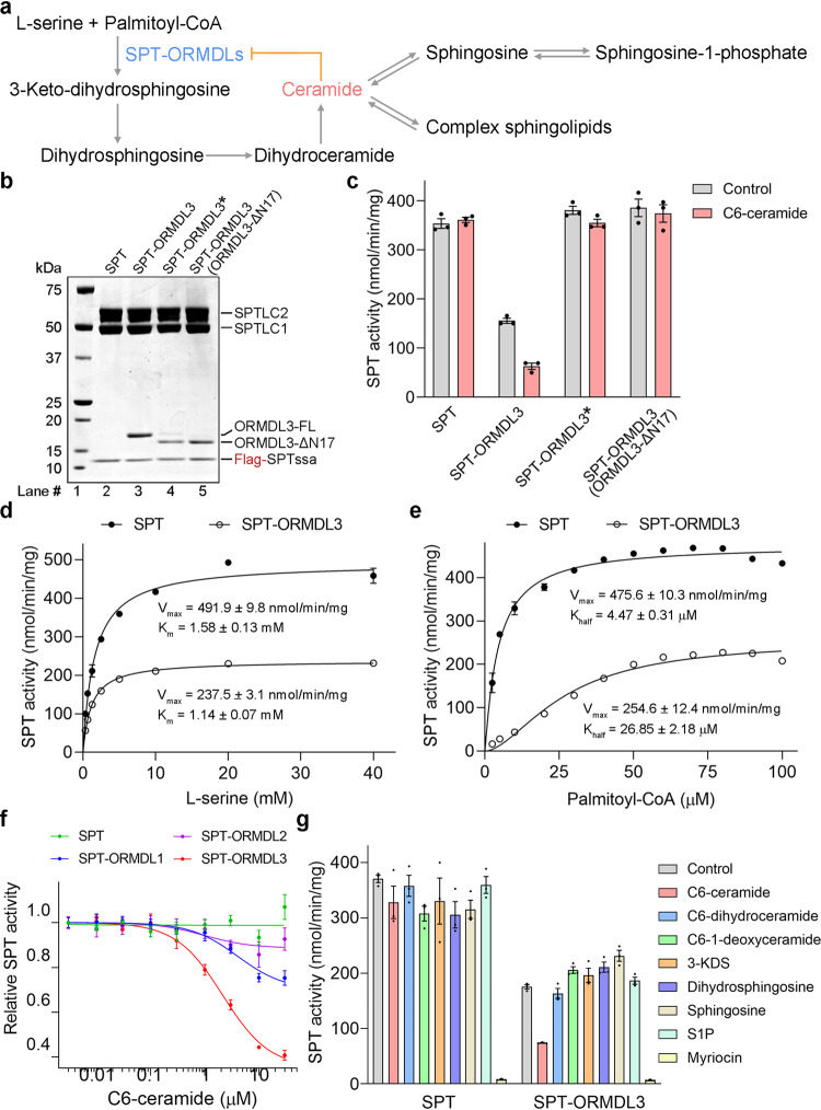
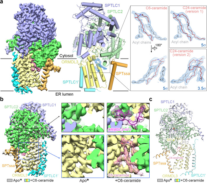
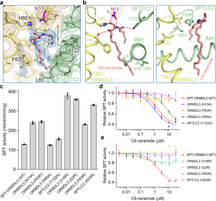
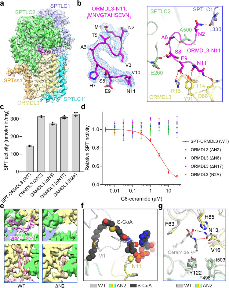
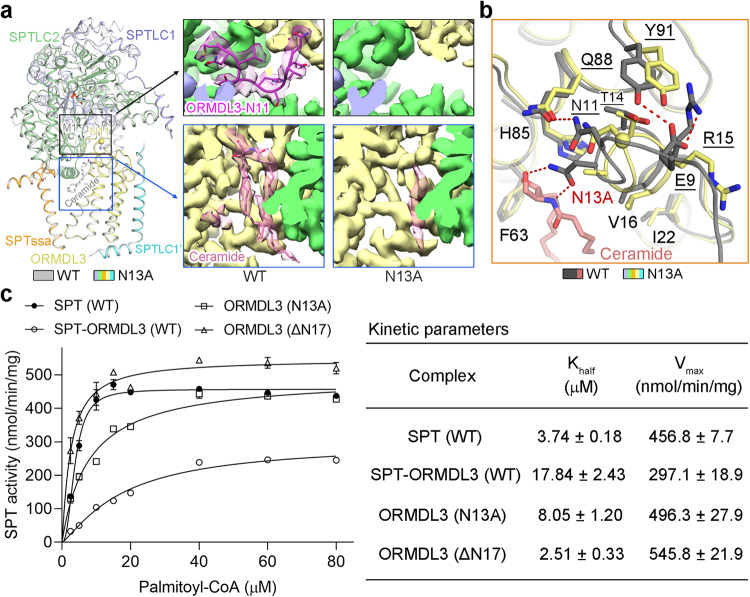
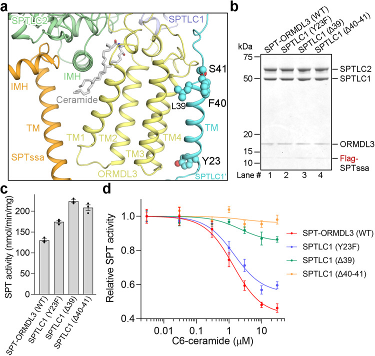
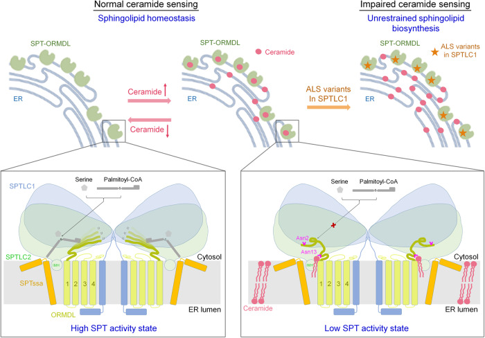
The ORM/ORMDL family proteins function as regulatory subunits of the serine palmitoyltransferase (SPT) complex, which is the initiating and rate-limiting enzyme in sphingolipid biosynthesis. This complex is tightly regulated by cellular sphingolipid levels, but the sphingolipid sensing mechanism is unknown. Here we show that purified human SPT-ORMDL complexes are inhibited by the central sphingolipid metabolite ceramide. We have solved the cryo-EM structure of the SPT-ORMDL3 complex in a ceramide-bound state. Structure-guided mutational analyses reveal the essential function of this ceramide binding site for the suppression of SPT activity. Structural studies indicate that ceramide can induce and lock the N-terminus of ORMDL3 into an inhibitory conformation. Furthermore, we demonstrate that childhood amyotrophic lateral sclerosis (ALS) variants in the SPTLC1 subunit cause impaired ceramide sensing in the SPT-ORMDL3 mutants. Our work elucidates the molecular basis of ceramide sensing by the SPT-ORMDL complex for establishing sphingolipid homeostasis and indicates an important role of impaired ceramide sensing in disease development.


Conflict of interest statement

The authors declare no competing interests.

Figures

Fig. 1
Fig. 1. Purified human SPT-ORMDL complexes can be inhibited by C6-ceramide.
a Overview of the de novo sphingolipid biosynthetic pathway and the homeostatic regulation of SPT activity by ceramide in an ORMDL-dependent manner. b, c Coomassie blue-stained SDS-PAGE gel (b) and the SPT activity (c) of purified SPT and various SPT-ORMDL3 complexes. ORMDL3* stands for the ORMDL3 construct from our previous study (ref. ), which appears to express a mixture of minor full-length ORMDL3 protein (ORMDL3-FL) and dominant N-terminus truncated ORMDL3 protein. The newly made SPT-ORMDL3 (ORMDL3-FL) complex could be inhibited by C6-ceramide, whereas the SPT, SPT-ORMDL3*, and SPT-ORMDL3 (ORMDL3-ΔN17) complexes had no response to C6-ceramide. Data in (c) are presented as mean values ± SEM of three independent experiments. d SPT activity versus L-serine concentration measured using SPT or SPT-ORMDL3 complex. Data are presented as mean values ± SEM of three independent experiments. The data points were fitted with a Michaelis-Menten equation. e SPT activity versus palmitoyl-CoA concentration for SPT and SPT-ORMDL3 complex. Data are presented as mean values ± SEM of three independent experiments. The data points were fitted with an allosteric sigmoidal equation. f The inhibition curves of human SPT and SPT-ORMDL complexes by C6-ceramide in the in vitro SPT activity assay. Data are presented as mean values ± SEM of three independent experiments. All the estimated IC50 values were summarized in Supplementary Table 1. g Selective inhibition of SPT-ORMDL3 by C6-ceramide among various sphingolipid species or analogs. Data are presented as mean values ± SEM of three independent experiments. The different sphingolipid species or analogs were applied at 10 μM in each assay. Source data are provided as a Source Data file.
Fig. 2
Fig. 2. Structure determination of the C6-ceramide-bound SPT-ORMDL3 complex.
a The cryo-EM map and overall structure of the monomeric C6-ceramide-bound SPT-ORMDL3 complex. SPTLC1 and SPTLC2 are shown in light blue and light green, respectively; SPTssa and ORMDL3 are shown in orange and light yellow, respectively; SPTLC1’ is shown in cyan; the lipid-like density is shown in pink (left panel) or blue (right panels). Insets: The lipid-like density, shown in blue meshes, were contoured at 5σ or 3.5σ. C6-ceramide and C24-ceramide are shown as pink sticks; the acyl chains are shown as gray sticks. b, c Comparison of the EM maps (b) and structures (c) of the apo SPT-ORMDL3* complex and C6-ceramide-bound SPT-ORMDL3 complex. Apo* stands for the apo SPT-ORMDL3* complex used in our previous study. The apo SPT-ORMDL3* complex (EMD-30080 and PDB 6M4O) is colored gray. The C6-ceramide-bound SPT-ORMDL3 complex is colored based on the subunits. The ORMDL3-N11 (the 11 residues at the N-terminus) and C6-ceramide in the C6-ceramide-bound SPT-ORMDL3 complex are shown as sticks, and the corresponding maps are colored semi-transparent magenta and pink, respectively (b, zoomed-in view, right). In contrast, the densities for the ORMDL3-N11 and C6-ceramide are not visible in the apo SPT-ORMDL3* complex (b, zoomed-in view, left). Met1 and Pro12, the resolved N-terminus of ORMDL3 and ORMDL3*, are shown as yellow and gray spheres, respectively (c).
Fig. 3
Fig. 3. The ceramide binding site in SPT-ORMDL3 complex.
a A close-up view of the density map for the ceramide binding site contoured at 6σ. b Detailed views of the ceramide binding site. Residues involved in the interactions are shown as sticks. All potential polar interactions are indicated by red dashed lines. c Functional characterization of the key residues involved in ceramide binding by SPT activity assay. Data are presented as mean values ± SEM of three independent experiments. d, e Inhibition curves of the SPT-ORMDL3 ceramide-binding variants by C6-ceramide. Curves for the variants mediating the polar interactions and hydrophobic interactions were presented in (d) and (e), respectively. Data are presented as mean values ± SEM of three independent experiments. Source data are provided as a Source Data file.
Fig. 4
Fig. 4. The N-terminus of ORMDL3 is essential for SPT inhibition.
a The resolved N-terminus of ORMDL3 in the C6-ceramide-bound SPT-ORMDL3 map. The ORMDL3-N11 (the 11 residues at the N-terminus) are highlighted in magenta; all the other parts are shown in the same coloring scheme as above. The maps are shown as semi-transparent. b Close-up views of the density map and coordination of ORMDL3-N11. The electron densities for ORMDL3-N11, shown as blue mesh, were contoured at 6σ. The residues that form polar interactions with ORMDL3-N11 in SPTLC1, SPTLC2, and ORMDL3 are shown as sticks, and the potential polar interactions are indicated by red dashed lines. c Functional characterization of ORMDL3 N-terminal deletion variants and N2A variant by SPT activity assay. Data are presented as mean values ± SEM of three independent experiments. d The loss of C6-ceramide-mediated inhibition of SPT activity for the ORMDL3 N-terminal deletion variants and N2A variant. Data are presented as mean values ± SEM of three independent experiments. e Comparison of the EM maps for ORMDL3-N11 (upper) and ceramide (lower) in the apo wild-type (WT) SPT-ORMDL3 complex and the ORMDL3-ΔN2 mutant. The ORMDL3-N11 is no longer visible in the ORMDL3-ΔN2 mutant. The ceramide-like density largely disappeared in the EM map of the ORMDL3-ΔN2 mutant. f Release of the N-terminus of ORMDL3 for substrate binding in the ORMDL3-ΔN2 mutant. The ORMDL3-ΔN2 mutant structure was superimposed with the C6-ceramide-bound WT SPT-ORMDL3 structure and S-CoA-bound SPT-ORMDL3* structure (PDB 7CQK). S-CoA, short for S-(2-oxoheptadecyl)-CoA and a nonreactive analog of palmitoyl-CoA, is displayed as black spheres. The C6-ceramide-bound SPT-ORMDL3 structure is colored gray, and the ORMDL3-ΔN2 mutant structure is colored based on the subunits as above. Asn11, the resolved N-terminus of ORMDL3-ΔN2, is shown as a yellow sphere. g Conformational changes of the ceramide-binding site in the ORMDL3-ΔN2 mutant. The ORMDL3-ΔN2 mutant structure (colored based on the subunits) was superimposed with the C6-ceramide-bound WT SPT-ORMDL3 structure (gray). Source data are provided as a Source Data file.
Fig. 5
Fig. 5. Ceramide induces and locks the N-terminus of ORMDL3 into an inhibition conformation and the enzymatic kinetics of two SPT-ORMDL3 variants.
a Superposition of the apo WT SPT-ORMDL3 structure (gray) and the ORMDL3-N13A mutant structure (colored based on the subunits). Met1 and Asn11, the resolved N-terminus of WT ORMDL3 and ORMDL3-N13A, respectively, are shown as spheres. Insets: The EM maps for ORMDL3-N11 (upper) and ceramide-like density (lower) in the apo WT SPT-ORMDL3 and ORMDL3-N13A mutant structures. The ORMDL3-N11 is no longer visible in the ORMDL3-N13A mutant. The ceramide-like density largely disappeared in the EM map of the ORMDL3-N13A mutant. b Conformational changes of the ceramide-binding site and the ORMDL3-N11 coordinating residues in the ORMDL3-N13A mutant structure. The ORMDL3-N13A mutant structure (colored based on the subunits) was superimposed with the C6-ceramide-bound WT SPT-ORMDL3 structure (gray). The residues involved in ORMDL3-N11 coordination are underscored. c Enzymatic kinetics of the ORMDL3-N13A and ORMDL3-ΔN17 variants compared to those of the WT SPT and SPT-ORMDL3 complexes. Data are presented as mean values ± SEM of three independent experiments. Source data are provided as a Source Data file.
Fig. 6
Fig. 6. The impaired ceramide sensing by ALS-associated SPTLC1 variants.
a A close-up view of the ALS-associated SPTLC1 variants. b The SPTLC1 ALS variants exhibit little effect on ORMDL3 binding. The SPT-ORMDL3 complexes bearing ALS variants, purified by size exclusion chromatography, were visualized by Coomassie-blue stained SDS-PAGE gel. c Functional characterization of the ALS variants by SPT activity assay. Data are presented as mean values ± SEM of three independent experiments. d The impaired C6-ceramide-mediated SPT-ORMDL3 activity inhibition for the ALS variants. Data are presented as mean values ± SEM of three independent experiments. Source data are provided as a Source Data file.
Fig. 7
Fig. 7. A working model for the homeostatic regulation of SPT-ORMDL complex by ceramide and biological implications of the ALS-associated SPTLC1 variants.
At low ceramide levels, the ORMDL N-terminus remains flexible, thus allowing the entry of substrate for SPT catalysis. The constant reaction of SPT-ORMDL would lead to the accumulation of ceramide, which would inhibit SPT-ORMDL by locking the N-terminus of ORMDL3 into an inhibition conformation. The fixed ORMDL N-terminus would block the entry of substrate for further catalysis. The Asn2 in ORMDL is important for maintaining the inactive conformation of the SPT-ORMDL complex. The Asn13 in ORMDL is critical for ceramide binding. The ALS-associated SPTLC1 variants, located near the transmembrane interface between SPTLC1 and ORMDL, impair the ceramide sensing of the SPT-ORMDL mutants. The consequently unregulated SPT activity of ALS variants would result in unrestrained sphingolipid biosynthesis.

Similar articles

Cited by

References

    1. Breslow DK, et al. Orm family proteins mediate sphingolipid homeostasis. Nature. 2010;463:1048–U1065. - PMC - PubMed
    1. Han SM, Lone MA, Schneiter R, Chang A. Orm1 and Orm2 are conserved endoplasmic reticulum membrane proteins regulating lipid homeostasis and protein quality control. Proc. Natl. Acad. Sci. USA. 2010;107:5851–5856. - PMC - PubMed
    1. Siow DL, Wattenberg BW. Mammalian ORMDL proteins mediate the feedback response in ceramide biosynthesis. J. Biol. Chem. 2012;287:40198–40204. - PMC - PubMed
    1. Siow D, Sunkara M, Dunn TM, Morris AJ, Wattenberg B. ORMDL/serine palmitoyltransferase stoichiometry determines effects of ORMDL3 expression on sphingolipid biosynthesis. J. lipid Res. 2015;56:898–908. - PMC - PubMed
    1. Davis DL, Gable K, Suemitsu J, Dunn TM, Wattenberg BW. The ORMDL/Orm-serine palmitoyltransferase (SPT) complex is directly regulated by ceramide: Reconstitution of SPT regulation in isolated membranes. J. Biol. Chem. 2019;294:5146–5156. - PMC - PubMed

Publication types