. 2023 Jun 12;14(1):3318.
doi: 10.1038/s41467-023-39051-x.

Characterization of p38α autophosphorylation inhibitors that target the non-canonical activation pathway

Affiliations

Characterization of p38α autophosphorylation inhibitors that target the non-canonical activation pathway

Lorena González et al. Nat Commun. .

Abstract

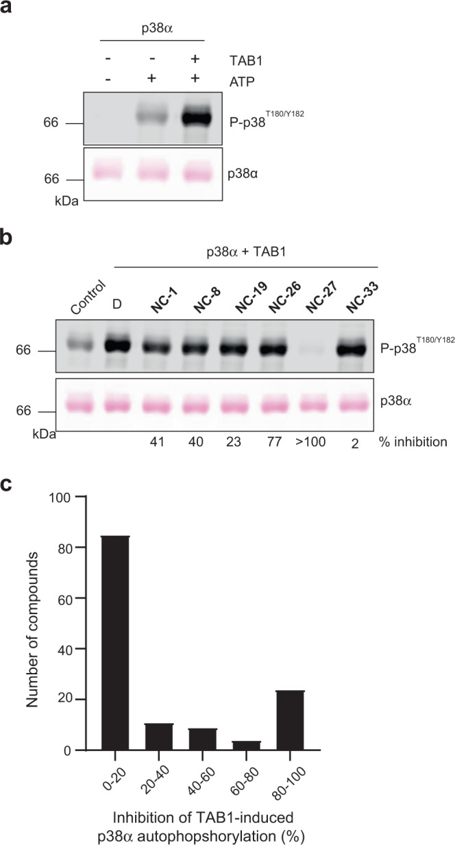
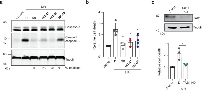
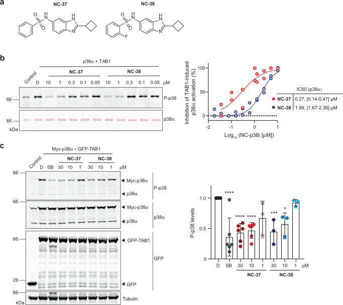
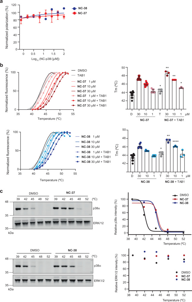
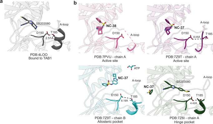
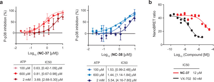
p38α is a versatile protein kinase that can control numerous processes and plays important roles in the cellular responses to stress. Dysregulation of p38α signaling has been linked to several diseases including inflammation, immune disorders and cancer, suggesting that targeting p38α could be therapeutically beneficial. Over the last two decades, numerous p38α inhibitors have been developed, which showed promising effects in pre-clinical studies but results from clinical trials have been disappointing, fueling the interest in the generation of alternative mechanisms of p38α modulation. Here, we report the in silico identification of compounds that we refer to as non-canonical p38α inhibitors (NC-p38i). By combining biochemical and structural analyses, we show that NC-p38i efficiently inhibit p38α autophosphorylation but weakly affect the activity of the canonical pathway. Our results demonstrate how the structural plasticity of p38α can be leveraged to develop therapeutic opportunities targeting a subset of the functions regulated by this pathway.


Conflict of interest statement

IRB Barcelona, UB, ICREA, BSC-CNS, and Nostrum Biodiscovery have filed the patent application WO2020120576 - P38Α AUTOPHOSPHORYLATION INHIBITORS. L.G., L.D., A.I., R.S., M.O., and A.R.N. are named inventors on this application, but the patent was abandoned. The rest of the authors declare no competing interests.

Figures

Fig. 1
Fig. 1. Identification of compounds that inhibit TAB1-induced p38α autophosphorylation.
a Purified GST-p38α protein (1.5 μM, 2 μg in 20 μl) was incubated in autophosphorylation buffer with TAB1386–414 peptide (15 μM) and ATP (600 μM), as indicated. After 2 h at 37 °C, samples were analyzed by Ponceau staining and immunoblotting. Results are representative from n = 3 experiments. b Purified GST-p38α was incubated with TAB1386–414 peptide and ATP in the presence of DMSO (D) or the indicated compounds at 30 μM, and were processed as in (a). Control, GST-p38α, and ATP without TAB1. Results are representative from n = 3 experiments. c Histogram showing the average inhibitory activity of the 133 compounds analyzed at ≥10 μM in the TAB1-induced p38α autophosphorylation assay. Source data are provided as a Source data file.
Fig. 2
Fig. 2. Identification of compounds that inhibit ischemia-reperfusion-induced cell death.
a H9c2 cells were untreated (Control) or treated for 2 h with simulated ischemia followed by 4 h of reperfusion (SIR), in the presence of DMSO (D), the ATP-competitive inhibitor SB203580 (10 μM) or the indicated compounds at 30 μM. The compounds were added 24 h before and maintained during the treatment. Total cell lysates were analyzed by immunoblotting. Cleaved caspase-3 levels were quantified and normalized to DMSO-treated cells to calculate the percentage of inhibition. Results are representative from n ≥ 3 experiments for compounds NC-37, NC-38, and NC-60. All the samples were analyzed in the same immunoblotting membrane and the dashed line indicates removed samples. b H9c2 cells were treated with the indicated compounds as in (a) and then were subjected to SIR, stained with Annexin V/Propidium Iodide (PI), and analyzed by flow cytometry. Cell death values were standardized giving the value of 1 to the control in each experiment. Data are shown as the mean ± SD (Control, D and NC-38, n = 4; SB and NC-37, n = 3). Two-sided unpaired t-test was used to compare the relative cell death in samples treated with the different inhibitors versus D. *p < 0.05. c H9c2 cells were untreated or transfected with TAB1 siRNA (TAB1 KD) and then were subjected to SIR as in (a). Cells were stained with Annexin V/PI and analyzed by flow cytometry to determine cell death values, which were standardized giving the value of 1 to the control in each experiment. Data are shown as the mean ± SD (n = 3 experiments). Two-sided unpaired t-test was used to compare the relative cell death in TAB1 KD versus D. *p < 0.05. TAB1 downregulation was analyzed by immunoblotting using total cell lysates. Source data are provided as a Source data file.
Fig. 3
Fig. 3. NC-p38i compounds inhibit TAB1-induced p38α autophosphorylation.
a Structure of compounds NC-37 and NC-38. b Purified GST-p38α protein (1.5 μM, 2 μg in 20 μl) was incubated in autophosphorylation buffer with TAB1386–414 peptide (15 μM) and ATP (600 μM) in the presence of DMSO (D) or the compounds NC-37 and NC-38 at the indicated concentrations. Control, GST-p38α and ATP without TAB1. After 2 h at 37 °C, samples were analyzed by Ponceau staining and immunoblotting with antibodies anti-phospho-p38 (T180/Y182). Values were normalized to the phosphorylated p38α levels in the DMSO sample. Data were fitted using a nonlinear regression fit model (Graphpad Prism) to determine the IC50s, with 95% confidence intervals indicated in brackets. n = 2 experiments. c HEK293T cells were co-transfected with GFP-TAB1 and myc-p38α and then were treated with SB203580 (SB, 10 μM), DMSO (D) or compounds NC-37 and NC-38 at the indicated concentrations for 6 h. Control, cells co-transfected with GFP and myc-p38α. The histogram shows the quantification of p38α phosphorylation levels normalized to the DMSO-treated cells. Data are shown as the mean ± SD (D, n = 8; SB 30 μM and NC-37 10 μM, n = 6; NC-37 1 μM and all concentrations of NC-38, n = 3). One-way ANOVA test was used to compare the P-p38 levels in samples treated with the different inhibitors versus D. *p < 0.05, ***p < 0.001, and ****p < 0.0001. Source data are provided as a Source data file.
Fig. 4
Fig. 4. NC-p38i compounds inhibit p38α autophosphorylation but not the canonical pathway.
a, b Purified GST-p38α (2 μg) was incubated with ATP (600 μM) (a) or with constitutively active MKK6DD (0.5 μg) (b) in 20 μl of kinase buffer and in the presence of DMSO (D) or the compounds NC-37 and NC-38 (10 μM). After 30 min at 37 °C, samples were analyzed by Ponceau staining and immunoblotting with antibodies anti-phospho-p38 (T180/Y182). Control, GST-p38α without ATP (a) or MKK6 (b). Results are representative from n = 3 experiments. c GST-p38α activated by MKK6 as in (b) (200 ng) was incubated with the substrates GST-MK2 or GST-ATF2 (0.5 μg) in 20 μl of kinase buffer and in the presence of DMSO (D), PH797804 (PH, 2 μM) or the compounds NC-37 and NC-38 (10 μM). After 30 min at 30 °C, samples were analyzed by immunoblotting with antibodies against MK2 phospho-T334 or ATF2 phospho-T71. Control, GST-MK2 or GST-ATF2 without active GST-p38α. The histogram shows the percentage of phosphorylation inhibition using only the stronger band for the quantification in both cases. Results are shown as mean ± SD from n = 3 experiments. Two-sided unpaired t-test was used to compare the P-MK2/P-ATF2 inhibition in samples treated with the different inhibitors. *p < 0.05 and **p < 0.01. Source data are provided as a Source data file.
Fig. 5
Fig. 5. NC-p38i compounds are specific for p38α.
a Percentage of inhibition on the catalytic activity of 97 human protein kinases incubated with compounds NC-37 or NC-38 (10 μM). Kinases were assayed at the ATP concentration of their apparent Km or below. Two different protocols were used to assay p38α kinase activity referred to as p38α and p38α (Direct). Results are shown as the mean of n = 1 done in duplicates. b, c The ability of compound NC-38 (10 μM) to displace a chemical probe that binds to the active site of 468 human protein kinases was analyzed (b). The three kinases that show less than 50% recovery in the presence of NC-38 were re-analyzed (c) in duplicates and using a range of concentrations, and only the binding to p38α was confirmed. Source data are provided as a Source data file.
Fig. 6
Fig. 6. NC-p38i compounds bind to and stabilize p38α without inhibiting its interaction with TAB1.
a FITC-labeled peptide TAB1386-414 (10 nM) was incubated for 1 h with purified GST-p38α (5.6 μM) and with increasing concentrations of compounds NC-37 and NC-38 (0.75–100 μM), as indicated. The FP signal was monitored as a readout of protein interaction. Experiments were done in triplicates, and results are shown as mean ± SD (NC-37, n = 3) or the mean (NC-38, n = 2). b Representative denaturation curves of GST-p38α in the presence of compounds NC-37 and NC-38 at the indicated concentrations alone or in combination with the TAB1386-414 peptide (15 μM). DMSO was used as control. Curves were generated by normalizing fluorescence values, considering the lowest value as 0 and highest value as 100% in each condition. Results are representative from n = 3 experiments, except for NC-38 1 and 30 µM (n = 2). The histograms (right panels) show the calculated Tm values. Two-sided unpaired t-test was used to compare the Tm of GST-p38α incubated with TAB1 (T) versus DMSO control (D), and NC-37 or NC-38 alone versus NC-37 or NC-38 combined with TAB1. Results are shown as mean ± SD (D, n = 13; NC-37 30 µM, n = 9; NC-37 10 µM, n = 8; NC-37 1 µM, and NC-38 30 µM and 10 µM, n = 7; NC-38 1 µM, n = 6; T, n = 4; all concentrations of NC-37 and NC-38 + TAB1, n = 3). *p < 0.05, **p < 0.01 and ****p < 0.0001. c KBM7 cells were treated with DMSO or compounds NC-37 and NC-38 (30 μM) for 2 h and then heated in a temperature range from 39 °C to 52 °C to induce protein unfolding and aggregation. Cell lysates were analyzed by immunoblotting, and the indicated bands were quantified and represented in the graphics. n = 1. Source data are provided as a Source data file.
Fig. 7
Fig. 7. Schematic representation of p38α structures.
a Complex of p38α and TAB1 (PDB:4LOO) and a representative p38α structure determined in this work (PDB:7Z6I) displaying the different structural and functional regions characteristic of p38α, and indicating the ligand binding sites occupied by TAB1 and the ATP binding region highlighted in yellow. b The three binding pockets of NC-p38i identified in the crystals are represented on the p38α surface.
Fig. 8
Fig. 8. Structural characterization of the binding of NC-p38i compounds to p38α.
a Crystal structures of p38α in complex with SB203580 (located in the active site) and compound NC-37 bound to the hinge pocket. b p38α complexes bound to NC-37 (dark blue, PBD:7Z9T) and NC-38 (yellow, PBD:7PVU) bound to the active site. Both complexes are highly similar. Specific residues involved in the interaction are shown for the NC-38 complex (compound represented in gray, the fluorine atom is shown in green). c The p38α complex bound to NC-37 (chain B) shows the compound bound to the allosteric pocket adjacent to the active site. Residues involved in the binding are highlighted. The X-ray data collection and refinement statistics are shown in Supplementary Table 4. d All complexes of p38α with NC-p38i show the DFG motif in the active conformation (DFG-in). Compounds are labeled in the figure for clarity.
Fig. 9
Fig. 9. NC-p38i compounds disrupt the formation of a hydrogen bond between Thr185 and Asp150 in p38α.
a Crystal structure of the p38α-TAB1 complex showing the hydrogen bond (2.5 Å) formed between Thr185 and Asp150 that is required for TAB1-induced autophosphorylation of p38α. b Crystal structures of the three p38α-NC-p38i complexes presented in this manuscript showing that Thr185 and Asp150 are either too far apart or not properly positioned for the formation of the critical hydrogen bond. Distances are indicated in Å.
Fig. 10
Fig. 10. NC-p38i compounds behave as weak ATP-competitive inhibitors.
a Inhibition curves of p38α autophosphorylation by different concentrations of compounds NC-37 and NC-38 (0.035 to 10 μM) at the indicated ATP concentrations. Results are shown as mean ± SD (n = 3 experiments). Data were fitted using a nonlinear regression fit model (Graphpad Prism) to determine the IC50s, with 95% confidence intervals indicated in brackets. b HEK293T cells expressing NanoLuc-fused p38α were incubated with a cell-permeable energy transfer probe, which generates a BRET signal upon binding to the ATP site of p38α. Cells were incubated with different concentrations of compound NC-37 or the ATP-competitive inhibitor VX-702 as a control, and the NanoBRET ratio was measured to calculate the IC50. Results are shown for two technical repeats from a representative experiment. In total two biologically independent experiments were performed and gave similar results. Source data are provided as a Source data file.

References

    1. Wang Z, et al. The structure of mitogen-activated protein kinase p38 at 2.1-A resolution. Proc. Natl Acad. Sci. USA. 1997;94:2327–2332. doi: 10.1073/pnas.94.6.2327. - DOI - PMC - PubMed
    1. Canovas B, Nebreda AR. Diversity and versatility of p38 kinase signalling in health and disease. Nat. Rev. Mol. Cell Biol. 2021;22:346–366. doi: 10.1038/s41580-020-00322-w. - DOI - PMC - PubMed
    1. Ge B, et al. MAPKK-independent activation of p38alpha mediated by TAB1-dependent autophosphorylation of p38alpha. Science. 2002;295:1291–1294. doi: 10.1126/science.1067289. - DOI - PubMed
    1. Ge B, et al. TAB1beta (transforming growth factor-beta-activated protein kinase 1-binding protein 1beta), a novel splicing variant of TAB1 that interacts with p38alpha but not TAK1. J. Biol. Chem. 2003;278:2286–2293. doi: 10.1074/jbc.M210918200. - DOI - PubMed
    1. DeNicola GF, et al. Mechanism and consequence of the autoactivation of p38alpha mitogen-activated protein kinase promoted by TAB1. Nat. Struct. Mol. Biol. 2013;20:1182–1190. doi: 10.1038/nsmb.2668. - DOI - PMC - PubMed

Publication types