. 2023 Jun 12;13(1):9510.
doi: 10.1038/s41598-023-36698-w.

Quantifying the shear modulus of the adductor longus muscle during hip joint motion using shear wave elastography

Affiliations

Quantifying the shear modulus of the adductor longus muscle during hip joint motion using shear wave elastography

Takuya Kato et al. Sci Rep. .

Abstract

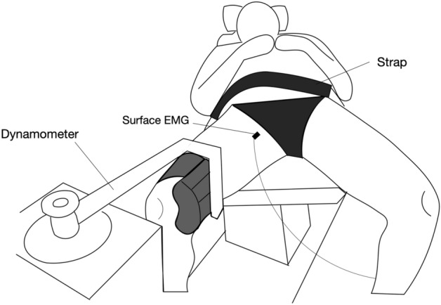
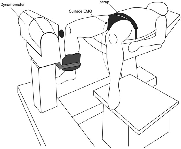
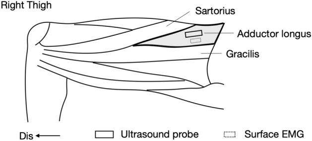
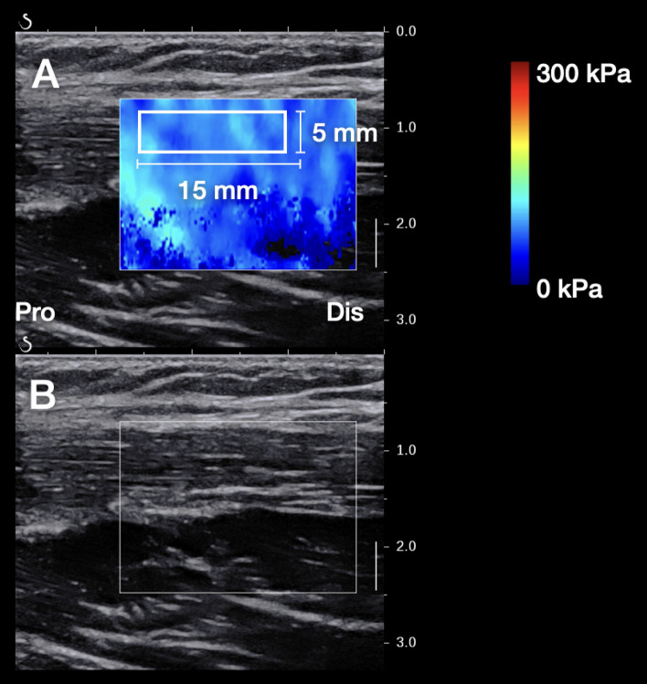
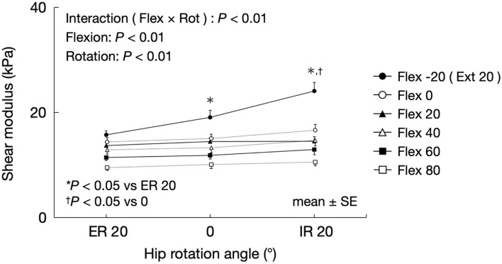
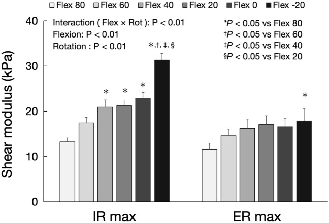
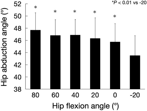
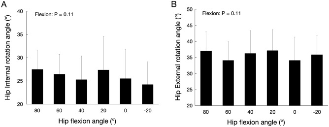
The present study aims to assess the effect of the hip flexion angle on the shear modulus of the adductor longus (AL) muscle associated with passive hip abduction and rotation. Sixteen men participated in the study. For the hip abduction task, the hip flexion angles used were - 20, 0, 20, 40, 60, and 80°, and the hip abduction angles were 0, 10, 20, 30, and 40°. For the hip rotation task, the hip flexion angles used were - 20, 0, 20, 40, 60, and 80°, hip abduction angles were 0 and 40°, and hip rotation angles were 20° internal rotation, 0° rotation, and 20° external rotation. The shear modulus at 20° extension was significantly higher than that at 80° flexion for the 10, 20, 30 and 40° hip abduction (i.e., P < 0.05). The shear modulus at 20° internal rotation and 20° extension was significantly higher than that at 0° rotation and 20° external rotation, regardless of the hip abduction angle (i.e., P < 0.05). The mechanical stress of the AL muscle associated with hip abduction was higher in the extended position. Furthermore, the mechanical stress could increase with internal rotation only at the hip-extended position.


Conflict of interest statement

The authors declare no competing interests.

Figures

Figure 1
Figure 1
Experimental setup of the hip abduction task.
Figure 2
Figure 2
Experimental setup of the hip roration task.
Figure 3
Figure 3
Location of the ultrasound probe and surface EMG electrode. Dis distal.
Figure 4
Figure 4
Typical examples of the SWE image (A) and B-mode image (B). SWE shear wave elastography, Pro proxima, Dis Distal.
Figure 5
Figure 5
Changes in the shear modulus of the AL muscle associated with hip abduction in each hip flexion angle. AL adductor longus, Flex flexion, Abd abduction.
Figure 6
Figure 6
Shear modulus of the AL muscle at the maximum hip abduction angle for each hip flexion angle. AL adductor longus.
Figure 7
Figure 7
Changes in the shear modulus of the AL muscle associated with hip rotation for each hip flexion angle. AL adductor longus, Flex flexion, Rot rotation, ER external rotation, IR internal rotation.
Figure 8
Figure 8
Shear modulus of the AL muscle at the maximum hip external and internal rotation angles for each hip flexion angle. AL adductor longus, Flex flexion, Rot rotation, ER max maximum external rotation, IR max maximum internal rotation.
Figure 9
Figure 9
Changes in passive hip adduction torque associated with hip abduction for each hip flexion angle. Flex flexion, Abd abduction.
Figure 10
Figure 10
Relationship between shear modulus of the AL muscle and passive hip adduction torque for each hip flexion angle. AL adductor longus, Flex flexion.
Figure 11
Figure 11
Maximum hip abduction angle for each hip flexion angle.
Figure 12
Figure 12
Maximum hip internal (A) and external (B) rotation angles at 40° hip abduction for each hip flexion angle.

References

    1. Ekstrand J, et al. Time before return to play for the most common injuries in professional football: A 16-year follow-up of the UEFA Elite Club Injury Study. Br. J. Sports Med. 2020;54:421–426. doi: 10.1136/bjsports-2019-100666. - DOI - PMC - PubMed
    1. Ekstrand J, Hagglund M, Walden M. Epidemiology of muscle injuries in professional football (soccer) Am. J. Sports Med. 2011;39:1226–1232. doi: 10.1177/0363546510395879. - DOI - PubMed
    1. Holmich P. Long-standing groin pain in sportspeople falls into three primary patterns, a “clinical entity” approach: A prospective study of 207 patients. Br. J. Sports Med. 2007;41:247–252. doi: 10.1136/bjsm.2006.033373. - DOI - PMC - PubMed
    1. Taylor R, et al. Multidisciplinary assessment of 100 athletes with groin pain using the Doha agreement: High prevalence of adductor-related groin pain in conjunction with multiple causes. Clin. J. Sport Med. 2018;28:364–369. doi: 10.1097/JSM.0000000000000469. - DOI - PubMed
    1. Serner A, et al. Diagnosis of acute groin injuries: A prospective study of 110 athletes. Am. J. Sports Med. 2015;43:1857–1864. doi: 10.1177/0363546515585123. - DOI - PubMed

Publication types