Review
. 2023 Nov;20(11):723-737.
doi: 10.1038/s41569-023-00886-y. Epub 2023 Jun 12.

Interplay of hypoxia-inducible factors and oxygen therapy in cardiovascular medicine

Affiliations
Review

Interplay of hypoxia-inducible factors and oxygen therapy in cardiovascular medicine

Yafen Liang et al. Nat Rev Cardiol. 2023 Nov.

Abstract

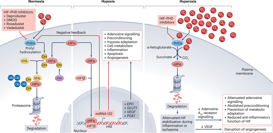
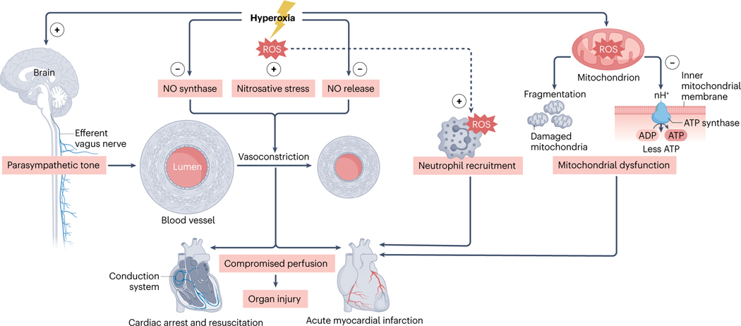
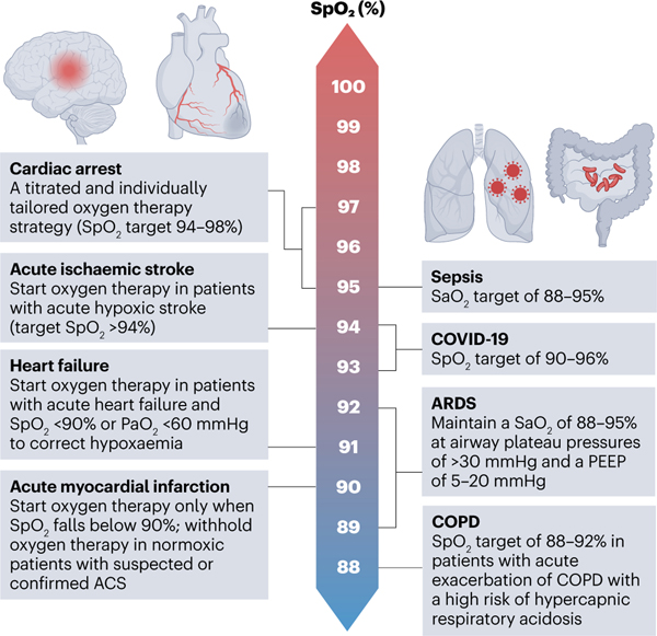
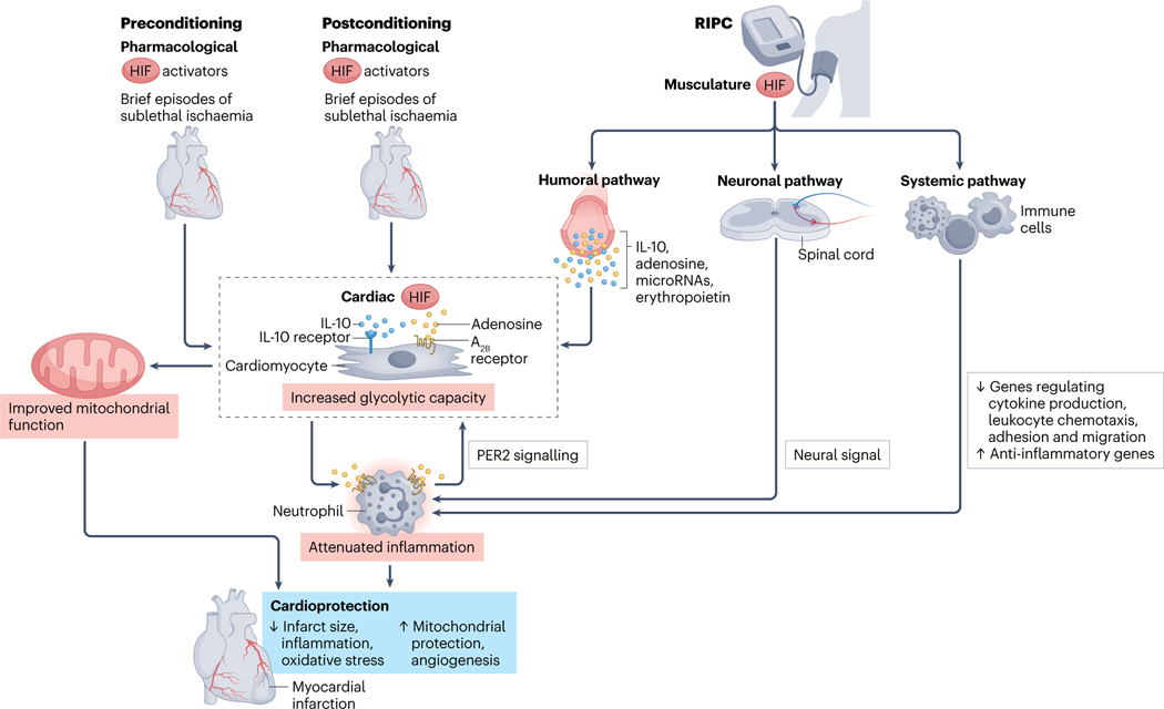
Mammals have evolved to adapt to differences in oxygen availability. Although systemic oxygen homeostasis relies on respiratory and circulatory responses, cellular adaptation to hypoxia involves the transcription factor hypoxia-inducible factor (HIF). Given that many cardiovascular diseases involve some degree of systemic or local tissue hypoxia, oxygen therapy has been used liberally over many decades for the treatment of cardiovascular disorders. However, preclinical research has revealed the detrimental effects of excessive use of oxygen therapy, including the generation of toxic oxygen radicals or attenuation of endogenous protection by HIFs. In addition, investigators in clinical trials conducted in the past decade have questioned the excessive use of oxygen therapy and have identified specific cardiovascular diseases in which a more conservative approach to oxygen therapy could be beneficial compared with a more liberal approach. In this Review, we provide numerous perspectives on systemic and molecular oxygen homeostasis and the pathophysiological consequences of excessive oxygen use. In addition, we provide an overview of findings from clinical studies on oxygen therapy for myocardial ischaemia, cardiac arrest, heart failure and cardiac surgery. These clinical studies have prompted a shift from liberal oxygen supplementation to a more conservative and vigilant approach to oxygen therapy. Furthermore, we discuss the alternative therapeutic strategies that target oxygen-sensing pathways, including various preconditioning approaches and pharmacological HIF activators, that can be used regardless of the level of oxygen therapy that a patient is already receiving.


Figures

Fig. 1 |
Fig. 1 |. Responses of HIFs to hypoxia or hyperoxia.
Under normoxic conditions, hydroxylation at two proline residues by prolyl hydroxylases (PHDs) promotes the association between hypoxia-inducible factor-α (HIFα) and the von Hippel–Lindau (VHL) gene product, leading to HIFα destruction via the ubiquitin–proteasome pathway,,. During hypoxia, this process is suppressed due to the lack of oxygen as a substrate for PHDs, allowing the HIFα subunit to escape proteolysis. After stabilization, HIFα dimerizes with HIF1β, and the heterodimer translocates to the nucleus to activate the transcription of target genes that contain hypoxia response elements (HREs) in their promoter. These target genes control essential physiological functions such as hypoxia adaptation, cell metabolism, inflammation, apoptosis and angiogenesis. Importantly, gene repression by HIFs frequently occurs as an indirect response that involves the transcriptional induction of HIF-dependent microRNAs (miRNAs) and the subsequent repression of target gene expression. Furthermore, miRNAs have also been shown to participate in a feedforward pathway via an increase in HIF responses through PHD1 repression to provide organ protection during ischaemia–reperfusion injury. HIF stabilization by HIF–PHD inhibitors (such as daprodustat, dimethyloxalylglycine, roxadustat and vadadustat) might be a potential therapeutic approach for acute cardiovascular disease,. In addition, HIF activity is also regulated by hydroxylation of a single conserved asparaginyl residue at the C-terminal transactivation domain by the oxygen-dependent asparaginyl hydroxylase factor inhibiting HIF1 (FIH1). During hyperoxia, high oxygen levels prevent the hypoxic inhibition of PHDs during conditions such as inflammation, ischaemia or metabolic imbalance. Attenuated HIF stabilization can dampen adaptive responses, such as angiogenic responses, or cardioprotection by adenosine generation and signalling (for example, through decreased adenosine A2A receptor signalling). As such, hyperoxia is associated with attenuated adenosine production and signalling, abolished preconditioning of the heart, suppression of adaptive metabolic responses, and reduced capacity of HIFs to dampen inflammation. Of note, given that HIF–PHD inhibitors will still be functional in stabilizing HIFs, even during hyperoxia, HIF activators can be considered as a cardioprotective strategy that works independently of the level of oxygen therapy that a patient receives. DMOG, dimethyloxalylglycine; OH, hydroxylation; Ub, ubiquitination; VEGF, vascular endothelial growth factor.
Fig. 2 |
Fig. 2 |. Pathophysiological effects of hyperoxia on the cardiovascular system.
Hyperoxia inhibits endothelial nitric oxide (NO) synthase and NO release from S-nitrosothiol, thereby reducing NO bioavailability and promoting vasoconstriction in cerebral and coronary arteries. Hyperoxia also increases the production of reactive oxygen species (ROS). An increase in ROS converts available NO to peroxynitrite, which leads to nitrosative stress. In addition, hyperoxia increases parasympathetic tone, resulting in a reduction in heart rate. As a consequence of this vasoconstrictive effect, hyperoxia reduces cardiac output, compromises organ perfusion and exacerbates organ injury. Moreover, ROS facilitate the migration of neutrophils to the blood–endothelial cell interface and the surrounding tissues, further exacerbating ischaemia–reperfusion injury,. Finally, hyperoxia can also directly mediate mitochondrial dysfunction at the cellular level, thereby contributing to cell death,,.
Fig. 3 |
Fig. 3 |. Oxygen targets during cardiovascular disease and acute illness.
Guidelines have recommended different oxygen targets for treating specific diseases on the basis of current clinical evidence. However, regardless of the disease entity, there is a general agreement to avoid hypoxia and excessive hyperoxia. In patients with acute myocardial infarction, the 2017 ESC guidelines recommend supplementing oxygen only when oxygen saturation (SpO2) levels fall below 90%. The 2015 AHA guidelines recommend withholding oxygen supplementation in normoxic patients with suspected or confirmed acute coronary syndrome (ACS) in the prehospital, emergency department and hospital settings, and a titrated and individualized oxygen therapy strategy for patients after cardiac arrest with a target SpO2 of 94–98% after the return of spontaneous circulation. For patients with heart failure, the 2021 ESC guidelines recommend oxygen therapy for patients with acute heart failure and SpO2 of <90% or partial pressure of oxygen (PaO2) of <60 mmHg to correct hypoxaemia, and that oxygen should not be used routinely in patients without hypoxaemia. For patients with acute ischaemic stroke, the 2018 AHA guidelines recommend providing oxygen therapy to maintain an oxygen saturation level of >94%. For patients with acute respiratory distress syndrome (ARDS), the ARDS Network recommends a target SpO2 level of 88–95% with an airway plateau pressure of <30 mmHg and a positive end-expiratory pressure (PEEP) of 5–20 mmHg (ref. 138). For patients with sepsis, the Surviving Sepsis Campaign guidelines recommend an arterial haemoglobin oxygen saturation (SaO2) target of 88–95%. In addition, for patients with ARDS related to COVID-19, a target SpO2 of between 90% and 96% is recommended. Finally, for patients with chronic obstructive pulmonary disease (COPD), the British Thoracic Society, the Global Initiative for Chronic Obstructive Lung Disease, the American Thoracic Society and the European Respiratory Society recommend titrating oxygen therapy to alleviate hypoxia while avoiding hyperoxia to maintain SaO2 levels of between 88% to 92% in patients with acute exacerbation of COPD and a high risk of hypercapnic respiratory acidosis,.
Fig. 4 |
Fig. 4 |. Molecular mechanisms of preconditioning, postconditioning and RIPC for cardioprotection.
During ischaemic preconditioning, short episodes of myocardial ischaemia provide cardioprotection during a subsequent prolonged ischaemic event, whereas ischaemic postconditioning is achieved by repetitive brief interruptions of coronary blood flow before final complete reperfusion. Remote ischaemic postconditioning (RIPC) can be achieved by repetitive cycles of ischaemia and reperfusion to a limb via inflation and subsequent deflation of a non-invasive blood-pressure cuff. All three experimental approaches target oxygen-sensing pathways and converge on the stabilization of hypoxia-inducible factors (HIFs), with the concomitant induction of crucial HIF target genes. For example, the extracellular signalling molecule adenosine and its subsequent activation of the adenosine A2B receptor are central to ischaemic preconditioning-mediated cardioprotection. Activation of the adenosine A2B receptor also stabilizes circadian protein homologue 2 (PER2), which results in amplified PER2 signalling and improved glycolytic capacity of the cardiomyocytes and ischaemic tolerance. This increase in ischaemic tolerance leads to myocardial protection via attenuated inflammation and oxidative stress, reduced myocardial infarct size, and improved mitochondrial function. RIPC can protect organs against ischaemia–reperfusion injury via three different pathways: the humoral, the neuronal and the systemic pathways. RIPC stabilizes HIF1α in the peripheral musculature, leading to the elevated expression of the HIF target gene IL10. The release of IL-10 from the remote tissue exposed to hypoxia-induced ischaemia subsequently activates the IL-10 receptor on cardiomyocytes and protects the heart against ischaemic injury. Other mediators involved in this humoral pathway include adenosine, erythropoietin and various microRNAs. Second, RIPC activates the production of autacoids, such as adenosine and bradykinin, in the remote preconditioned organ, which stimulates afferent nerves and relays the neural signal to the myocardium via the efferent nerve fibres. Denervation of the neural pathway in the remote organ abolishes RIPC protection,. Third, RIPC provokes a systemic response by suppressing genes involved in regulating leukocyte chemotaxis and cytokine production, adhesion, and migration and upregulating anti-inflammatory genes. Pharmacological HIF activators or prolyl hydroxylase inhibitors have been shown to have similar preconditioning and cardioprotective effects to those of ischaemic preconditioning.

Similar articles

Cited by

References

    1. Eltzschig HK & Carmeliet P. Hypoxia and inflammation. N. Engl. J. Med. 364, 656–665 (2011). - PMC - PubMed
    1. Steele C. Severe angina pectoris relieved by oxygen inhalations.Br. Med. J. 2, 1568 (1900).
    1. Heffner JE The story of oxygen. Respir. Care 58, 18–31 (2013). - PubMed
    1. Shultz SM & Hartmann PM George E Holtzapple (1862–1946) and oxygen therapy for lobar pneumonia: the first reported case (1887) and a review of the contemporary literature to 1899. J. Med. Biogr. 13, 201–206 (2005). - PubMed
    1. Severinghaus JW Eight sages over five centuries share oxygen’s discovery. Adv. Physiol. Educ. 40, 370–376 (2016). - PubMed

Publication types