. 2023 Jul;8(7):1318-1329.
doi: 10.1038/s41564-023-01411-0. Epub 2023 Jun 12.

Mechanism of D-alanine transfer to teichoic acids shows how bacteria acylate cell envelope polymers

Affiliations

Mechanism of D-alanine transfer to teichoic acids shows how bacteria acylate cell envelope polymers

Bailey J Schultz et al. Nat Microbiol. 2023 Jul.

Abstract

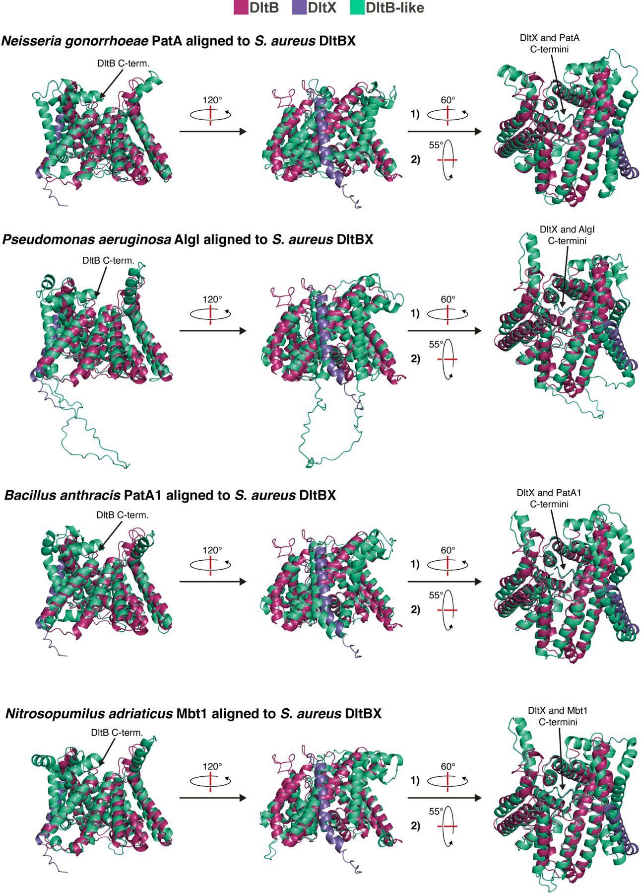
Bacterial cell envelope polymers are often modified with acyl esters that modulate physiology, enhance pathogenesis and provide antibiotic resistance. Here, using the D-alanylation of lipoteichoic acid (Dlt) pathway as a paradigm, we have identified a widespread strategy for how acylation of cell envelope polymers occurs. In this strategy, a membrane-bound O-acyltransferase (MBOAT) protein transfers an acyl group from an intracellular thioester onto the tyrosine of an extracytoplasmic C-terminal hexapeptide motif. This motif shuttles the acyl group to a serine on a separate transferase that moves the cargo to its destination. In the Dlt pathway, here studied in Staphylococcus aureus and Streptococcus thermophilus, the C-terminal 'acyl shuttle' motif that forms the crucial pathway intermediate is found on a transmembrane microprotein that holds the MBOAT protein and the other transferase together in a complex. In other systems, found in both Gram-negative and Gram-positive bacteria as well as some archaea, the motif is fused to the MBOAT protein, which interacts directly with the other transferase. The conserved chemistry uncovered here is widely used for acylation throughout the prokaryotic world.


Figures

Extended Data Fig. 1
Extended Data Fig. 1. S. aureus strains without dltX cannot survive on tunicamycin but still produce LTAs.
a, Chemical structures of three repeat units of Staphylococcus aureus LTA (left) and one repeat unit of Streptococcus pneumoniae LTA (right). d-Alanine esters are shown in blue. b, Full set of controls for the spot titer assay shown in Figure 1c. S. aureus strains were grown on tryptic soy agar (TSA) plates containing the indicated compounds: dimethyl sulfoxide (DMSO; 1.25 μL per 1.00 mL of TSA), isopropyl β-D-1-thiogalactopyranoside (IPTG; 1.00 mM), and/or tunicamycin (tunic.; 1.0 μg/mL). Tunic. inhibits WTA biosynthesis, IPTG was used to induce expression of the indicated plasmid-borne cassette, and DMSO was used as a vehicle control. The image of the plate with tunicamycin and IPTG has been reproduced here for clarity. The plasmid harboring dltXABCD is leaky thus resulting in growth of the sixth strain even in the absence of IPTG. c, An α-LTA Western blot indicates that all of the strains tested in Figure 1c and b produce LTAs at roughly equivalent levels to wild-type S. aureus. The strain in Lane 2 (4S5) is a mutant that lacks LtaS and contains a suppressor mutation that permits growth in the absence of LTAs. It does not produce any LTAs. The LTAs themselves run as a smear from ~15–37 kDa. d, Spot titer assay results for a partially different set of S. aureus strains. Here, the complementation constructs are under anhydrotetracycline (aTc)-inducible control, and the overall cassettes are genomically integrated. The TSA plates here contained DMSO, tunic., and aTc as indicated, with the concentrations of DMSO and tunic. being the same as for b and that for aTc being 0.4 μM. In these strains, the dltXABCD complementation construct does not appear leaky. e, SDS-PAGE autoradiography of LTAs from S. aureus cells grown in the presence of d-[14C]alanine again shows that dltX is required for LTA d-alanylation. Here, the same set of strains from d was used. f, An α-LTA Western blot indicates that all strains tested in d and e produce LTAs at roughly equivalent levels to wild-type S. aureus.
Extended Data Fig. 2
Extended Data Fig. 2. DltX is a predicted bitopic transmembrane protein with an extracellular C-terminus and is required for DltD to copurify with DltB.
a, (top) The TOPCONS web server for consensus-based membrane protein topology prediction was used to predict the topology of S. aureus DltX. The sequence of S. aureus DltX is shown with the predicted localization of each individual residue indicated beneath. (bottom) An AlphaFold2 model of S. aureus DltX generated using ColabFold. This specific model is from an overall model of DltX with DltD and was selected here because it illustrates the topology of the protein well. We note that in the DltDX model, DltX’s predicted topology is in agreement with the known topology of DltD. b, An α-myc Western blot indicates that a similar amount of myc-StDltD was present in the solubilized membranes loaded onto the TALON immobilized metal affinity chromatography (IMAC) resin during purifications of StDltB-His10 (and interactors) from E. coli cells expressing StdltB and StdltD, with or without StdltX. A similar amount of myc-StDltD was also present in the flow-through from the resin in the two samples. However, no myc-StDltD could be observed in the IMAC elution fraction from the sample derived from E. coli cells that did not express StdltX.
Extended Data Fig. 3
Extended Data Fig. 3. The predicted S. aureus DltBDX structure is a high-confidence prediction.
a, (top) The top-ranked AlphaFold2 model of the S. aureus DltBDX complex, with proteins colored as in Fig. 2c, is a high-confidence model. The predicted local Distance Difference Test (plDDT) is a metric of model confidence, with plDDT values > 90 representing very high confidence, 70–90 representing moderate confidence, 50–70 representing low confidence, and < 50 representing very low confidence. The graph in the upper right shows the plDDT values at each residue for all five models. The numbering on the x-axis here corresponds to the residue number for the concatenated sequence of the complex (i.e., the sequence of DltX concatenated N-terminally to the sequence of DltB, and then that overall sequence concatenated N-terminally to the sequence of DltD). The rightmost of the two models underneath the “Rank 1” heading shows a graphical depiction of the plDDT values with a color gradient scale (dark blue representing a plDDT value of 100, and red representing a plDDT value of 0). (bottom) Each of the other four models is highly similar to the top-ranked model. Five models were generated by ColabFold, and the models were ranked by predicted template modeling (pTM) scores. b, The relative positions between the three proteins are high-confidence based on the Predicted Aligned Error (PAE). Here, the value at each (x, y) point in the graphs indicates the “expected position error at residue x if the predicted and true structures were aligned on residue y” (https://alphafold.ebi.ac.uk/faq). Low values (blue) indicate low expected position error. Again, the numbering on the x-axis corresponds to the residue number for the concatenated sequence of the complex. Numbering on the y-axis (not shown) would place “0” at the top of the axis. c, Views of the surface representation of the S. aureus DltBDX AlphaFold2 model.
Extended Data Fig. 4
Extended Data Fig. 4. The C-terminal motif of DltX, and particularly its invariant tyrosine residue, is required for S. aureus growth on tunicamycin.
a, Full set of controls for the spot titer assay shown in Figure 3b. S. aureus strains were grown on TSA plates containing the indicated compounds: DMSO at 1.25 μL per 1.00 mL of TSA, IPTG at 1.00 mM, anhydrotetracycline (aTc) at 0.4 μM, and tunicamycin at 1.0 μg/mL. Tunic. inhibits wall teichoic acid biosynthesis, IPTG was used to induce expression of the indicated plasmid-borne cassette (here, null refers to a strain with an empty IPTG-inducible cassette in the plasmid), aTc was used to induce expression of the indicated chromosomally integrated cassette, and DMSO was used as a vehicle control for the tunic. The image of the plate with tunic., IPTG, and aTc has been reproduced here for clarity. flag-dltX∆motif encodes for an N-terminally FLAG-tagged DltX variant lacking the last six amino acids. b, Full set of controls for the spot titer assay shown in Figure 3c. Compounds were used at the same concentrations as above, for the same reasons as above. Here, the bracketed sequences in the superscripts of the aTc-inducible cassettes represent the C-terminal motif sequence of the given DltX variant. Red letters denote changes from the wild-type sequence.
Extended Data Fig. 5
Extended Data Fig. 5. Length alterations at the C-terminus of DltX are not well-tolerated.
a, Full set of controls for the spot titer assay shown in Figure 3d. S. aureus strains were grown on TSA plates containing the indicated compounds: DMSO at 1.25 μL per 1.00 mL of TSA, IPTG at 1.00 mM, anhydrotetracycline (aTc) at 0.4 μM, and tunicamycin at 1.0 μg/mL. Tunic. inhibits wall teichoic acid biosynthesis, IPTG was used to induce expression of the indicated plasmid-borne cassette (here, null refers to a strain with an empty IPTG-inducible cassette in the plasmid), aTc was used to induce expression of the indicated chromosomally integrated cassette, and DMSO was used as a vehicle control for the tunic. The image of the plate with tunic., IPTG, and aTc has been reproduced here for clarity. Here, the bracketed sequences in the superscripts of the aTc-inducible cassettes represent the C-terminal motif sequence of the given DltX variant. Red letters denote changes from the wild-type sequence, and a red hyphen denotes the deletion of a residue. b, A spot titer assay shows that, of all the DltX mutants we have tested that do not allow for growth of a dltX-null strain on tunicamycin when expressed at low levels (from a chromosomally integrated aTc-inducible cassette), only the DltX mutant with an alanine added onto the C-terminus can complement when expressed at high levels (from an IPTG-inducible cassette on a medium copy number plasmid). Here, dltX* indicates that the plasmid encodes for a variant of DltX with the C-terminal sequence indicated in the “DltX C-term.” column. Again, red letters denote changes from the wild-type sequence, and a red hyphen denotes the deletion of a residue. Both dltXwt (wild-type) and all dltX* here encode an N-terminal FLAG tag.
Extended Data Fig. 6
Extended Data Fig. 6. The S. aureus DltBX and DltDX predicted structures are high-confidence predictions.
a, The top-ranked AlphaFold2 model of the S. aureus DltBX complex is a high-confidence model. The predicted local Distance Difference Test (plDDT) is a metric of model confidence, with plDDT values > 90 representing very high confidence, 70–90 representing moderate confidence, 50–70 representing low confidence, and < 50 representing very low confidence. The graph in the upper right shows the plDDT values at each residue for five models. The numbering on the x-axis corresponds to the residue number for the concatenated sequence of the complex (i.e., the sequence of DltB concatenated N-terminally to the sequence of DltX). The rightmost of the two full model images depicts the plDDT values with a color gradient scale (dark blue representing a plDDT value of 100, and red representing a value of 0). The zoom-in image shows how the DltX C-terminal motif fits into the DltB tunnel. b, The relative positions between the proteins are high-confidence based on the Predicted Aligned Error (PAE). Here, the value at each (x, y) point in the graphs indicates the “expected position error at residue x if the predicted and true structures were aligned on residue y” (https://alphafold.ebi.ac.uk/faq). Low values (blue) indicate low expected position error. Again, the numbering on the x-axis corresponds to the residue number for the concatenated sequence of the complex. Numbering on the y-axis (not shown) would place “0” at the top of the axis. c, The top-ranked AlphaFold2 model of the S. aureus DltDX complex is a high-confidence model. The rightmost of the two full model images depicts the plDDT values with a color gradient scale matching that found in a. The zoom-in image shows how the DltX C-terminal motif fits into the DltD active site region. The graph in the lower right shows the plDDT values at each residue for five models. The numbering on the x-axis here parallels that in a. d, The relative positions between the proteins are high-confidence based on the PAE. The numbering on the x- and y-axes parallels the numbering in b.
Extended Data Fig. 7
Extended Data Fig. 7. DltB, DltD, and DltX levels are comparable between samples in the DltD d-alanylation in vitro reconstitution assay, and active forms of all three proteins plus DltA and DltC allow for rapid d-alanylation of DltD.
a, A Coomassie-stained SDS-PAGE gel from an in vitro reconstitution experiment run in parallel to the one shown in Fig. 4b but with cold d-alanine rather than d-[14C]alanine. Lane 4 contains all five active proteins, whereas Lanes 3, 5, and 6 are missing the indicated protein(s). Lanes 7–9 contain four active proteins and the indicated mutant. b, A Coomassie-stained SDS-PAGE gel (run using a tris-glycine gel rather than a bis-tris gel in a) of purified StDltBDX complex, specifically containing wild-type DltB, inactive DltX (Y56F), and wild-type DltD, provided as a reference for comparison to the samples in a. This suggests the impurity below DltB in a may be a degradation product. c, SDS-PAGE autoradiograph from a time course of the in vitro reconstitution with DltA, DltC, and DltBDX. For the “10 sec.” time point, the DltBDX complex was added to the DltC charging reaction, the sample was mixed by pipetting up and down several times, and then immediately 4x XT sample buffer was added and mixed followed by flash-freezing of the sample in liquid nitrogen. For all other time points, after the DltBDX complex was added to the DltC charging reaction, samples were incubated at 30 ℃ for the indicated amount of time before addition of SDS-PAGE loading buffer and immediate flash freezing in liquid nitrogen.
Extended Data Fig. 8
Extended Data Fig. 8. DltB, DltD, and DltX levels are comparable between samples in the DltX d-alanylation in vitro reconstitution assay, and d-alanylation of DltXY56K is position-specific.
a, A Coomassie-stained SDS-PAGE gel from an in vitro reconstitution experiment run in parallel to the one shown in Fig. 4c but with cold d-alanine rather than d-[14C]alanine. Lanes 2 and 3 contain wild-type (wt) 1x or 3xFLAG-DltX, while Lanes 4 and 5 contain 1x or 3xFLAG-DltXY56K. b, A Coomassie-stained SDS-PAGE gel (run using a tris-glycine gel rather than a bis-tris gel in a) of purified StDltBDX complex, specifically containing wild-type DltB, DltXN57K, and wild-type DltD, provided as a reference for comparison to the samples in a. This suggests that the background smearing in the Coomassie-stained gels in a and c (right) is a result of the high-pH reaction conditions combined with the specific gel type used (see Supplementary Fig. 2a for further evidence of this). Additionally, the impurity below DltB in a and c (right) may be a degradation product. We note that the mobility of 1xFLAG-DltX in the bis-tris gel here is different from that in the tris-glycine gel in panel a, but the bands were confirmed by Western blot (See Supplementary Figs. 2b–c). c, (left) SDS-PAGE autoradiography of in vitro reconstitution results shows that the DltXY56K mutant is d-alanylated in a position specific manner. All lanes contain active DltA, DltC, and DltBDX complex, with the DltX in the complex being the variant with the C-terminal sequence indicated by the key. These reconstitutions were run alongside those shown in Fig. 4c and a, and the samples were run on the same gel and thus imaged together. (right) A Coomassie-stained SDS-PAGE gel from an in vitro reconstitution run in parallel to the one shown in c (left) but with cold d-alanine rather than d-[14C]alanine. Again, all lanes contain active DltA, DltC, and DltBDX complex, with the DltX in the complex being the variant with the C-terminal sequence indicated by the key. In the key, blue coloring indicates the lysine, and red coloring indicates the tyrosine-to-phenylalanine swap.
Extended Data Fig. 9
Extended Data Fig. 9. A C-terminal motif reminiscent of DltX’s is found at the C-termini of MBOAT proteins that partner with DltD-like proteins in various prokaryotic cell envelope polymer acylation pathways.
a, Graphical depictions illustrate the genomic co-localization of genes encoding MBOAT proteins (green) and DltD-like proteins (blue) in bacterial cell envelope polymer acylation systems, as well as a system of unknown function found in some archaea. The chemical structures show a portion of the relevant polymer for each system, with the modification (acetylation in each of these cases), in blue,,. The lower right depicts a potential polymer acylation system of unknown function found in the archaeal species Nitrosopumilus adriaticus. We have designated the genes mbt1 for MBOAT1 and mbtp for MBOAT Partner. b, AlphaFold2 models of the two-protein complexes encoded by the genes from a. The models are all shown cytosolic side-down. c, Sequence logo generated by EVcouplings with N. gonorrhoeae PatA as the query sequence showing the conservation of the final 51 amino acid positions of an alignment of bacterial cell envelope acylation-associated MBOAT proteins (as well as similar MBOAT proteins of unknown function), with the overall consensus sequence and corresponding NgPatA sequence shown underneath. The height of each individual letter represents the degree of conservation of that specific amino acid at the given position. The final transmembrane (TM) helix indicated is a prediction based on AlphaFold. d, Active sites of the MBOAT proteins of three of the systems shown in this figure (the corresponding active site for NgPatA is found in Fig. 4f) from AlphaFold2 models of the MBOAT proteins alone. These images are all shown with an “aerial” view of the active site (looking toward the cytosol from the extracytoplasmic region). The catalytic histidine known to be required for all studied MBOAT proteins is shown in cyan, and the invariant tyrosine present in this class of MBOAT proteins is shown in purple.
Extended Data Fig. 10
Extended Data Fig. 10. The MBOAT proteins in non-Dlt pathway cell envelope acylation systems have additional C-terminal helices that allow the C-terminal motif of these proteins to nestle into the active site.
Alignments of predicted structures of MBOAT proteins from bacterial cell envelope polymer acetylation systems (and a system of unknown function in archaea) with the predicted structure of the DltBX complex show that the non-DltB MBOAT proteins likely share a similar architecture to DltB but with a C-terminal extension of several additional helices. All of the predicted structures here were generated using the ColabFold implementation of AlphaFold2 and aligned using the PyMOL “super” command.
Figure 1.
Figure 1.. dltX is required for lipoteichoic acid d-alanylation in S. aureus.
a, Schematic of the current model for lipoteichoic acid (LTA) d-alanylation, which involves four proteins. DltA attaches d-alanine (D-Ala; blue circle with a “plus” sign) onto the phosphopantetheinyl arm of DltC. DltC binds to the MBOAT protein DltB. DltB and DltD are required for LTA d-alanylation, but their roles are unclear. A question mark denotes a possible pathway intermediate. Not depicted here is wall teichoic acid (WTA) d-alanylation. Whether the Dlt pathway proteins can directly d-alanylate WTAs is unclear; however, d-alanylated LTAs are thought to serve as the primary d-alanyl donors to WTAs,,,. b, dltX is a small gene that is conserved across dlt and dlt-like operons from a variety of organisms. Sa, Staphylococcus aureus; Bt, Bacillus thuringiensis; Ef, Enterococcus faecalis; Cd, Clostridioides difficile; Bp, Bordetella pertussis; El, Eggerthella lenta. c, (left) A spot titer assay shows that dltX is required for S. aureus growth on tunicamycin* (1.0 μg/mL), an inhibitor of wall teichoic acid biosynthesis that kills S. aureus strains lacking the capacity for LTA d-alanylation. In addition to tunicamycin, plates contained isopropyl β-D-1-thiogalactopyranoside (IPTG; 1.0 mM) to induce expression of the indicated plasmid-borne cassette. (right) SDS-PAGE autoradiography of LTAs from S. aureus cells grown in the presence of d-[14C]alanine shows that dltX is required for LTA d-alanylation. Additional controls for both experiments in c can be found in Extended Data Fig. 1. *MraY is a secondary target of tunicamycin that is inhibited at much higher concentrations than those used here,.
Figure 2.
Figure 2.. DltX interacts with DltB and DltD as part of a three-member membrane protein complex.
a, A co-immunoprecipitation (co-IP) from S. aureus identified DltB and DltD as DltX interaction partners. N-terminally FLAG-tagged GFP was expressed in the control strain and immunoprecipitated. b, The Streptococcus thermophilus homolog of DltD (StDltD) co-purified from E. coli with StDltB-His10 only in the presence of StDltX. The two SDS-PAGE gel sections come from images of two different gels imaged with the same set-up and settings. c, An AlphaFold2 model, generated using ColabFold, of a SaDltBDX heterotrimer shows DltX interacting extensively with both DltB and DltD through its transmembrane helix.
Figure 3.
Figure 3.. DltX has a highly conserved C-terminal motif that is critical for lipoteichoic acid d-alanylation but not for complex formation.
a, The C-terminus of DltX contains a highly conserved six-amino acid motif featuring an invariant tyrosine four residues from the C-terminus. A single asterisk denotes a high degree of conservation of similar residues at the given position (86.6% N at position 4, 89.2% E/D at position 5). Two asterisks denote very high conservation (100% F/Y at position 1, 97.4% I/V/L at position 2). Three asterisks denote invariance (100% Y at position 3) or near-invariance (99.9% F at position 6, with only one sequence containing a different residue—a Y). Sa, Staphylococcus aureus; Bs, Bacillus subtilis; Sp, Streptococcus pneumoniae; Ef, Enterococcus faecalis; Lm, Leuconostoc mesenteroides; Cd, Clostridioides difficile; Ll, Lactococcus lactis; Lc, Loigolactobacillus coryniformis. b, (top) A spot titer assay* shows that a DltX variant lacking the C-terminal motif (DltX∆motif) cannot rescue growth of a dltX-null S. aureus strain on tunicamycin. (bottom) FLAG-DltX∆motif co-immunoprecipitates DltB and DltD from S. aureus. c, A spot titer assay* testing growth on tunicamycin of strains with DltX C-terminal substitution mutants shows that conservative substitutions are tolerated at every position except that of the invariant tyrosine. d, A spot titer assay* testing growth of strains on tunicamycin shows that removal of the C-terminal phenylalanine on DltX or insertion of residues before or after it is not tolerated. e, An AlphaFold2 model of SaDltBX demonstrates that the C-terminal motif of DltX binds in the tunnel of DltB with the invariant tyrosine of DltX positioned near DltB’s active site histidine (cyan). f, An AlphaFold2 model of SaDltDX shows that the C-terminal motif of DltX binds near the active site of DltD, with the invariant tyrosine of DltX positioned near DltD’s active site serine. *In addition to tunicamycin (1.0 μg/mL), the plates contained IPTG (1.0 mM) to induce expression of plasmid-borne dltABCD and anhydrotetracycline (aTc; 0.4 μM) to induce expression of chromosomally integrated dltX. Results of relevant control experiments (spotting on plates without antibiotic/inducer) can be found in Extended Data Figs. 4a, 4b, and 5a.
Figure 4.
Figure 4.. The invariant tyrosine of DltX is required for d-alanylation of DltD, and this chemistry is conserved across many bacterial cell envelope polymer acylation systems.
a, Schematic of the in vitro reconstitution set-up in which DltA and phosphopantetheinylated DltC are pre-incubated with ATP and d-[14C]alanine before addition of the micelle-embedded DltBDX complex. b, SDS-PAGE autoradiography of in vitro reconstitution samples shows that DltD is d-alanylated and that this requires the invariant tyrosine of DltX. Lane 2 contains all five active proteins, whereas lanes 1, 3, and 4 are missing the indicated protein(s). Lanes 5–7 contain four active proteins and the indicated mutant. Inactive DltB: H336A; Inactive DltX: Y56F; Inactive DltD: S73A. c, SDS-PAGE autoradiography of an in vitro reconstitution system in which DltXY56K is used to stably capture the d-Ala-DltX intermediate. Lanes 1 and 2 contain wild-type (wt) 1x or 3xFLAG-DltX, while Lanes 3 and 4 contain 1x or 3xFLAG-DltXY56K. d, Schematic of our proposed model for lipoteichoic acid (LTA) d-alanylation. Motif residue numbering corresponds to the S. thermophilus sequence. e, (top left) Partial schematics of alginate and secondary cell wall polysaccharide acetylation pathways in Pseudomonas aeruginosa (Pa) and Bacillus anthracis (Ba), respectively, showing the MBOAT proteins AlgI and PatA1—with their C-terminal amino acids featured—and the transferases AlgJ and PatB1. (bottom) A similar motif to that of DltX is found at the C-termini of MBOAT proteins in widely distributed pathways for cell envelope polymer decoration in bacteria and pathways of unknown function in archaea. A single asterisk denotes a high degree of conservation at a given position (86.6% F/Y at position 4, 90% Q/R/N/K at position 5). Two asterisks denote very high conservation (98.5% F/Y at position 1, 97% I/L/V at position 2, and 100.0% F/Y at position 6). Three asterisks denote near-invariance (>99.9% Y at position 3). Peptidoglycan acetylation: Ng, Neisseria gonorrhoeae; Hp, Helicobacter pylori; and Cj, Campylobacter jejuni. Cellulose acetylation: Pf, Pseudomonas fluorescens. Unknown function: Nitrosopumilus adriaticus, Na (archaea). f, An AlphaFold2 model of NgPatA predicts that the C-terminal motif binds in the protein’s tunnel with the invariant tyrosine positioned near the active site histidine.

Similar articles

Cited by

References

    1. Silhavy TJ, Kahne D & Walker S The bacterial cell envelope. Cold Spring Harb. Perspect. Biol 2, a000414 (2010). - PMC - PubMed
    1. Percy MG & Gründling A Lipoteichoic acid synthesis and function in Gram-positive bacteria. Annu. Rev. Microbiol 68, 81–100 (2014). - PubMed
    1. Wecke J, Madela K & Fischer W The absence of d-alanine from lipoteichoic acid and wall teichoic acid alters surface charge, enhances autolysis and increases susceptibility to methicillin in Bacillus subtilis. Microbiology 143, 2953–2960 (1997). - PubMed
    1. Blackburn NT & Clarke AJ Characterization of soluble and membrane-bound family 3 lytic transglycosylases from Pseudomonas aeruginosa. Biochemistry 41, 1001–1013 (2002). - PubMed
    1. Lunderberg JM et al. Bacillus anthracis acetyltransferases PatA1 and PatA2 modify the secondary cell wall polysaccharide and affect the assembly of S-layer proteins. J. Bacteriol 195, 977–989 (2013). - PMC - PubMed

Publication types

LinkOut - more resources