. 2023 Jul;15(7):1012-1021.
doi: 10.1038/s41557-023-01210-4. Epub 2023 Jun 12.

Transient water wires mediate selective proton transport in designed channel proteins

Affiliations

Transient water wires mediate selective proton transport in designed channel proteins

Huong T Kratochvil et al. Nat Chem. 2023 Jul.

Abstract

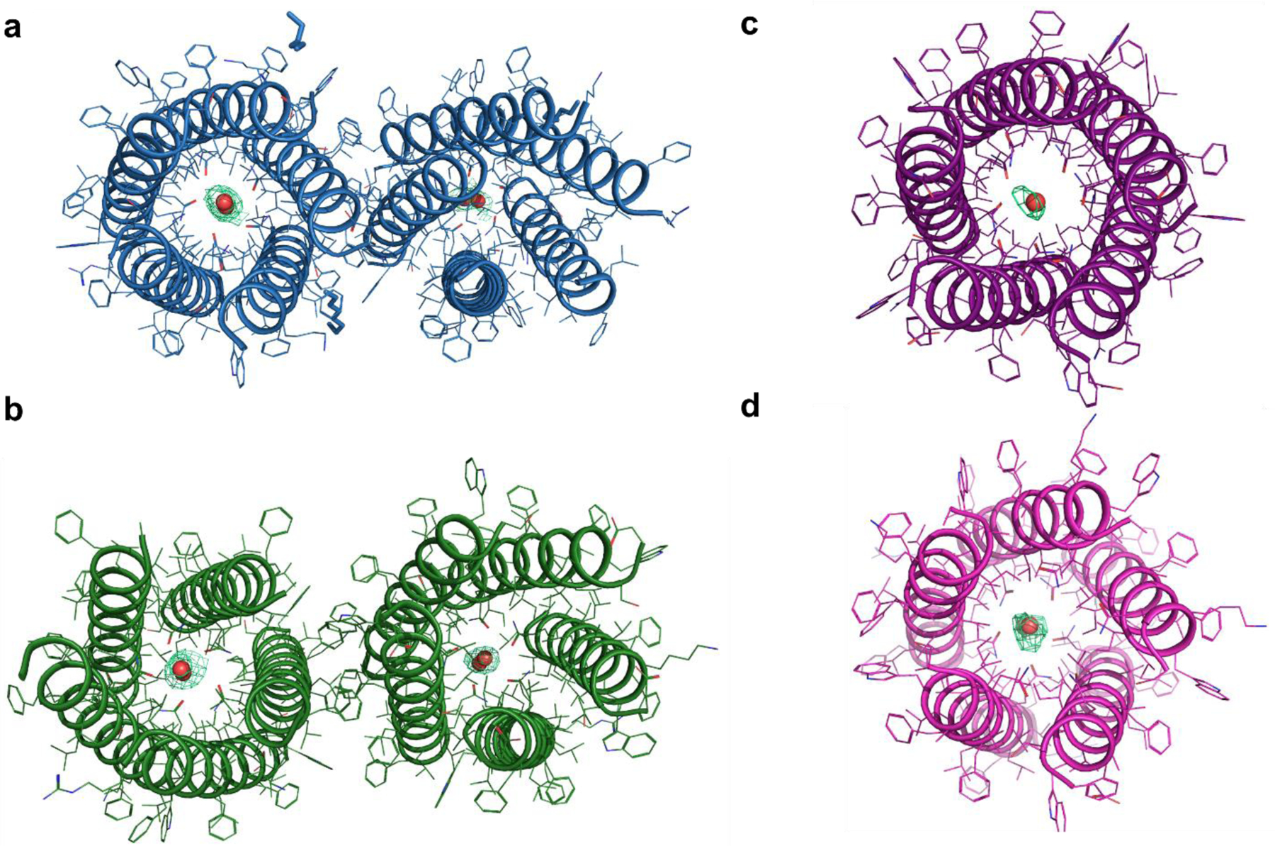
Selective proton transport through proteins is essential for forming and using proton gradients in cells. Protons are conducted along hydrogen-bonded 'wires' of water molecules and polar side chains, which, somewhat surprisingly, are often interrupted by dry apolar stretches in the conduction pathways, inferred from static protein structures. Here we hypothesize that protons are conducted through such dry spots by forming transient water wires, often highly correlated with the presence of the excess protons in the water wire. To test this hypothesis, we performed molecular dynamics simulations to design transmembrane channels with stable water pockets interspersed by apolar segments capable of forming flickering water wires. The minimalist designed channels conduct protons at rates similar to viral proton channels, and they are at least 106-fold more selective for H+ over Na+. These studies inform the mechanisms of biological proton conduction and the principles for engineering proton-conductive materials.


Conflict of interest statement

Competing interests: The authors declare no competing interests.

Figures

Extended Data Fig. 1.
Extended Data Fig. 1.. Composite omit maps (2mFo-DFc) of designed proton channels.
Composite omit maps of the asymmetric unit for a, LQLL, b, LLQL, and one pentamer from the asymmetric unit for c, QQLL, and d, QLQL (shown only for the waters for clarity). All contours at σ = 1.0. Omit maps with simulated cartesian annealing were generated using Phenix, using methodology described in Hodel, et al.
Extended Data Fig. 2.
Extended Data Fig. 2.. Pulse diagram and water-edited 13C spectra, water buildup curves of membrane-bound LQLL and LLLL peptides.
a, Pulse diagram of water-edited 13C CP experiment. b, Representative water-edited 13C spectra of I13 in LQLL and LLLL, measured with 225 ms and 49 ms 1H mixing. The relative intensities of the 49 ms spectrum to the 225 ms spectrum are higher for LQLL Ile13 than LLLL Ile13, especially for the sidechain Cγ2 and Cδ carbons. c, Site-resolved water buildup curves for Ile13 in LQLL and LLLL. For all 13C sites, LQLL shows a faster water buildup than LLLL, consistent with water molecules in the pore lumen due to the Gln10 PLS.
Extended Data Fig. 3.
Extended Data Fig. 3.. Overview of proton flux measurements.
a, Full schematic for proton flux measurement including CCCP step, which is included to check vesicle leakiness and confirm proton selectivity. b, Chemical structures of key components of vesicle assay. c, Calibration curves for HPTS at ~5 μM in 12 solutions of 50 mM K2SO4, 30 mM K2HPO4 at different pH values for two plate readers used in data collection process. Unless stated, all data collected with instrument that generated the blue calibration curve. Fits used for downstream data processing shown for each of the two instruments with adjusted R-squared values of 0.9866 and 0.9998 for the left and right curves, respectively. Data for n = 3 independent samples shown as mean values +/− SD.
Extended Data Fig. 4.
Extended Data Fig. 4.. All proton flux assay data for long kinetics runs.
Long kinetics runs of about 5 hours total for a, empty, b, LLLL, and c, LQLL vesicles. Dotted lines denote times in experiment when valinomycin and CCCP were added to the samples. Three samples of the different conditions were measured in triplicate. These long-time measurements reveal that the vesicles are not significantly leaky to Na+, K+, or H+ and maintain their cargo and assembly over the entire course of the measurement. d, From the linear regression fits of the first 220 s following addition of valinomycin, all slopes (which give the initial rates (in M/s)) were used to calculate the mean and standard error. The one-way ANOVA analysis (with Dunn’s test) reveals that LLLL rates are not significantly different (p > 0.05, adjusted p = 0.4021) when compared to the control empty vesicles. LQLL rates, however, are statistically significant (p < 0.0001, p = 3.23E-5) when compared to the control empty vesicles using one-way ANOVA analysis with Dunn’s test. All data from n = 3 independent samples are shown as mean values +/− SD.
Extended Data Fig. 5.
Extended Data Fig. 5.. All proton flux assay data for QLLL vesicle samples.
Nine samples (each run in triplicate with shaded error bars shown) containing 1:500 peptide:lipid ratio; samples were run independently in the assay. a, pHin as a function of time throughout the measurement for each independent sample. b, Mean and standard deviation for data collection. c, Data prior to CCCP addition shows little change in pHin after addition of valinomycin. d, Fits for the initial 50 seconds following addition of valinomycin. From the linear regression fits, all slopes (which give the initial rates (in M/s)) were used to calculate mean and standard error presented in Fig. 6g and Supplementary Table S3.
Extended Data Fig. 6.
Extended Data Fig. 6.. All proton flux assay data for LLQL vesicle samples.
Eight samples (each run in triplicate with shaded error bars shown) containing 1:500 peptide:lipid ratio; samples were run independently in the assay. a, pHin as a function of time throughout the measurement for each independent sample. b, Mean and standard deviation for data collection. c, Data prior to CCCP addition shows significant change in pHin after addition of valinomycin. d, Fits for the initial 50 seconds following addition of valinomycin. From the linear regression fits, all slopes (which give the initial rates (in M/s)) were used to calculate mean and standard error presented in Fig. 6g and Supplementary Table S3.
Extended Data Fig. 7.
Extended Data Fig. 7.. All proton flux assay data for QLQL vesicle samples.
Seven samples (each run in triplicate with shaded error bars shown) containing 1:500 peptide:lipid ratio; samples were run independently in the assay. a, pHin as a function of time throughout the measurement for each independent sample. b, Mean and standard deviation for data collection. c, Data prior to CCCP addition shows significant change in pHin after addition of valinomycin. d, Fits for the initial 50 seconds following addition of valinomycin. From the linear regression fits, all slopes (which give the initial rates (in M/s)) were used to calculate mean and standard error presented in Fig. 6g and Supplementary Table S3.
Extended Data Fig. 8.
Extended Data Fig. 8.. Determining orientation of pentamers in vesicles.
a, HPLC trace of unreacted and reacted peptides following reaction with the highly polar, amine-reactive methyltetrazine 3-sulfo-N-hydroxysuccinimide ester (methyltetrazine sulfo-NHS, see Materials and Methods). The only amine-reactive groups are the N-terminus or the N-terminal lysine sidechain. Thus, only peptides in which N-terminus is exposed on the outside of the vesicle should react to the dye. b, HPLC traces of mixtures of different ratios of non-reacted and reacted peptides. c, Calibration curves for area under the curve for nonreacted and reacted peaks in the HPLC traces corresponding to the different mixtures in b. Data shown are for n = 2 independent experiments and shown as mean values +/− SD. d, Traces of three independent samples of LQLL pentamers from vesicles after reaction with methyltetrazine sulfo-NHS. e, Using the calibration curves in c, the area under the curve was determined for each sample. The data indicate that half the amines react, as expected from a random orientation of pentamers in the lipid vesicle.
Fig. 1.
Fig. 1.. Hypothesis of proton-selective transport along transient water wires.
a, Protons hop across dynamically rearranging hydrogen-bonded wires of water in the Grotthuss mechanism. b, In a wholly apolar channel, the pore remains devoid of water regardless of membrane polarization (ΔΨ), but a polar PLS, c, can mediate the flickering of neutral water in and out of a relatively short apolar sectors of the channel. d, The presence of a hydrated excess proton facilitates water wire formation and transport of an excess proton through the hydrophobic sector by Grotthuss shuttling.
Fig. 2.
Fig. 2.. De novo channels incorporate PLSs at key positions to modulate hydrophobic lengths.
a, The parent scaffold, LLLL (pdb 6mct), contains layers of interdigitating Leu (green) and Ile (blue) residues. b, Five Leu-to-Gln variants were designed. c, Models of the designed channels illustrates the different expected hydrophobic lengths (lexp) of the channels relative to the parent scaffold. d, SDS-PAGE revealed that these designs formed stable pentamers. e, Comparison of the longest hydrophobic length expected (lexp) and observed (lobs, determined by classical MD simulations; see Methods and Extended Data Table 1). Side views of models in Fig. 2, 3, 4 and 6 have the fifth helix removed for clarity.
Fig. 3.
Fig. 3.. Permeation of water into the designed channels correlates with the position of the luminal Gln residues.
MD simulations were analyzed using the Channel Annotation Package . Plots of pore water density versus time reveal no water permeation into the hydrophobic pore in a, LLLL or b, QLLL. Pentamers with Gln in the second and third layers of the channel, including c, LQLL, d, LLQL, e, QQLL, and f, QLQL, have strong water density around the polar Gln site and flickering water molecules in the shorter apolar segment leading up to the mutation. However, the longer apolar segment remains dehydrated. Fifth helix in all figures removed for clarity.
Fig. 4.
Fig. 4.. Crystal structures of the designed channels demonstrate the introduction of polar Gln residues mediates stable water pockets.
The X-ray structures of the designed channels are within < 0.4 Å rmsd of the original design template, a, LLLL (pdb 6mct, gray), b, QLLL (pdb 7udy), c, LQLL (pdb 7udz), d, LLQL (pdb 7udv), e, QQLL (pdb 7udw), and f, QLQL (pdb 7udx). Fifth helix in all figures removed for clarity. g, Water buildup curves for uniformly labeled 13C, 15NIle13, Ile6, and Gln10 in LLLL and LQLL peptides. Both the Ile6 and Ile13 sites in the LQLL sample show faster water buildup than the corresponding sites in the LLLL sample.
Fig. 5.
Fig. 5.. MS-RMD predicts that introduction of the PLS enables the formation of proton-conducive transient water wires.
a, 2D-PMF of LLLL shows high barrier when the proton is at Z’CEC = 0 Å, or the center of mass of the channel at the Ile13 alpha-carbons. Z’CEC in a-c are in units of Å. b, Addition of the Gln residue at +4 Å in LQLL shifts the barrier to the C-terminal side of the channel and decreases the barrier height by ~20 kcal/mol. c, The two lowest mean free energy paths (MFEPs, white solid and dashed lines), derived from string theory (see Methods) through the LQLL channel. Note the scale change on the color bar in 5b versus 5c. d, Snapshots along the two pathways for LQLL (from panel c) reveal the mechanism of proton-induced water wires mediating proton translocation. The most hydronium-like structures are highlighted in yellow. Only two helices are represented for clarity.
Fig. 6.
Fig. 6.. Designed channels selectively move protons across the membrane.
a, Schematic for proton flux assays using a vesicle-entrapped pH-sensitive fluorescent dye, 8-hydroxypyrene-1,3,6-trisulfonic acid (HPTS). At t=0 the pH is the same inside and outside of the vesicles. b, Following addition of valinomycin, the pHin of empty vesicles does not change, because there is no proton channel or carrier included. c, When a proton channel is present, like the influenza A M2 channel shown in this panel, addition of valinomycin enables the transport of protons down the electrochemical gradient created by the efflux of potassium. This results in a significant decrease in the pHin over time as protons move into the vesicle up a concentration gradient. Snapshots (fifth helix removed for clarity), and proton flux assays for d, LLLL, e, LQLL, and f, QQLL indicates that addition of polar Gln near the middle of the channel enables water permeation events into the pore which facilitates proton-selective transport. g, Change in the H+ concentration of representative samples upon addition of valinomycin at t=0. For control samples, Influenza A M2 showed significant (p < 0.0001) initial rates relative to Empty. QLLL initial rates are not significant (p = 0.3947) relative to LLLL. Fitting of the initial rates shows that LQLL, LLQL, QQLL, and QLQL have significant proton transport activity (p < 0.0001) relative to LLLL. All data in b-f presented as mean values +/− SD for n > 6 independent experiments. Data in g shown as mean values +/− SD (left) and +/− SEM (right) bars. Data analyzed using unpaired t-test for Empty vs. Influenza A M2 and ordinary one-way ANOVA for multiple comparisons (Dunn’s test) of LLLL vs. designed channels (see Supplementary Tables S4 and S5 for complete analysis).

Similar articles

Cited by

References

References and notes:

    1. Moriyama Y & Futai M H+-ATPase, a primary pump for accumulation of neurotransmitters, is a major constituent of brain synaptic vesicles. Biochem. Biophys. Res. Comm 173, 443–448, doi:10.1016/s0006-291x(05)81078-2 (1990). - DOI - PubMed
    1. Nishi T & Forgac M The vacuolar (H+)-ATPases--nature's most versatile proton pumps. Nat. Rev. Mol. Cell. Biol 3, 94–103, doi:10.1038/nrm729 (2002). - DOI - PubMed
    1. Mitchell P Coupling of phosphorylation to electron and hydrogen transfer by a chemiosmotic type of mechanism. Nature 191, 144–148, doi:10.1038/191144a0 (1961). - DOI - PubMed
    1. Nicholls DG Mitochondrial ion circuits. Essays Biochem 47, 25–35, doi:10.1042/bse0470025 (2010). - DOI - PubMed
    1. Diering GH & Numata M Endosomal pH in neuronal signaling and synaptic transmission: role of Na(+)/H(+) exchanger NHE5. Front. Physiol 4, 412, doi:10.3389/fphys.2013.00412 (2014). - DOI - PMC - PubMed

References for Materials and Methods

    1. Caffrey M & Cherezov V Crystallizing membrane proteins using lipidic mesophases. Nat. Prot 4, 706–731, doi:10.1038/nprot.2009.31 (2009). - DOI - PMC - PubMed
    1. Caffrey M Crystallizing membrane proteins for structure determination: use of lipidic mesophases. Annu. Rev. Biophys 38, 29–51, doi:10.1146/annurev.biophys.050708.133655 (2009). - DOI - PubMed
    1. Kabsch W XDS. Acta Crystallogr. D 66, 125–132, doi:10.1107/S0907444909047337 (2010). - DOI - PMC - PubMed
    1. Winn MD et al. Overview of the CCP4 suite and current developments. Acta Crystallogr. D 67, 235–242, doi:10.1107/S0907444910045749 (2011). - DOI - PMC - PubMed
    1. McCoy AJ et al. Phaser crystallographic software. J. Appl. Crystallogr 40, 658–674, doi:10.1107/S0021889807021206 (2007). - DOI - PMC - PubMed

Publication types