. 2023 Jun 12;20(1):141.
doi: 10.1186/s12974-023-02820-y.

Varicella-zoster virus proteome-wide T-cell screening demonstrates low prevalence of virus-specific CD8 T-cells in latently infected human trigeminal ganglia

Affiliations

Varicella-zoster virus proteome-wide T-cell screening demonstrates low prevalence of virus-specific CD8 T-cells in latently infected human trigeminal ganglia

Michiel van Gent et al. J Neuroinflammation. .

Abstract

Background: Trigeminal ganglia (TG) neurons are an important site of lifelong latent varicella-zoster virus (VZV) infection. Although VZV-specific T-cells are considered pivotal to control virus reactivation, their protective role at the site of latency remains uncharacterized.

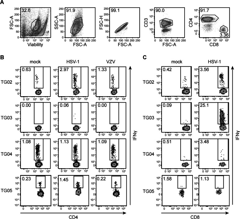
Methods: Paired blood and TG specimens were obtained from ten latent VZV-infected adults, of which nine were co-infected with herpes simplex virus type 1 (HSV-1). Short-term TG-derived T-cell lines (TG-TCL), generated by mitogenic stimulation of TG-derived T-cells, were probed for HSV-1- and VZV-specific T-cells using flow cytometry. We also performed VZV proteome-wide screening of TG-TCL to determine the fine antigenic specificity of VZV reactive T-cells. Finally, the relationship between T-cells and latent HSV-1 and VZV infections in TG was analyzed by reverse transcription quantitative PCR (RT-qPCR) and in situ analysis for T-cell proteins and latent viral transcripts.

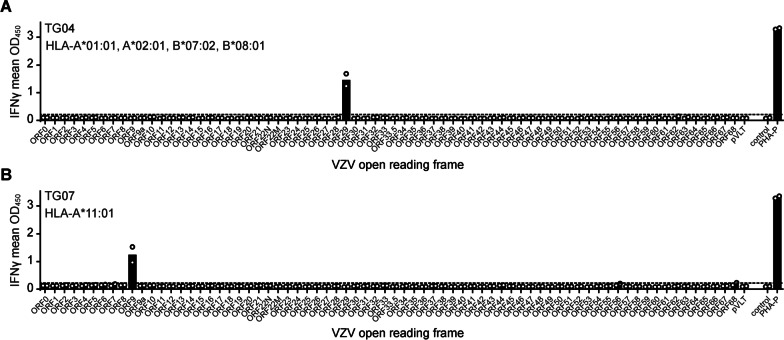
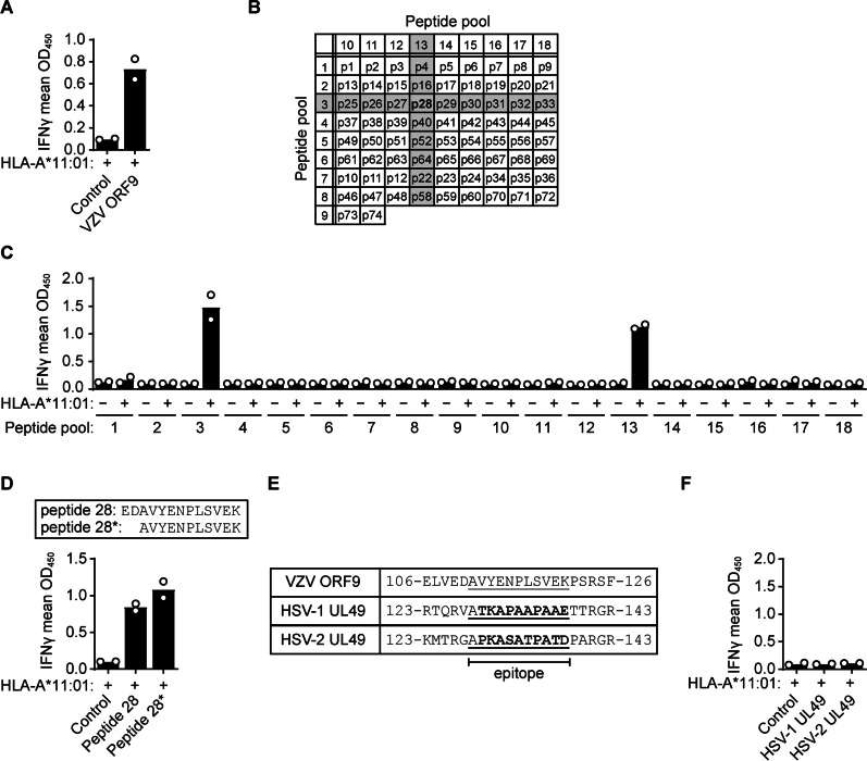
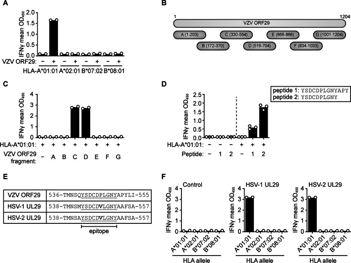
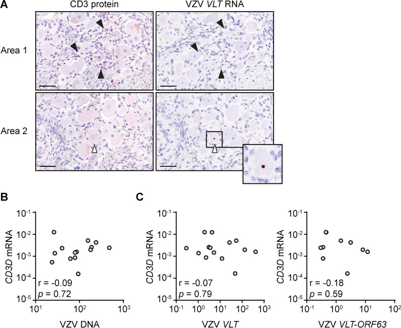
Results: VZV proteome-wide analysis of ten TG-TCL identified two VZV antigens recognized by CD8 T-cells in two separate subjects. The first was an HSV-1/VZV cross-reactive CD8 T-cell epitope, whereas the second TG harbored CD8 T-cells reactive with VZV specifically and not the homologous peptide in HSV-1. In silico analysis showed that HSV-1/VZV cross reactivity of TG-derived CD8 T-cells reactive with ten previously identified HSV-1 epitopes was unlikely, suggesting that HSV-1/VZV cross-reactive T-cells are not a common feature in dually infected TG. Finally, no association was detected between T-cell infiltration and VZV latency transcript abundance in TG by RT-qPCR or in situ analyses.

Conclusions: The low presence of VZV- compared to HSV-1-specific CD8 T-cells in human TG suggests that VZV reactive CD8 T-cells play a limited role in maintaining VZV latency.

Keywords: Herpes simplex virus; Human; Latency; T-cells; Trigeminal ganglion; Varicella-zoster virus.


Conflict of interest statement

The authors declare no competing interests.

Figures

Fig. 1
Fig. 1
HSV-1 and VZV responses of human TG-derived T-cell lines. A Gating strategy applied to determine T-cell reactivity, consisting of selection of viable cells followed by size exclusion and selection of single cells, selection of CD3+ T-cells, and finally selection of CD3+CD4+ and CD3+CD8+ T-cells. FSC-A, forward scatter—area; SSC-A, side scatter—area; FSC-H, forward scatter—height. B Flow cytometry analysis of IFNγ production by CD4 T-cells derived from TG donors TG02–TG05 following 6 h of co-culture with autologous B-LCL pulsed with lysates derived from mock, HSV-1, or VZV-infected ARPE-19 cells. C Flow cytometry analysis of IFNγ production by CD8 T-cells following 6 h of coculture with autologous mock- or HSV-1-infected B-LCL. Numbers indicate percentages of cells within the respective gate
Fig. 2
Fig. 2
VZV antigen specific response of human TG-derived CD8 T-cells. A, B TG-derived T-cell lines of donor TG04 (A) or TG07 (B) were screened for reactivity with artificial antigen presenting cells expressing the indicated subject-specific HLA class I allele(s) and individual VZV open reading frames or fragments thereof. For subject TG04, all 4 HLA alleles were co-transfected simultaneously, while for subject TG07, only HLA-A*11:01 was used. Empty vector (control) and phytohemagglutinin (PHA-P), were used as negative and positive controls, respectively. Levels of secreted IFNγ was determined by ELISA. Data are presented as the individual (circles) and mean (bars) OD450 values of two independent replicates. Horizontal dashed lines indicate the threshold of T-cell response set at twice the value of the empty vector control. Viral ORF nomenclature according to VZV reference strain Dumas (Genbank Accession number X04370)
Fig. 3
Fig. 3
TG-derived CD8 T-cells recognize an epitope in VZV ORF9 epitope in an HLA-A*11:01-dependent manner. A IFNγ secretion by TG07-derived T-cells following 24 h of incubation with Cos-7 cells co-transfected with a vector encoding HLA-A*11:01 and either an empty control vector or a vector encoding VZV ORF9. B Overview of the peptide composition of each ORF9 peptide pool used in panel C. Reactive pools are shaded. C IFNγ secretion by TG07-derived T-cells co-cultured with Cos-7 cells transfected with HLA-A*11:01 and pulsed with the indicated peptide pools, showing reactivity for two individual pools that overlap at peptide 28. D IFNγ secretion by TG07-derived T-cells following co-culture with HLA-A*11:01-transfected Cos-7 cells transfected and pulsed with ORF9 peptide 28 (amino acids 109–121, EDAVYENPLSVEK) or internal ORF9 peptide 28* (amino acids 111–121, AVYENPLSVEK). E Alignment of the VZV ORF9 epitope (AVYENPLSVEK, underlined) with the homologous regions of HSV-1 and HSV-2 UL49. Amino acids in bold differ between VZV and HSV-1/HSV-2; numbers indicate amino acid positions in the respective proteins. F IFNγ secretion by TG07-derived T-cells following co-culture with Cos-7 cells transfected with HLA-A*11:01 in combination with either an empty control vector or vectors encoding HSV-1 UL49 or HSV-2 UL49. Data are presented as the individual (circles) and mean (bars) OD450 values of at two independent replicates
Fig. 4
Fig. 4
TG-VZV reactive CD8 T-cells recognize an HSV/VZV cross-reactive epitope in an HLA-A*01:01-dependent manner. A IFNγ secretion by TG04-derived T-cells following incubation with Cos-7 cells transfected with the indicated subject-specific HLA class I allele together with an empty control vector (−) or a vector encoding VZV ORF29 (+). B Schematic of VZV ORF29 fragments used in (C) to map the T-cell epitope. Numbers indicate amino acid positions. C ELISA showing IFNγ secretion by TG04-derived T-cells following incubation with Cos-7 cells transfected with HLA-A*01:01 and an empty control vector (−) or vectors encoding the indicated VZV ORF29 fragments from (B). D IFNγ production by TG04-derived T-cells following coculture with Cos-7 cells transfected without (−) or with (+) HLA-A*01:01 and pulsed with the indicated ORF29 peptides or DMSO as a negative control (−). E Alignment of the VZV ORF29 epitope-containing region of VZV ORF29 with the homologous regions of HSV-1 UL29 and HSV-2 UL29 (underlined). Amino acid in bold differ between VZV, HSV-1, and HSV-2. Numbers indicate amino acid positions in the respective proteins. F IFNγ secretion by TG04-derived T-cells following co-culture with Cos-7 cells transfected to express the indicated HLA class I alleles in combination with either an empty control vector or vectors encoding full-length HSV-1 UL29 or HSV-2 UL29. Data are presented as the individual (circles) and mean (bars) OD450 values of 2 (A, C, F) or 3 (D) independent replicates
Fig. 5
Fig. 5
No correlation between T-cell infiltrates and VZV latency in human TG. A Consecutive TG sections were stained for CD3 by immunohistochemistry (red; left column) or VLT RNA by in situ hybridization (dark red, right column). Filled and open arrow heads indicate CD3+ cells and a VLT+ neuron, respectively; the inset shows a VLT+ neuron. Data is shown for two different areas (1 and 2) of one donor that are representative of five independent TG donors; scale bar: 50 µm. B, C Abundance of VZV DNA (B) or VZV VLT and VZV VLT-ORF63 RNA (C) as well as human CD3D transcripts in 18 TG specimens (described in [3]) as determined by q(RT)-PCR and presented as relative DNA/transcript levels normalized to single-copy gene HMBS or cellular housekeeping gene GAPDH. Spearman r and p values are indicated

References

    1. Knipe DMPMH, editor. Field’s Virology. 7. Philadelphia: Lipincott Williams & Wilkins; 2020.
    1. Ramachandran PS, Wilson MR, Catho G, et al. Meningitis caused by the live varicella vaccine virus: metagenomic next generation sequencing, immunology exome sequencing and cytokine multiplex profiling. Viruses. 2021;13:2286. doi: 10.3390/v13112286. - DOI - PMC - PubMed
    1. Depledge DP, Ouwendijk WJD, Sadaoka T, Braspenning SE, Mori Y, Cohrs RJ, Verjans GMGM, Breuer J. A spliced latency-associated VZV transcript maps antisense to the viral transactivator gene. Nat Commun. 2018;9:1167. doi: 10.1038/s41467-018-03569-2. - DOI - PMC - PubMed
    1. Ouwendijk WJD, Depledge DP, Rajbhandari L, Lenac Rovis T, Jonjic S, Breuer J, Venkatesan A, Verjans GMGM, Sadaoka T. Varicella-zoster virus VLT-ORF63 fusion transcript induces broad viral gene expression during reactivation from neuronal latency. Nat Commun. 2020;11:6324. doi: 10.1038/s41467-020-20031-4. - DOI - PMC - PubMed
    1. Zerboni L, Sobel RA, Lai M, Triglia R, Steain M, Abendroth A, Arvin A. Apparent expression of varicella-zoster virus proteins in latency resulting from reactivity of murine and rabbit antibodies with human blood group a determinants in sensory neurons. J Virol. 2012;86:578–583. doi: 10.1128/JVI.05950-11. - DOI - PMC - PubMed