. 2023 Jul 27;97(7):e0195722.
doi: 10.1128/jvi.01957-22. Epub 2023 Jun 13.

Herpes Simplex Virus-1 ICP27 Nuclear Export Signal Mutants Exhibit Cell Type-Dependent Deficits in Replication and ICP4 Expression

Affiliations

Herpes Simplex Virus-1 ICP27 Nuclear Export Signal Mutants Exhibit Cell Type-Dependent Deficits in Replication and ICP4 Expression

Leon Sylvester Sanders 3rd et al. J Virol. .

Abstract

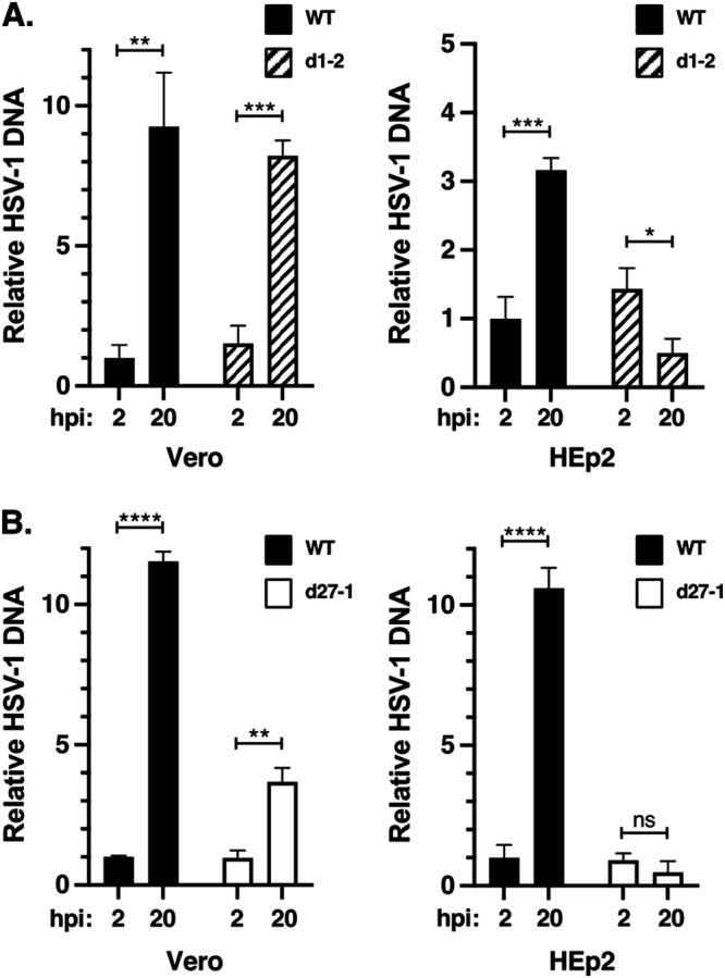
Herpes simplex virus type-1 (HSV-1) protein ICP27 is an essential immediate early (IE) protein that promotes the expression of viral early (E) and late (L) genes via multiple mechanisms. Our understanding of this complex regulatory protein has been greatly enhanced by the characterization of HSV-1 mutants bearing engineered alterations in the ICP27 gene. However, much of this analysis has been performed in interferon-deficient Vero monkey cells. Here, we assessed the replication of a panel of ICP27 mutants in several other cell types. Our analysis shows that mutants lacking ICP27's amino (N)-terminal nuclear export signal (NES) display a striking cell type-dependent growth phenotype, i.e., they grow semi-permissively in Vero and some other cells but are tightly blocked for replication in primary human fibroblasts and multiple human cell lines. This tight growth defect correlates with a failure of these mutants to replicate viral DNA. We also report that HSV-1 NES mutants are deficient in expressing the IE protein ICP4 at early times postinfection. Analysis of viral RNA levels suggests that this phenotype is due, at least in part, to a defect in the export of ICP4 mRNA to the cytoplasm. In combination, our results (i) show that ICP27's NES is critically important for HSV-1 replication in many human cells, and (ii) suggest that ICP27 plays a heretofore unappreciated role in the expression of ICP4. IMPORTANCE HSV-1 IE proteins drive productive HSV-1 replication. The major paradigm of IE gene induction, developed over many years, involves the parallel activation of the five IE genes by the viral tegument protein VP16, which recruits the host RNA polymerase II (RNAP II) to the IE gene promoters. Here, we provide evidence that ICP27 can enhance ICP4 expression early in infection. Because ICP4 is required for transcription of viral E and L genes, this finding may be relevant to understanding how HSV-1 enters and exits the latent state in neurons.

Keywords: HSV-1; ICP27; ICP4; molecular genetics; regulation of gene expression.


Conflict of interest statement

The authors declare no conflict of interest.

Figures

FIG 1
FIG 1
ICP27 mutant d1-2 exhibits cell type-dependent growth. (A) HSV-1 ICP27 mutants used in this study. At the top, a schematic diagram of the 512-residue ICP27 polypeptide is shown, highlighting known domains and functional sequences, including the nuclear export sequence (NES), acidic region, major nuclear localization signal (NLS), RGG-box RNA binding domain, and ICP27-homology domain (IHD). Below are line representations of mutant ICP27 polypeptides, with the numbers indicating the residues deleted (dLeu through d6-7) or mutated in point mutants (M11, M15, M16). In mutant n504R, the number corresponds to the C-terminal residue in the truncated protein, as a result of the insertion of a stop codon linker (inverted triangle). To the right are the mutants’ growth defects (log reductions) in Vero cells, according to previously published studies (15, 29, 45). (B) Growth of mutants in HeLa cells. HeLa cells were infected in biological duplicate at an MOI of 10 and incubated for 24h. The amount of viral progeny in each culture was determined by plaque assay of the harvested cell lysates on ICP27-complementing V27 cells. The bars denote mean titers; error bars denote SEMs. This experiment is representative of two independent experiments that each show d1-2 is unable to replicate to any extent in HeLa cells. (C) Direct comparison of WT and ICP27 mutant growth in HEp2 and Vero cells. Cells were infected in biological duplicate and analyzed as in (A). This experiment was performed one time. (D) Analysis of d1-2 growth in diverse monkey and human cell lines and primary cultures. The indicated cells were infected in biological triplicate at an MOI of 10 PFU/cell, incubated for 1 day, harvested, and titered on V27 cells. Statistical details are as in (A). The limit of detection in these experiments was 10 PFU/mL. This experiment was performed one time for most of the cell lines shown, but was performed twice for HeLa cells and >5 times for HEp2 cells. (E) Analysis of the effects of type-I interferon on d1-2 replication in Vero cells. Triplicate cultures of Vero cells were pretreated with 1,000 U of human α-IFN for 24 h, or mock pretreated, then infected with the viruses shown at an MOI of 10 PFU/cell. At 24 hpi, infections were terminated and replication was assessed by plaque assay of the lysates on appropriate complementing cells. For each viral infection, the fold inhibitory effect of type-I interferon treatment is indicated. The bars indicate mean titers and error bars denote SEMs. Statistical analyses were performed using the Student's t test. **, P < .01; ***, P < .001. This experiment was performed one time.
FIG 2
FIG 2
The ICP27 sequences associated with cell-type dependent replication map to the NES. (A) Mutations in ICP27 expression plasmids used in the analysis. The diagram shows the sequences at the N termini of WT ICP27 or various mutant proteins; dashes denote deletions and bold letters denote amino acid mutations. The ICP27 NES and acidic region are indicated. At the right, the block arrows denote HSV-1 ICP27 mutants that possess the indicated sequence alterations. (B) Viral complementation analyses. HEp-2 and Vero cells were transfected in biological triplicate with WT or ICP27 mutant plasmids. After 1 day, cells were infected with d27-1 and incubated for 24 h before being harvested. Cell lysates were titered on V27 cells to determine viral growth, and the mean titers of the mutant plasmid transfection samples were divided by the mean titer of the WT ICP27 plasmid transfection samples (and multiplied by 100 to express as a percentage). The values shown represent the means of two to four independent experiments for each plasmid, and error bars denote SEMs. (C) Relative complementation in Vero versus HEp2 cells. To determine this, the relative complementation of each plasmid in Vero versus Hep2 cells was determined for each of the two to four independent experiments. The means were then determined; error bars show SEMs.
FIG 3
FIG 3
ICP27 mutants d1-2 and d27-1 can replicate their DNA in Vero but not HEp2 cells. Vero or HEp2 were infected in biological triplicate at an MOI of 10 PFU/cell with WT HSV-1 and ICP27 mutant d1-2 (A) or d27-1 (B). Total infected cell DNA was isolated at 2 and 20 hpi and the HSV-1-specific DNA at each time point was quantitated by qPCR. The data were normalized to a single-copy cellular gene to correct for differences in cell recovery. Vero and HEp2 data were generated in different qPCR runs, so the results are shown in separate graphs. For each analysis, the level of WT HSV-1 DNA at 2 hpi was assigned the value of 1.0. The bars denote means, and error bars show SEMs. Statistical analyses were performed using the Student's t test. *, P < .05; **, P < .01; ***, P < .001; ****, P < .0001; ns (not significant), P > 0.05. This experiment is representative of two independent experiments that each show that d1-2 and d27-1 are able to replicate their DNA to some extent in Vero cells but not at all in HEp2 cells.
FIG 4
FIG 4
ICP4 expression is deficient in d1-2-infected cells. Vero or HEp2 cells were infected at an MOI of 10 PFU/cell as indicated and total proteins were harvested at 4 and 8 hpi. Protein accumulation was assessed by immunoblotting. Lanes showing abnormally low expression of ICP4 are indicated with an asterisk. Data shown are from a single infection, but the ICP4 results are representative of those obtained in numerous (>8) similar immunoblotting experiments.
FIG 5
FIG 5
Quantitative analysis of ICP4 expression in ICP27 mutant infections. HEp2 or Vero cells were infected in biological triplicate as shown, and total protein extracts were prepared at 4 hpi. ICP4 and ALYREF levels were determined by immunoblotting; the resulting data were analyzed using ImageJ software. To correct for differences in cell recovery, ICP4 signals were normalized to those of the ALYREF loading control on the same filter. The bars denote means, and error bars show SEMs. Statistical analyses were performed using the Student's t test. *, P < 0.05; **, P < 0.01; ns (not significant), P > 0.05. This comparative quantitative analysis of ICP4 expression was performed one time.
FIG 6
FIG 6
ICP4 expression is deficient in a subset of HSV-1 ICP27 mutants. Single cultures of HEp2 cells were mock infected or infected with the viruses indicated. Total proteins were isolated at 4 hpi and analyzed by immunoblotting for IE proteins ICP4 and ICP0, and β-actin as a loading control (left part of figure). Relative ICP4 levels were quantitated by ImageJ analysis (right part of figure); data were corrected for cell recovery by normalizing to β-actin signals. (A) Analysis of ICP27 N-terminal mutants; (B) Analysis of ICP27 NES mutants; (C) Analysis of ICP27 C-terminal mutants. Each of these experiments was performed one time; however, three independent experiments confirmed that N-terminal mutant dLeu is deficient in ICP4 expression at 4 hpi in HEp2 cells.
FIG 7
FIG 7
Ectopic expression of ICP4 fails to complement d1-2 in HEp2 cells. Hep2 cells were transfected in quintuplicate with ICP4 or ICP27 expression plasmids, or with pUC19 as a negative control. One day later, the cells were mock infected or infected with d1-2 or WT HSV-1 at an MOI of 10. At both 4 and 8 hpi, one of the replicate cultures were prepared for protein analysis, and the remaining three replicates were harvested at 24 hpi and subjected to plaque assay on V27 cells. (A) Immunoblot analysis. (B) Titer of infections. The expression plasmids transfected in each culture are indicated. The means of triplicate infections are shown; error bars denote SEMs. The limit of detection in the plaque assays was 10 PFU/mL. This experiment was performed one time.
FIG 8
FIG 8
The d1-2 mutant is trans-dominant. (A) Co-infection assay to assess the effects of d27-1 and d1-2 on WT HSV-1 infection. Triplicate cultures of HEp2 cells were infected with WT HSV-1 at an MOI of 1 PFU/cell, minus or plus co-infection with d27-1 or d1-2 at an MOI of 10 PFU/cell. At 24 hpi, infections were harvested and cell lysates were subjected to plaque assay on Vero cells to assess the replication of WT HSV-1. Note that d1-2 produces minute plaques under these conditions (29) which were not scored. (B) Co-infection assay to assess the trans-dominance of d1-2, n504R, and M11. The experiment was carried out as in (A), except that the WT HSV-1 infection alone was omitted. For both (A) and (B), the means of triplicate infections are shown; error bars show SEMs. Statistical analyses were performed using the Student's t test. *, P < .05; **, P < .01; ***, P < .001; ***, P < .0001; ns (not significant), P > 0.05. This experiment was performed one time.
FIG 9
FIG 9
ICP4 mRNA expression in ICP27 mutant infections. Vero or HEp2 cells were infected with the viruses indicated at an MOI of 10 PFU/cell, and infected cell mRNA was isolated at 3 hpi and quantitated by RT-qPCR as described in Materials and Methods. Three identical independent experiments were performed; the data shown are derived from these experiments. (A) Analysis of total cellular RNA. (B) Analysis of cytoplasmic and nuclear RNA fractions. For both (A) and (B), viral mRNA levels were normalized to 18S rRNA levels in the same samples, and are expressed as fold change compared to the WT HSV-1 infection, which was assigned the value of 1.0.
FIG 10
FIG 10
Models for the role of ICP27 in the early expression of ICP4 mRNA in cells infected by WT HSV-1 (A) or d1-2 (B). See text for details.

References

    1. Xu F, Sternberg MR, Kottiri BJ, McQuillan GM, Lee FK, Nahmias AJ, Berman SM, Markowitz LE. 2006. Trends in herpes simplex virus type 1 and type 2 seroprevalence in the United States. JAMA 296:964–973. doi:10.1001/jama.296.8.964. - DOI - PubMed
    1. Roizman B, Knipe DM, Whitley RJ. 2007. Herpes simplex viruses, p 2501–2601. In Knipe DM, Howley PM (ed), Fields virology, 5th ed, vol 2. Lippincott, Williams & Wilkins, Philadelphia, PA.
    1. Wald A, Corey L. 2007. Persistence in the population: epidemiology, transmission, chapter 36. In Arvin A, Campadelli-Fiume G, Mocarski E, Moore PS, Roizman B, Whitley R, Yamanishi K (ed), Human herpesviruses: biology, therapy, and immunoprophylaxis. Cambridge University Press, Cambridge, UK. - PubMed
    1. Marcocci ME, Napoletani G, Protto V, Kolesova O, Piacentini R, Li Puma DD, Lomonte P, Grassi C, Palamara AT, De Chiara G. 2020. Herpes simplex virus-1 in the brain: the dark side of a sneaky infection. Trends Microbiol 28:808–820. doi:10.1016/j.tim.2020.03.003. - DOI - PubMed
    1. Pinninti SG, Kimberlin DW. 2018. Neonatal herpes simplex virus infections. Semin Perinatol 42:168–175. doi:10.1053/j.semperi.2018.02.004. - DOI - PubMed

Publication types

LinkOut - more resources