

RENEB Inter-Laboratory Comparison 2021: Inter-Assay Comparison of Eight Dosimetry Assays
- PMID: 37310880
- PMCID: PMC10508307
- DOI: 10.1667/RADE-22-00207.1
RENEB Inter-Laboratory Comparison 2021: Inter-Assay Comparison of Eight Dosimetry Assays
Abstract
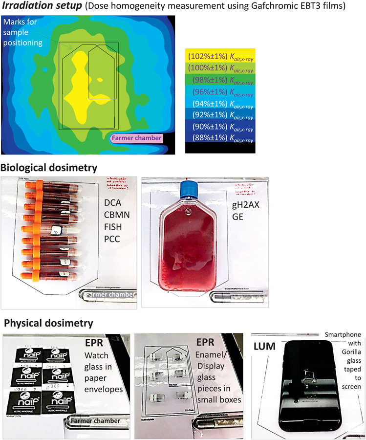
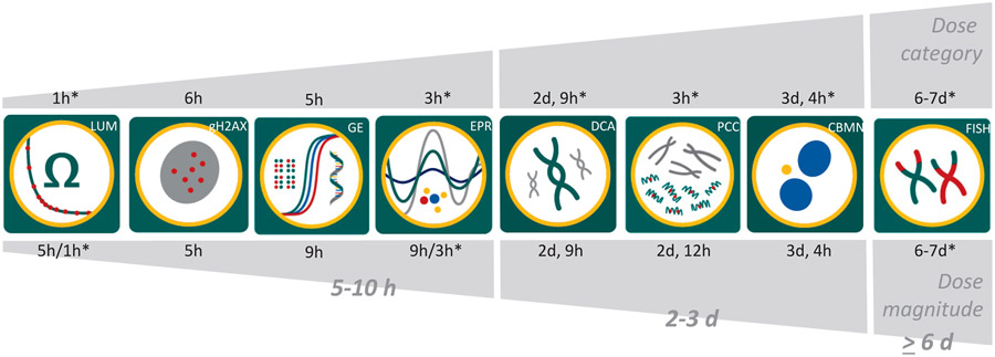
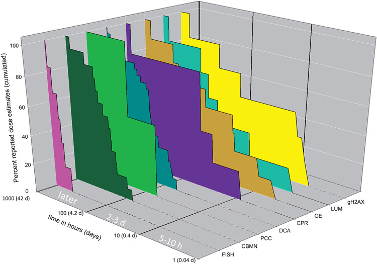
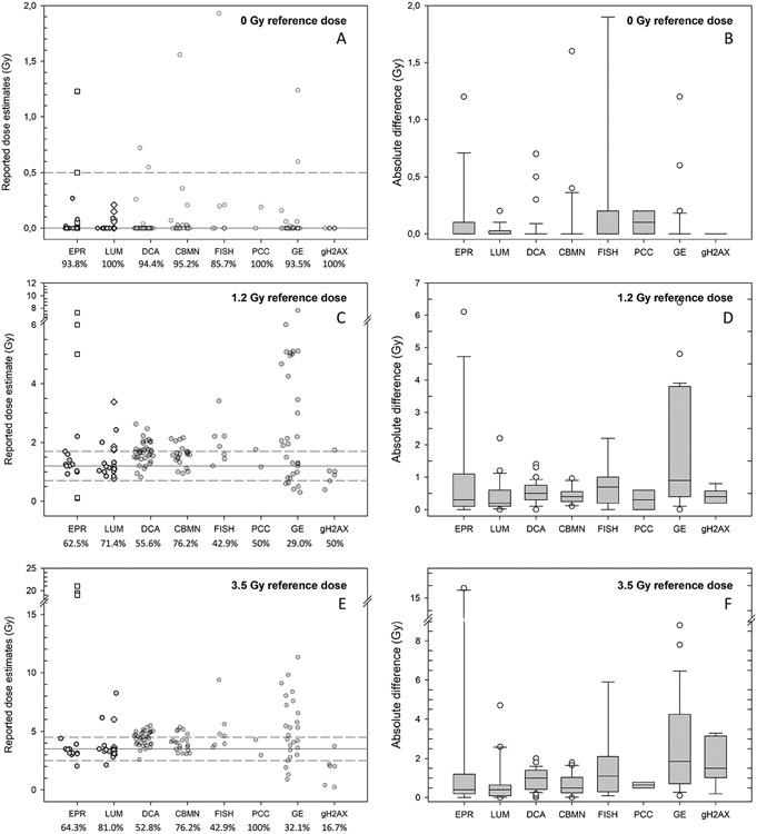
Tools for radiation exposure reconstruction are required to support the medical management of radiation victims in radiological or nuclear incidents. Different biological and physical dosimetry assays can be used for various exposure scenarios to estimate the dose of ionizing radiation a person has absorbed. Regular validation of the techniques through inter-laboratory comparisons (ILC) is essential to guarantee high quality results. In the current RENEB inter-laboratory comparison, the performance quality of established cytogenetic assays [dicentric chromosome assay (DCA), cytokinesis-block micronucleus assay (CBMN), stable chromosomal translocation assay (FISH) and premature chromosome condensation assay (PCC)] was tested in comparison to molecular biological assays [gamma-H2AX foci (gH2AX), gene expression (GE)] and physical dosimetry-based assays [electron paramagnetic resonance (EPR), optically or thermally stimulated luminescence (LUM)]. Three blinded coded samples (e.g., blood, enamel or mobiles) were exposed to 0, 1.2 or 3.5 Gy X-ray reference doses (240 kVp, 1 Gy/min). These doses roughly correspond to clinically relevant groups of unexposed to low exposed (0-1 Gy), moderately exposed (1-2 Gy, no severe acute health effects expected) and highly exposed individuals (>2 Gy, requiring early intensive medical care). In the frame of the current RENEB inter-laboratory comparison, samples were sent to 86 specialized teams in 46 organizations from 27 nations for dose estimation and identification of three clinically relevant groups. The time for sending early crude reports and more precise reports was documented for each laboratory and assay where possible. The quality of dose estimates was analyzed with three different levels of granularity, 1. by calculating the frequency of correctly reported clinically relevant dose categories, 2. by determining the number of dose estimates within the uncertainty intervals recommended for triage dosimetry (±0.5 Gy or ±1.0 Gy for doses <2.5 Gy or >2.5 Gy), and 3. by calculating the absolute difference (AD) of estimated doses relative to the reference doses. In total, 554 dose estimates were submitted within the 6-week period given before the exercise was closed. For samples processed with the highest priority, earliest dose estimates/categories were reported within 5-10 h of receipt for GE, gH2AX, LUM, EPR, 2-3 days for DCA, CBMN and within 6-7 days for the FISH assay. For the unirradiated control sample, the categorization in the correct clinically relevant group (0-1 Gy) as well as the allocation to the triage uncertainty interval was, with the exception of a few outliers, successfully performed for all assays. For the 3.5 Gy sample the percentage of correct classifications to the clinically relevant group (≥2 Gy) was between 89-100% for all assays, with the exception of gH2AX. For the 1.2 Gy sample, an exact allocation to the clinically relevant group was more difficult and 0-50% or 0-48% of the estimates were wrongly classified into the lowest or highest dose categories, respectively. For the irradiated samples, the correct allocation to the triage uncertainty intervals varied considerably between assays for the 1.2 Gy (29-76%) and 3.5 Gy (17-100%) samples. While a systematic shift towards higher doses was observed for the cytogenetic-based assays, extreme outliers exceeding the reference doses 2-6 fold were observed for EPR, FISH and GE assays. These outliers were related to a particular material examined (tooth enamel for EPR assay, reported as kerma in enamel, but when converted into the proper quantity, i.e. to kerma in air, expected dose estimates could be recalculated in most cases), the level of experience of the teams (FISH) and methodological uncertainties (GE). This was the first RENEB ILC where everything, from blood sampling to irradiation and shipment of the samples, was organized and realized at the same institution, for several biological and physical retrospective dosimetry assays. Almost all assays appeared comparably applicable for the identification of unexposed and highly exposed individuals and the allocation of medical relevant groups, with the latter requiring medical support for the acute radiation scenario simulated in this exercise. However, extreme outliers or a systematic shift of dose estimates have been observed for some assays. Possible reasons will be discussed in the assay specific papers of this special issue. In summary, this ILC clearly demonstrates the need to conduct regular exercises to identify research needs, but also to identify technical problems and to optimize the design of future ILCs.
Figures
Similar articles
-
RENEB Inter-Laboratory Comparison 2021: The Gene Expression Assay.Radiat Res. 2023 Jun 1;199(6):598-615. doi: 10.1667/RADE-22-00206.1. Radiat Res. 2023. PMID: 37057982 Free PMC article.
-
RENEB Inter-Laboratory Comparison 2021: The Dicentric Chromosome Assay.Radiat Res. 2023 Jun 1;199(6):556-570. doi: 10.1667/RADE-22-00202.1. Radiat Res. 2023. PMID: 37018160
-
RENEB Inter-Laboratory Comparison 2021: The Gamma-H2AX Foci Assay.Radiat Res. 2023 Jun 1;199(6):591-597. doi: 10.1667/RADE-22-00205.1. Radiat Res. 2023. PMID: 37057975
-
Overview of physical dosimetry methods for triage application integrated in the new European network RENEB.Int J Radiat Biol. 2017 Jan;93(1):65-74. doi: 10.1080/09553002.2016.1221545. Epub 2016 Sep 12. Int J Radiat Biol. 2017. PMID: 27584947 Review.
-
Review of reconstruction of radiation incident air kerma by measurement of absorbed dose in tooth enamel with EPR.Radiat Prot Dosimetry. 2012 Mar;149(1):71-8. doi: 10.1093/rpd/ncr446. Epub 2011 Nov 28. Radiat Prot Dosimetry. 2012. PMID: 22128353 Review.
Cited by
-
In silico analysis of radiation-induced double-strand breaks by internal ex vivo irradiation of lymphocytes for 45 alpha- and beta/gamma-emitting radionuclides.EJNMMI Res. 2025 Mar 10;15(1):21. doi: 10.1186/s13550-025-01214-w. EJNMMI Res. 2025. PMID: 40063302 Free PMC article.
-
Centers for Medical Countermeasures against Radiation Consortium: Past, Present, and Beyond.Radiat Res. 2025 Jun 27:10.1667/RADE-24-00275.1. doi: 10.1667/RADE-24-00275.1. Online ahead of print. Radiat Res. 2025. PMID: 40571995 Free PMC article.
-
Application of dicentric chromosome assay for evaluation of radioprotective effect.Biol Methods Protoc. 2025 Aug 5;10(1):bpaf058. doi: 10.1093/biomethods/bpaf058. eCollection 2025. Biol Methods Protoc. 2025. PMID: 40809867 Free PMC article. Review.
-
Modelling the In Vivo and Ex Vivo DNA Damage Response after Internal Irradiation of Blood from Patients with Thyroid Cancer.Int J Mol Sci. 2024 May 17;25(10):5493. doi: 10.3390/ijms25105493. Int J Mol Sci. 2024. PMID: 38791531 Free PMC article.
-
DNA Damage and Repair in PBMCs after Internal Ex Vivo Irradiation with [223Ra]RaCl2 and [177Lu]LuCl3 Mixtures.Int J Mol Sci. 2024 Aug 7;25(16):8629. doi: 10.3390/ijms25168629. Int J Mol Sci. 2024. PMID: 39201316 Free PMC article.
References
-
- Waselenko JK, MacVittie TJ, Blakely WF, Pesik N, Wiley AL, Dickerson WE, et al. Medical management of the acute radiation syndrome: recommendations of the Strategic National Stockpile Radiation Working Group. Ann Intern Med. 2004; 140(12):1037–51. 10.7326/0003-4819-140-12-200406150-00015. - DOI - PubMed
Publication types
MeSH terms
Grants and funding
LinkOut - more resources
Full Text Sources
Miscellaneous
