. 2023 Aug:164:114997.
doi: 10.1016/j.biopha.2023.114997. Epub 2023 Jun 8.

β-Cyclodextrins as affordable antivirals to treat coronavirus infection

Affiliations

β-Cyclodextrins as affordable antivirals to treat coronavirus infection

Dalia Raïch-Regué et al. Biomed Pharmacother. 2023 Aug.

Abstract

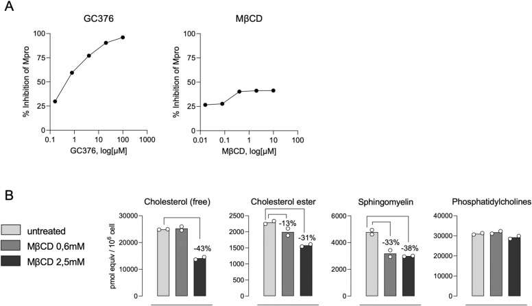
The SARS-CoV-2 pandemic made evident that there are only a few drugs against coronavirus. Here we aimed to identify a cost-effective antiviral with broad spectrum activity and high safety profile. Starting from a list of 116 drug candidates, we used molecular modelling tools to rank the 44 most promising inhibitors. Next, we tested their efficacy as antivirals against α and β coronaviruses, such as the HCoV-229E and SARS-CoV-2 variants. Four drugs, OSW-1, U18666A, hydroxypropyl-β-cyclodextrin (HβCD) and phytol, showed in vitro antiviral activity against HCoV-229E and SARS-CoV-2. The mechanism of action of these compounds was studied by transmission electron microscopy and by fusion assays measuring SARS-CoV-2 pseudoviral entry into target cells. Entry was inhibited by HβCD and U18666A, yet only HβCD inhibited SARS-CoV-2 replication in the pulmonary Calu-3 cells. Compared to the other cyclodextrins, β-cyclodextrins were the most potent inhibitors, which interfered with viral fusion via cholesterol depletion. β-cyclodextrins also prevented infection in a human nasal epithelium model ex vivo and had a prophylactic effect in the nasal epithelium of hamsters in vivo. All accumulated data point to β-cyclodextrins as promising broad-spectrum antivirals against different SARS-CoV-2 variants and distant alphacoronaviruses. Given the wide use of β-cyclodextrins for drug encapsulation and their high safety profile in humans, our results support their clinical testing as prophylactic antivirals.

Keywords: Antiviral; COVID-19; Coronavirus; Cyclodextrin; Drug repurposing; SARS-CoV-2; β-cyclodextrin.


Conflict of interest statement

Declaration of Competing Interest The authors declare the following financial interests/personal relationships which may be considered as potential competing interests: CR, IFC, MS, RT, PO-G, AF-O, SF-S, NI-U, DP-Z, JM-B, DR-R, JPC-C, JAG and EN-D are inventors of a patent application for use of cyclodextrins to treat viral infection (PCT/EP2023/057735). The authors declare no other related competing interest.

Figures

ga1
Graphical abstract
Fig. 1
Fig. 1
Virtual screening and molecular dynamics for selecting antiviral drugs. (A) Schematic representation of our library of compounds; experimental structures for all selected viral targets displayed in cartoons. (B) Chemical structures for the best-ranked compounds. (C) Poses adopted by the best-ranked compounds (targets are sketched in grey cartoons and surface); the associated root mean-square deviation (RMSD) in angstroms (Å). For the sake of clarity, the same color scheme has been adopted: black for OSW-1, red for U18666A, green for wortmannin, blue for phytol and purple for β-CD.
Fig. 2
Fig. 2
Antiviral activity of compounds tested against HCoV-229E. MRC-5 cells were absorbed with HCoV-229E at MOI of 0.1 PFUs/cell for 1 h, exposed to increasing concentrations of the drug for 24 h and processed by immunofluorescence with antibodies specific for the HCoV-229E N protein and with an anti-rabbit secondary antibody conjugated with Alexa fluor 488 (green). Nuclei were labeled with DAPI (blue). Images were collected with an epifluorescence microscope. (A) Representative pictures of the immunofluorescence assay. Scale bars, 100 µm. (B) Dose-response curves (red lines) of OSW-1, HβCD, U18666A, Phytol, Mdivi-1, FLI 06, Baclogen and CI976 were determined by nonlinear regression. Data is shown as mean ± S.E.M. of 3 biological replicates. Cytotoxic effect on MRC-5 cells exposed to increasing concentrations of drugs in the absence of virus is also shown (black lines).
Fig. 3
Fig. 3
Antiviral activity of drugs against SARS-CoV-2. (A) Cytopathic effect on Vero E6 cells exposed to 200 TCID50/mL of SARS-CoV-2 in the presence of increasing concentrations of OSW-1 (1 – 1,3 ×10-5 µM), HβCD (20 – 0,00026 mM), U18666A (20 – 2,6 ×10-4 µM) and Phytol (100 – 0,78 µM). Non-linear fit to a variable response curve from one representative experiment out of three with two replicates is shown (red lines), excluding data from drug concentrations with associated toxicity. Cytotoxic effect with the same drug concentrations in the absence of virus is also shown (black lines). The IC50 value is indicated on each graph. IC50 for OSW-1 could not be calculated because 100 % inhibition was not obtained with this compound. (B) Cytopathic effect on Vero E6 cells exposed to 200 TCID50/mL of different variants of concern of SARS-CoV-2 as described in A, and in presence of Remdesivir.
Fig. 4
Fig. 4
TEM of Vero E6 cells infected with SARS-CoV-2. Ultrathin sections of cells infected with SARS-CoV-2 at MOI of 0.02 PFU/cell and 48 hpi. (A) Overview of a cell with clusters of DMVs (asterisks) near the nucleus (N). (B) High magnification of a group of early DMVs with electron-dense content. (C) Cluster of late DMVs with fibrillar content. (D) Single membrane vesicles with viral particles in their lumen (white arrowheads). (E) Complex vacuole (CV) with viral particles (black arrowheads) in their lumen. The inset shows a higher magnification of one of the viral particles inside the CV. (F) Viral particles (black arrowheads) at the plasma membrane (P). M, mitochondrion, Scale bars, 1 µm in A, 200 in B-F.
Fig. 5
Fig. 5
TEM of Vero E6 cells infected with SARS-CoV-2 and effects of HβCD. Cells infected with SARS-CoV-2 at MOI of 0.02 PFU/cell were incubated with 0.16 mM (A-D) or 20 mM HβCD (E-I) and prepared for TEM at 48 hpi. (A) Overview of a cell with a cluster of DMVs (asterisk). (B) Group of DMVs with alteration of the inner membrane (arrows) and a single membrane vesicle with a viral particle (white arrowhead) in close vicinity. (C) Complex vacuole (CV) with viral particles in the lumen. The inset shows a viral particle at higher magnification. (D) Viral particles (black arrowheads) at the plasma membrane (P). (E) Overview of a cell. (F) DMV with a group of single membrane vesicles containing viral particles (white arrowheads) in close vicinity. (G) Group of DMVs with alteration of the inner membrane (arrow). (H) Complex vacuole with viral particles (arrowheads) in the lumen. (I) Viral particles (black arrowheads) at the plasma membrane (P). N, nucleus. Scale bars, 1 µm in A and E; 200 nm in B-D, F-I.
Fig. 6
Fig. 6
Drug inhibition of pseudovirus entry in ACE2–293 T cells and of SARS-CoV-2 infection in pulmonary cells. (A) Relative viral entry of SARS-CoV-2 pseudoviruses in the presence of the indicated drugs in ACE2 expressing HEK-293 T cells. Cells were exposed to fixed amounts of SARS-CoV-2 Spike lentiviruses in the presence of decreasing drug concentrations. Values show luciferase expression of the reporter lentiviruses pseudotyped with SARS-CoV-2, normalized to the luciferase expression of mock-treated cells (set at 100 %). Mean and standard deviation from two experiments with two replicates each are represented, excluding cytotoxic values. (B) Relative viral replication of SARS-CoV-2 assessed on CaLu-3 cells in the presence of the indicated drugs. After 24 h of adding virus and drugs at the indicated concentrations, cells were washed and compounds were added at the same final concentration for an additional 48 h. Then supernatants were tested for viral release by detecting SARS-CoV-2 nucleocapsid concentration by ELISA. Values are normalized to the nucleocapsid concentration by mock-treated cells (set at 100 %), which reached 5716 ± 2237 pg/mL (mean ± SD). Mean and standard deviation from four experiments are represented, excluding cytotoxic values.
Fig. 7
Fig. 7
Members of the Cyclodextrin family inhibit SARS-CoV-2 infection in pulmonary cells. (A) Relative viral entry of SARS-CoV-2 pseudoviruses in the presence of the indicated cyclodextrins in ACE2 expressing HEK-293 T cells. Cells were exposed to fixed amounts of SARS-CoV-2 Spike lentiviruses in the presence of decreasing drug concentrations. Values show luciferase expression of the reporter lentiviruses pseudotyped with SARS-CoV-2, normalized to the luciferase expressi % on of mock-treated cells (set at 100 %). Mean and standard deviation from two experiments with two replicates each are represented, excluding cytotoxic values. (B) Relative viral replication of D614G (C) or Omicron (D) SARS-CoV-2 variant was assessed on CaLu-3 cells in the presence of the indicated cyclodextrins. After 24 h of adding virus and drugs at the indicated concentrations, cells were washed and compounds were added at the same final concentration for additional 48 h. Then supernatants were tested for viral release by detecting SARS-CoV-2 nucleocapsid concentration by ELISA. Values are normalized to the nucleocapsid concentration by mock-treated cells (set at 100 %), which reached 5716 ± 2237 pg/mL (mean ± SD). Mean and standard deviation from three experiments are represented, excluding cytotoxic values.
Fig. 8
Fig. 8
Cyclodextrins inhibit SARS-CoV-2 replication by interfering with viral fusion via cholesterol depletion. (A) Mpro activity measured in presence of increasing concentrations of methyl-β-CD (MβCD) or GC376 as positive inhibitor. Results are represented as the percentage of inhibition of Mpro activity in the absence of drugs. (B) Lipidomic measurement of plasma membranes from Calu3 cells treated or not with MβCD for 2 h at 37ºC and 5 % CO2.
Fig. 9
Fig. 9
Methyl-β-cyclodextrin inhibit SARS-CoV-2 replication in a human nasal epithelial (HNE) model. (A) Schematic representation of the HNE model used, showing the apical side, the basal medium and the cells cultured in the air-liquid interphase. (B) SARS-CoV-2 replication in the HNE model in the presence of 2.5 mM MβCD either on the apical side (red) or on the basal medium (orange), or 2.5 uM Remdesivir (green) on the basal medium, without drugs (blue), or in the absence of virus (black). SARS-CoV-2 was added to the apical side for 1 h, extensively washed afterwards, and nucleocapsid concentration was measured by ELISA at 24, 48 and 72 hpi. (C) The results of two independent experiments following the same procedure explained in ‘B’ and measured at 72 hpi.
Fig. 10
Fig. 10
Methyl-β-cyclodextrin nasal application inhibits SARS-CoV-2 replication in a hamster model. Hamsters were intranasally administered with or without MβCD at 50 mM and challenged with a SARS-CoV-2 Nanoluciferase reporter virus. Control uninfected animals were also assayed. (A-B) At 1- or 2-days post-infection, nasal turbinates and lungs collected from euthanized animals were lysed with the Nano-glo Luciferase system (Promega) and luminescence was measured with a plate reader in relative light units (RLUs). (C-J) At 1- or 2-days post-infection, nasal turbinates and lungs collected from euthanized animals were analysed for (C-D) viral RNA presence in inverted CTs by qPCR, (E-F) by viral infectivity, (G-H) by immunohistochemistry to detect the NP of SARS-CoV-2 and (I-J) by histopathological observation in haematoxylin and eosin-stained sections.

References

    1. Agarwal A., Rochwerg B., Lamontagne F., Siemieniuk R.A.C., Agoritsas T., Askie L., Lytvyn L., Leo Y.-S., Macdonald H., Zeng L., Amin W., Barragan F.A.J., Bausch F.J., Burhan E., Calfee C.S., Cecconi M., Chanda D., Dat V.Q., De Sutter A., Du B., Geduld H., Gee P., Harley N., Hashmi M., Hunt B., Jehan F., Kabra S.K., Kanda S., Kim Y.-J., Kissoon N., Krishna S., Kuppalli K., Kwizera A., Lisboa T., Mahaka I., Manai H., Mino G., Nsutebu E., Preller J., Pshenichnaya N., Qadir N., Sabzwari S., Sarin R., Shankar-Hari M., Sharland M., Shen Y., Ranganathan S.S., Souza J.P., Stegemann M., Swanstrom R., Ugarte S., Venkatapuram S., Vuyiseka D., Wijewickrama A., Maguire B., Zeraatkar D., Bartoszko J.J., Ge L., Brignardello-Petersen R., Owen A., Guyatt G., Diaz J., Kawano-Dourado L., Jacobs M., Vandvik P.O. A living WHO guideline on drugs for covid-19. BMJ. 2020:m3379. doi: 10.1136/bmj.m3379. - DOI - PubMed
    1. Benet S., Blanch-Lombarte O., Ainsua-Enrich E., Pedreño-Lopez N., Muñoz-Basagoiti J., Raïch-Regué D., Perez-Zsolt D., Peña R., Jiménez E., de la Concepción M.L.R., Ávila C., Cedeño S., Escribà T., Romero-Martín L., Alarcón-Soto Y., Rodriguez-Lozano G.F., Miranda C., González S., Bailón L., Blanco J., Massanella M., Brander C., Clotet B., Paredes R., Esteve M., Izquierdo-Useros N., Carrillo J., Prado J.G., Moltó J., Mothe B. Limited humoral and specific T-cell responses after SARS-CoV-2 vaccination in PLWH with poor immune reconstitution. J. Infect. Dis. 2022 doi: 10.1093/infdis/jiac406. - DOI - PMC - PubMed
    1. Berman H.M., Battistuz T., Bhat T.N., Bluhm W.F., Bourne P.E., Burkhardt K., Feng Z., Gilliland G.L., Iype L., Jain S., Fagan P., Marvin J., Padilla D., Ravichandran V., Schneider B., Thanki N., Weissig H., Westbrook J.D., Zardecki C. The protein data bank. Acta Crystallogr. Sect. D. Biol. Crystallogr. 2002;58:899–907. doi: 10.1107/S0907444902003451. - DOI - PubMed
    1. Bezerra B.B., Silva G.P.D., da, Coelho S.V.A., Correa I.A., Souza M.R.M., de, Macedo K.V.G., Matos B.M., Tanuri A., Matassoli F.L., Costa L.J., da, Hildreth J.E.K., Arruda L.B. de. Hydroxypropyl-beta-cyclodextrin (HP-BCD) inhibits SARS-CoV-2 replication and virus-induced inflammatory cytokines. Antivir. Res. 2022;205 doi: 10.1016/j.antiviral.2022.105373. - DOI - PMC - PubMed
    1. Bowers K.J., Chow E., Xu H., Dror R.O., Eastwood M.P., Gregersen B.A., Klepeis J.L., Istvan Kolossvary M., Moraes A., Sacerdoti F.D., Salmon J.K., Shan Y., Shaw D.E. Proceedings of the 2006 ACM/IEEE Conference on Supercomputing (SC ’06) Association for Computing Machinery; 2006. Scalable algorithms for molecular dynamics simulations on commodity clusters. - DOI

Supplementary concepts