. 2023 Jul 6;83(13):2206-2221.e11.
doi: 10.1016/j.molcel.2023.05.022. Epub 2023 Jun 12.

Histone H3 lysine 27 crotonylation mediates gene transcriptional repression in chromatin

Affiliations

Histone H3 lysine 27 crotonylation mediates gene transcriptional repression in chromatin

Nan Liu et al. Mol Cell. .

Abstract

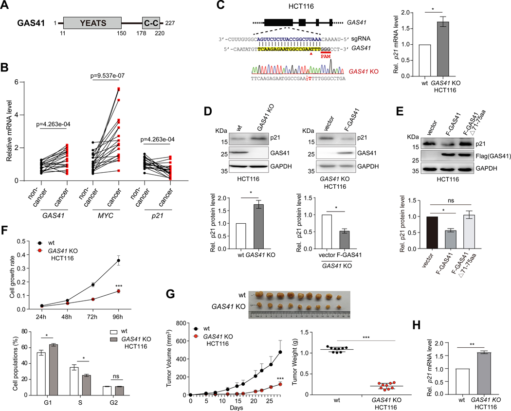
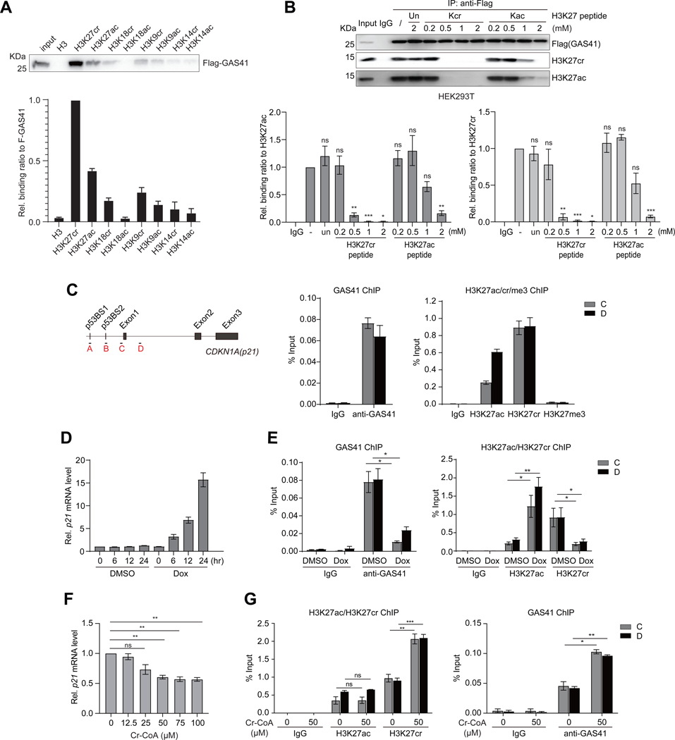
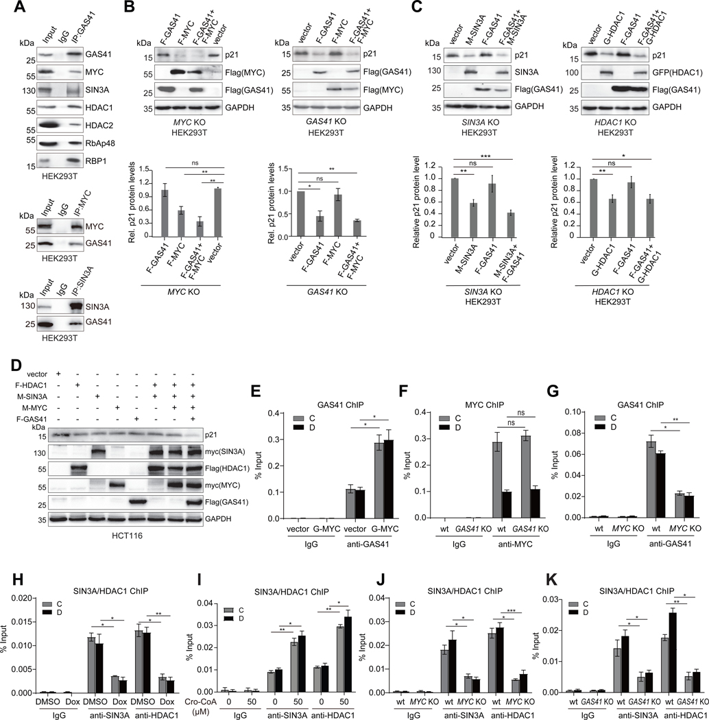
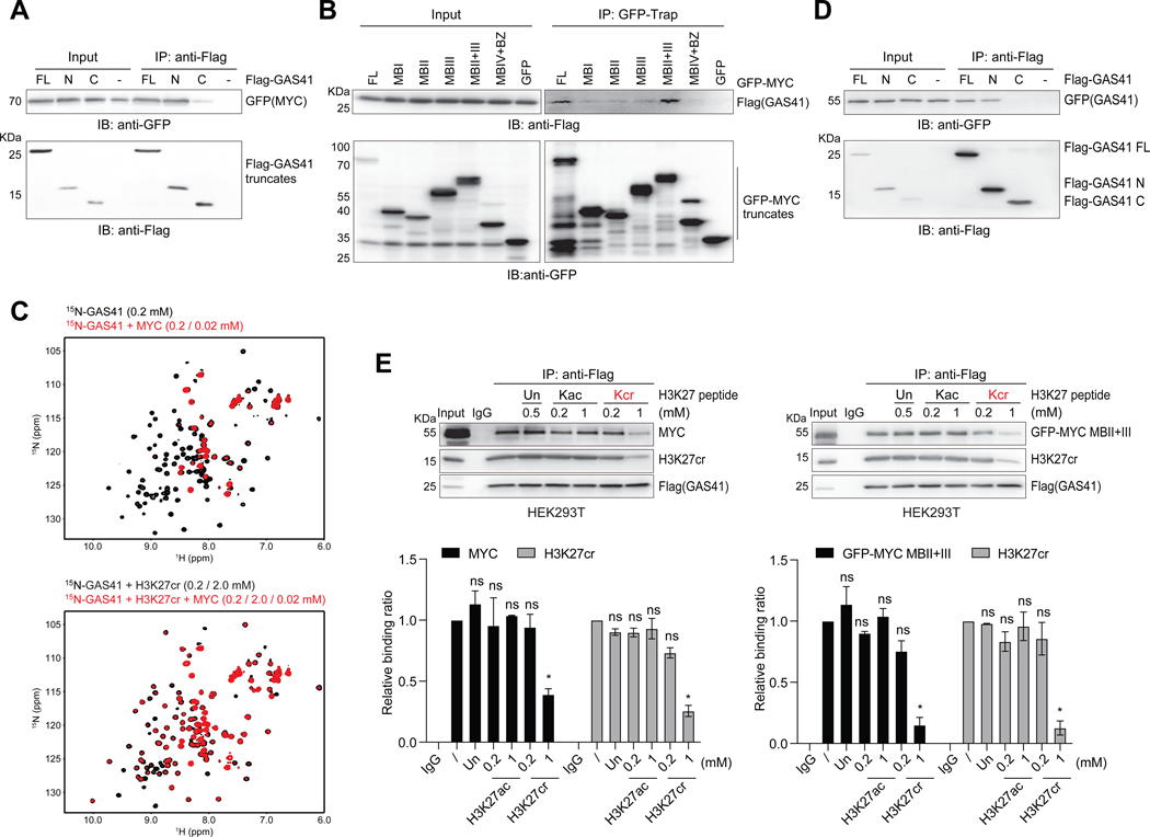
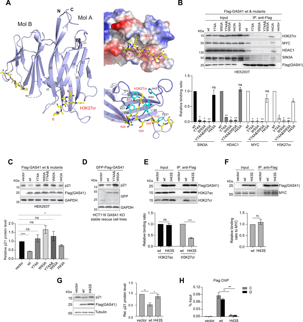
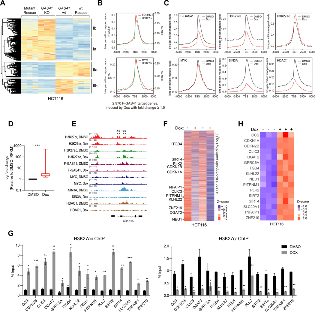
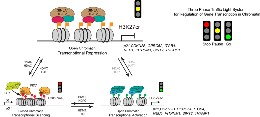
Histone lysine acylation, including acetylation and crotonylation, plays a pivotal role in gene transcription in health and diseases. However, our understanding of histone lysine acylation has been limited to gene transcriptional activation. Here, we report that histone H3 lysine 27 crotonylation (H3K27cr) directs gene transcriptional repression rather than activation. Specifically, H3K27cr in chromatin is selectively recognized by the YEATS domain of GAS41 in complex with SIN3A-HDAC1 co-repressors. Proto-oncogenic transcription factor MYC recruits GAS41/SIN3A-HDAC1 complex to repress genes in chromatin, including cell-cycle inhibitor p21. GAS41 knockout or H3K27cr-binding depletion results in p21 de-repression, cell-cycle arrest, and tumor growth inhibition in mice, explaining a causal relationship between GAS41 and MYC gene amplification and p21 downregulation in colorectal cancer. Our study suggests that H3K27 crotonylation signifies a previously unrecognized, distinct chromatin state for gene transcriptional repression in contrast to H3K27 trimethylation for transcriptional silencing and H3K27 acetylation for transcriptional activation.

Keywords: GAS41; YEATS domain; crystal structure; gene transcriptional repression; histone lysine crotonylation.


Conflict of interest statement

Declaration of interests M.-M.Z. is a scientific founder, director, and shareholder of Parkside Scientific Inc.

Figures

Figure 1.
Figure 1.. GAS41 contributes to p21 repression in tumor cell proliferation
(A) Scheme depicting protein functional domain organization of GAS41, consisting of the YEATS domain at the N-terminus and the coiled-coil motif at the C-terminus. (B) Before-after plot showing mRNA expression levels of GAS41, p21 and MYC in 21 pairs of cancer vs. adjacent non-cancerous tissues of human colorectal cancer patients. GAS41, MYC or p21 expression level in non-cancer tissue of #1 was set as 1. Statistical significance (p value, as indicated) was calculated with Wilcoxon signed-rank test (paired samples Wilcoxon test). (C) Scheme depicting guide RNA design targeting GAS41 start codon for generating HCT116 GAS41 knockout (KO) cells using CRISPR/Cas9 method. Right, RT-qPCR analysis of p21 expression in wild type (wt) and HCT116 GAS41 KO cells. n=3. (D) Western blot analysis and quantification of p21 and GAS41 expression in HCT116 GAS41 KO cells with or without transfection of Flag-GAS41 using antibodies as indicated. n=3. (E) Western blot analysis and quantification of p21 expression in HCT116 cells transiently transfected with a Flag vector or a vector expressing Flag-GAS41 wt or YEATS domain mutant (Δ71–75). n=3. (F) Cell growth rate of HCT116 wt and GAS41 KO cells as assessed by MTT assay. Shown were mean ± s.e.m. from 6 technical repeats. Lower, cell cycle analysis of wt and HCT116 GAS41 KO cells by fluorescence-activated cell sorter analysis. Relative distributions of cells in different cell cycle phases, as analyzed using FlowJo software. n=3. (G) Effect of GAS41 knockout on tumor volume and weight in mice. 1×10⁶ HCT116 wt or GAS41 KO cells were injected into the mammary fat pad of NOD-SCID mice. The tumor volume and weight were recorded for 29 days. Left, mean + s.d, Right, mean ± s.d. n=9. (H) RT-qPCR analysis of p21 expression in the tumors derived from HCT116 wt and GAS41 KO cells in the mouse xenograft study. n=3. KO stands for knockout, wt, wild type, and F-GAS41, Flag-GAS41. All error bars throughout this study represent mean ± s.e.m. from indicated (n) biological repeats and statistical significance was calculated using two-tailed paired Student’s t-test unless noted otherwise. *P < 0.05, **P < 0.01, ***P < 0.001, ns, not significant. See also Figure S1.
Figure 2.
Figure 2.. GAS41 regulates transcriptional repression of p21 mediated by H3K27cr
(A) Western blots analysis and quantitation of ectopically expressed Flag-GAS41 in HCT116 cells binding to a set of biotinylated histone H3 peptides derived from H3 (residues 1–25, biotin-ARTKQTARKSTGGKAPRKQLATKAA), H3K27cr/ac (residues 21–33, biotin-ATKAAR-Kcr/Kac-S-APATG), H3K18cr/ac (residues 12–24, biotin-GGKAPR-Kcr/Kac-QLATKA), H3K9cr/ac (residues 3–15, biotin-TKQTAR-Kcr/Kac-SSGGKA), and H3K14cr/ac (residues 1–25, biotin-ARTKQTARKS-TGG-Kcr/Kac-APRKQLATKAA). n=3. (B) Western blot analysis and quantitation of Flag-GAS41 binding to nucleosomal H3K27cr or H3K27ac in the lysate of HEK293T cells transiently transfected with Flag-GAS41 in the presence of unmodified histone H3K27, H3K27cr or H3K27ac peptide at concentrations as indicated. n=3. (C) Scheme illustrating primer positions at p21 gene locus used in the study. Right, ChIP-qPCR analyses of endogenous GAS41(left), and histone H3K27cr, H3K27ac, or H3K27me3 (right) enrichment at p21 locus in HEK293T cells. n=3. (D) mRNA transcript levels of p21 in HEK293T cells treated with Dox for 0, 6, 12, and 24 hours, respectively, n=3. (E) ChIP-qPCR analyses showing changes of endogenous GAS41 (left), and H3K27ac or H3K27cr (right) enrichment at p21 locus in HEK293T cells treated with Dox for 24 hours, n=3. (F) qPCR analysis of p21 expression in HEK293T cells treated with crotonyl-CoA at concentrations as indicated for 24 hours. n=3. (G) ChIP-qPCR analysis of H3K27ac, H3K27cr (left), and endogenous GAS41 (right) enrichment at p21 locus in HEK293T cells with 24-hour crotonyl-CoA (50 μM) treatment. n=3. See also Figure S2 and Figure S3.
Figure 3.
Figure 3.. GAS41 works with MYC and SIN3A/HDAC1 co-repressors for p21 repression
(A) Co-immunoprecipitation (Co-IP) of endogenous GAS41 and immunoblotting (IB) with specific antibodies assessing GAS41 association with MYC, SIN3A, HDAC1/2, RbAP48 and RBP1 in HEK293T cells (upper). Lower, reverse Co-IP showing endogenous MYC and SIN3A interactions with GAS41, respectively, in HEK293T cells. (B) Western blot analysis and quantification of p21 expression in HEK293T MYC KO (left) and GAS41 KO (right) cells co-transfected with F-GAS41 and F-MYC, n=6 for MYC KO, n=3 for GAS41 KO. (C) Western blot analysis and quantification of p21 expression in HEK293T SIN3A KO cells co-transfected with F-GAS41 and M-SIN3A (left), and HDAC1 KO cells co-transfected with F-GAS41 and G-HDAC1 (right). n=5 for SIN3A KO, and n=6 for HDAC1 KO. (D) Western blot analysis of p21 expression in HCT116 cells co-transfected with F-HDAC1, M-SIN3A, M-MYC, and F-GAS41. (E) ChIP-qPCR analysis of GAS41 enrichment at p21 locus in HEK293T cells transiently co-transfected with GFP empty vector, or GFP-MYC. n=3. (F) ChIP-qPCR analysis of endogenous MYC enrichment at p21 locus in HEK293T wt and GAS41 KO cells. n=3. (G) ChIP-qPCR analysis of GAS41 enrichment at p21 locus in HEK293T wt and MYC KO cells. n=3. (H) ChIP-qPCR analysis of SIN3A and HDAC1 enrichment at p21 locus in HEK293T cells before and after 24-hour Dox treatment. n=3. (I) ChIP-qPCR analysis of SIN3A/HDAC1 enrichment at p21 locus in HEK293T cells with 24-hour crotonyl-CoA (50 μM) treatment. n=3 (J) ChIP-qPCR analysis of SIN3A and HDAC1 enrichment at p21 locus in HEK293T wt and MYC KO cells. n=3. (K)ChIP-qPCR analysis of SIN3A and HDAC enrichment at p21 locus in HEK293T wt and GAS41 KO cells. n=3. F-GAS41 stands for Flag-GAS41, F-MYC for Flag-MYC, F-HDAC1 for Flag-HDAC1, G-HDAC1 for GFP-HDAC1, M-SIN3A for myc-SIN3A, and M-MYC for myc-MYC. See also Figure S4 and Figure S5.
Figure 4.
Figure 4.. GAS41 YEATS domain interactions with H3K27cr and MYC
(A) Co-IP of Flag-GAS41 full-length (FL; residues 1–227), N-terminal segment (N; residues 1–165) or C-terminal segment (C; residues 148–227) and IB with Flag and GFP antibodies assessing GAS41 interaction with MYC in HEK293T cells co-transfected with Flag-GAS41 and GFP-MYC. (B) Co-IP of GFP-MYC and its various segments and IB with GFP and Flag antibodies in Flag-GAS41 and GFP-MYC co-transfected HEK293T cells. (C) GAS41 YEATS domain binding to MYC (residues 1–262) (upper), or in the presence of the H3K27cr peptide (lower), as assessed by 2D 1H-15N HSQC spectra of the GAS41 YEATS domain. (D) GAS41 dimerization, as assessed by Flag-GAS41 and GFP-GAS41 interaction in HEK293T cells, co-transfected with GFP-GAS41 and Flag-GAS41 of full-length, N- or C-terminal segment constructs, respectively. Cell lysate was subjected to Flag IP and followed by IB with antibodies against GFP. (E) Western blot analysis and quantification of H3K27ac or H3K27cr peptide competition against Flag-GAS41 binding to MYC and H3K27cr in HEK293T cells. Varying amounts of H3K27, H3K27cr or H3K27ac peptide, as indicated, was added to the lysate of HEK293T cells, transfected with either Flag-GAS41 alone or together with GFP-MYC MBII+III, and followed by Flag IP and IB with antibodies against Flag, MYC, GFP, and H3K27cr. n=2. See also Figure S6 and Table S1.
Figure 5.
Figure 5.. Molecular basis of GAS41 YEATS domain interactions with H3K27cr and MYC for p21 repression
(A) Crystal structure of GAS41 YEATS domain (blue) in complex with H3K27cr peptide (yellow) as shown in a dimer (molecules A and B). Right, Electrostatic potential surface representation of GAS41 YEATS domain bound to H3K27cr peptide (upper), and the detailed structural basis of GAS41 YEATS domain recognition of the H3K27cr peptide (lower), as depicted in molecule A. Side chains of key residues engaged in protein/peptide interactions are color-coded by atom type, and a hydrogen bond is indicated by a dashed line (red). (B) Effects of mutations of acyl-lysine binding residues on GAS41 binding to H3K27cr, MYC and SIN3A-HDAC co-repressor complex proteins. The lysate of HEK293T cells transfected with Flag-GAS41 wt or mutants was subjected to Flag IP and followed by IB with antibodies against Flag, H3K27cr, MYC, SIN3A and HDAC1, respectively. n=2. (C) Effects of mutations of acyl-lysine binding residues in GAS41 on p21 expression in HEK293T cells transfected Flag-GAS41 wt or corresponding mutants. Quantification is shown as indicated. n=3. (D) Western blot analysis of p21 expression in HCT116 GAS41 KO cells stably expressing Flag-GAS41 wt, Y74A/W93A mutant, or empty vector. (E, F) Effects of H43S mutation on GAS41 binding to H3K27ac, H3K27cr and MYC. The lysate of HCT116 cells transfected with Flag-GAS41 wt and H43S was subjected to Flag IP and followed by IB with antibodies against Flag, H3K27ac, H3K27cr and MYC respectively. n=3. (G) Western blot analysis of p21 expression in HCT116 expressing Flag-GAS41 wt, H43S mutant, or empty vector. n=4. (H) ChIP-qPCR analysis showing Flag-GAS41 occupancy at p21 locus in HCT116 cells expressing Flag-GAS41 wt and H43S, n=3. See also Figure S7, Figure S8, and Table S2.
Figure 6.
Figure 6.. H3K27cr regulates gene transcriptional repression in chromatin
(A) Heatmap of RNA-seq counts of HCT116 GAS41 wt, GAS41 KO, wt rescue, and Y74A/W93A mutant rescue cells. Genes selected are significantly changed (P <0.1) and ranked by log2[fold change] >=1.5 between wild-type and GAS41 KO samples. Counts are represented as z-scores between all samples for each gene. Genes responded to wt or mutant rescue are grouped into 2 categories whose transcription is either fully or partially repressed by (Ia & Ib), or positively dependent fully or partially upon GAS41 (IIa & IIb). (B) ChIP-seq peaks of H3K27cr vs. Flag-GAS41 (upper), and H3K27cr vs. MYC (lower) (average of 2–3 samples), centered at the TSS (±5 kb) of 2,970 GAS41 target genes in HCT116 cells that showed transcriptional induction upon Dox treatment with fold change ≥ 1.5. Reads were normalized to bins per million mapped reads. (C) ChIP-seq peaks of Flag-GAS41, MYC, SIN3A, HDAC1, H3K27cr and H3K27ac (average of 2–3 samples), centered at the TSS (±5 kb) of the 2,970 GAS41 target genes in HCT116 cells before and after Dox treatment. Reads were normalized to bins per million mapped reads. (D) Box-and-whisker plot (min to max) of RNA-seq data showing fold change of all genes that are stimulated upon Dox treatment in HCT116 cells and bound by Flag-GAS41 within ±5 kb TSS (total 2,970 genes). *** p<0.001. (E) Genome browser view of H3K27cr, H3K27ac, Flag-GAS41, MYC, SIN3A and HDAC1 ChIP-seq peaks at the p21(CDKN1A) locus in DMSO and Dox treated HCT116 cells. (F) Heatmap of H3K27cr peak intensities at H3K27cr peaks as identified by ChIP-seq in DMSO and Dox treated HCT116 cells, with each replicate sample represented as one column. (G) ChIP-qPCR analysis of H3K27ac and H3K27cr occupancy at the gene loci of CCS, CDKN2B, CLIC3, DGAT2, GPRC5A, ITGB4, KLHL22, NEU1, PITPNM1, PLK2, SIRT2, SIRT4, SLC20A1, TNFAIP1 and ZNF219 in HCT116 cells before and after Dox treatment. Values are normalized to DMSO control group. n=3. (H) Heatmap of RNA-seq datasets of triplicate samples of HCT116 cells showing selected genes that fulfill the following criteria: differentially expressed in GAS41 KO cells, targeted by GAS41 as identified by ChIP-seq, and showed decreased H3K27cr occupancy upon Dox treatment as shown by ChIP-seq. See also Figure S9, Figure S10 and Table S3.
Figure 7.
Figure 7.. H3K27cr represents a distinct chromatin state for gene transcriptional repression
Schematic illustration of a “Three-phase traffic light system” model describing three different H3K27 modifications as distinct chromatin states for gene transcription, i.e. H3K27me3 (Stop) marks for transcriptional silencing, H3K27cr (Pause) for transcriptional repression, and H3K27ac for transcriptional activation (Go). As illustrated for p21 and other GAS41 target genes such as those validated in Figure 6H, H3K27cr-directed gene transcriptional repression operates through selective recognition of H3K27cr by the YEATS domain of GAS41 in complex with MYC and SIN3A/HDAC1 co-repressor complex in chromatin.

References

    1. Tan M, Luo H, Lee S, Jin F, Yang JS, Montellier E, Buchou T, Cheng Z, Rousseaux S, Rajagopal N, et al. (2011). Identification of 67 Histone Marks and Histone Lysine Crotonylation as a New Type of Histone Modification. Cell 146, 1016–1028. 10.1016/j.cell.2011.08.008. - DOI - PMC - PubMed
    1. Dhalluin C, Carlson JE, Zeng L, He C, Aggarwal AK, and Zhou MM (1999). Structure and ligand of a histone acetyltransferase bromodomain. Nature 399, 491–496. 10.1038/20974. - DOI - PubMed
    1. Zaware N, and Zhou MM (2019). Bromodomain biology and drug discovery. Nat Struct Mol Biol 26, 870–879. 10.1038/s41594-019-0309-8. - DOI - PMC - PubMed
    1. Schulze JM, Wang AY, and Kobor MS (2010). Reading chromatin: insights from yeast into YEATS domain structure and function. Epigenetics : official journal of the DNA Methylation Society 5, 573–577. - PMC - PubMed
    1. Li Y, Wen H, Xi Y, Tanaka K, Wang H, Peng D, Ren Y, Jin Q, Dent SY, Li W, et al. (2014). AF9 YEATS domain links histone acetylation to DOT1L-mediated H3K79 methylation. Cell 159, 558–571. 10.1016/j.cell.2014.09.049. - DOI - PMC - PubMed