

BACH1 promotes lung adenocarcinoma cell metastasis through transcriptional activation of ITGA2
- PMID: 37311571
- PMCID: PMC10475762
- DOI: 10.1111/cas.15884
BACH1 promotes lung adenocarcinoma cell metastasis through transcriptional activation of ITGA2
Abstract
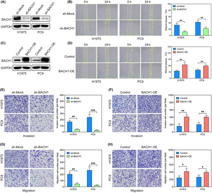
BACH1 plays an important role in promoting cancer. This study aims to further verify the relationship between the expression level of BACH1 in lung adenocarcinoma prognosis, as well as the influence of BACH1 expression on lung adenocarcinoma and the potential mechanism. The expression level of BACH1 in lung adenocarcinoma and its relationship with prognosis was evaluated by lung adenocarcinoma tissue microarray analysis combined with bioinformatics approaches. Gene knockdown and overexpression were used to investigate the functions and molecular mechanisms of BACH1 in lung adenocarcinoma cells. The regulatory downstream pathways and target genes of BACH1 in lung adenocarcinoma cells were explored by bioinformatics and RNA sequencing data analysis, real-time PCR, western blot analysis, and cell immunofluorescence and cell adhesion assays. Chromatin immunoprecipitation and dual-luciferase reporter assays were carried out to verify the target gene binding site. In the present study, BACH1 is abnormally highly expressed in lung adenocarcinoma tissues, and high BACH1 expression is negatively correlated with patient prognosis. BACH1 promotes the migration and invasion of lung adenocarcinoma cells. Mechanistically, BACH1 directly binds to the upstream sequence of the ITGA2 promoter to promote ITGA2 expression, and the BACH1-ITGA2 axis is involved in cytoskeletal regulation in lung adenocarcinoma cells by activating the FAK-RAC1-PAK signaling pathway. Our results indicated that BACH1 positively regulates the expression of ITGA2 through a transcriptional mechanism, thereby activating the FAK-RAC1-PAK signaling pathway to participate in the formation of the cytoskeleton in tumor cells and then promoting the migration and invasion of tumor cells.
Keywords: BACH1; ITGA2; cytoskeleton; lung adenocarcinoma; metastasis.
© 2023 The Authors. Cancer Science published by John Wiley & Sons Australia, Ltd on behalf of Japanese Cancer Association.
Conflict of interest statement
The authors declare no conflict of interest.
Figures


Similar articles
-
Silencing of BACH1 inhibits invasion and migration of prostate cancer cells by altering metastasis-related gene expression.Artif Cells Nanomed Biotechnol. 2018 Nov;46(7):1495-1504. doi: 10.1080/21691401.2017.1374284. Epub 2017 Sep 11. Artif Cells Nanomed Biotechnol. 2018. PMID: 28889753
-
BTB and CNC homology 1 (Bach1) induces lung cancer stem cell phenotypes by stimulating CD44 expression.Respir Res. 2021 Dec 23;22(1):320. doi: 10.1186/s12931-021-01918-2. Respir Res. 2021. PMID: 34949193 Free PMC article.
-
Galectin-3 activates TLR4/NF-κB signaling to promote lung adenocarcinoma cell proliferation through activating lncRNA-NEAT1 expression.BMC Cancer. 2018 May 22;18(1):580. doi: 10.1186/s12885-018-4461-z. BMC Cancer. 2018. PMID: 29788922 Free PMC article.
-
A Novel Therapeutic Target, BACH1, Regulates Cancer Metabolism.Cells. 2021 Mar 12;10(3):634. doi: 10.3390/cells10030634. Cells. 2021. PMID: 33809182 Free PMC article. Review.
-
The Multifaceted Roles of BACH1 in Disease: Implications for Biological Functions and Therapeutic Applications.Adv Sci (Weinh). 2025 Mar;12(10):e2412850. doi: 10.1002/advs.202412850. Epub 2025 Jan 30. Adv Sci (Weinh). 2025. PMID: 39887888 Free PMC article. Review.
Cited by
-
Identification and Evaluation of Hub Long Non-Coding RNAs and mRNAs in PM2.5-Induced Lung Cell Injury.Int J Mol Sci. 2025 Jan 22;26(3):911. doi: 10.3390/ijms26030911. Int J Mol Sci. 2025. PMID: 39940682 Free PMC article.
-
Multiomics analysis uncovers host-microbiota interactions regulate hybrid vigor traits in geese.Poult Sci. 2025 Aug;104(8):105289. doi: 10.1016/j.psj.2025.105289. Epub 2025 May 10. Poult Sci. 2025. PMID: 40393267 Free PMC article.
-
p-Coumaric acid alleviates neuronal damage in ischemic stroke mice by promoting BACH1 nuclear export and degradation.Acta Pharmacol Sin. 2025 Aug;46(8):2136-2150. doi: 10.1038/s41401-025-01510-0. Epub 2025 Mar 14. Acta Pharmacol Sin. 2025. PMID: 40087473
-
A systems-level analysis of the mutually antagonistic roles of RKIP and BACH1 in dynamics of cancer cell plasticity.J R Soc Interface. 2023 Nov;20(208):20230389. doi: 10.1098/rsif.2023.0389. Epub 2023 Nov 15. J R Soc Interface. 2023. PMID: 37963558 Free PMC article.
References
-
- Bray F, Ferlay J, Soerjomataram I, Siegel RL, Torre LA, Jemal A. Global cancer statistics 2018: GLOBOCAN estimates of incidence and mortality worldwide for 36 cancers in 185 countries. CA Cancer J Clin. 2018;68:394‐424. - PubMed
-
- Siegel RL, Miller KD, Fuchs HE, Jemal A. Cancer statistics, 2021. CA Cancer J Clin. 2021;71:7‐33. - PubMed
MeSH terms
Substances
Grants and funding
LinkOut - more resources
Full Text Sources
Medical
Research Materials
Miscellaneous
