. 2023 Jun 28;15(25):30008-30028.
doi: 10.1021/acsami.3c07248. Epub 2023 Jun 13.

Cell-Membrane-Coated and Cell-Penetrating Peptide-Conjugated Trimagnetic Nanoparticles for Targeted Magnetic Hyperthermia of Prostate Cancer Cells

Affiliations

Cell-Membrane-Coated and Cell-Penetrating Peptide-Conjugated Trimagnetic Nanoparticles for Targeted Magnetic Hyperthermia of Prostate Cancer Cells

Valentin Nica et al. ACS Appl Mater Interfaces. .

Abstract

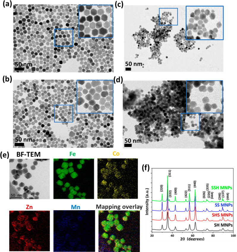
Prostate malignancy represents the second leading cause of cancer-specific death among the male population worldwide. Herein, enhanced intracellular magnetic fluid hyperthermia is applied in vitro to treat prostate cancer (PCa) cells with minimum invasiveness and toxicity and highly specific targeting. We designed and optimized novel shape-anisotropic magnetic core-shell-shell nanoparticles (i.e., trimagnetic nanoparticles - TMNPs) with significant magnetothermal conversion following an exchange coupling effect to an external alternating magnetic field (AMF). The functional properties of the best candidate in terms of heating efficiency (i.e., Fe3O4@Mn0.5Zn0.5Fe2O4@CoFe2O4) were exploited following surface decoration with PCa cell membranes (CM) and/or LN1 cell-penetrating peptide (CPP). We demonstrated that the combination of biomimetic dual CM-CPP targeting and AMF responsiveness significantly induces caspase 9-mediated apoptosis of PCa cells. Furthermore, a downregulation of the cell cycle progression markers and a decrease of the migration rate in surviving cells were observed in response to the TMNP-assisted magnetic hyperthermia, suggesting a reduction in cancer cell aggressiveness.

Keywords: cell membranes; cell-penetrating peptides; intracellular hyperthermia; prostate cancer; trimagnetic nanoparticles.


Conflict of interest statement

The authors declare no competing financial interest.

Figures

Figure 1
Figure 1
Schematic illustration of coating and functionalization procedures of BMNPs and TMNPs: Fe3O4@Co0.5Zn0.5Fe2O4, soft–hard (SH); Fe3O4@Co0.5Zn0.5Fe2O4@MnFe2O4, soft–hard–soft (SHS); Fe3O4@Mn0.5Zn0.5Fe2O4, soft–soft (SS); (c) Fe3O4@Mn0.5Zn0.5Fe2O4@CoFe2O4, soft–soft–hard (SSH).
Figure 2
Figure 2
Representative BF-TEM images of (a) SH, (b) SHS, (c) SS, and (d) SSH MNPs. The insets represent the magnified TEM image of corresponding samples. (e) BF-TEM image and corresponding EFTEM elemental maps for the SSH sample. EFTEM mapping demonstrates the presence of Fe, Zn, Mn, and Co. (f) X-ray diffraction patterns of pristine samples (SH, SHS, SS, and SSH MNPs).
Figure 3
Figure 3
(a) Magnetic curves at room temperature of pristine samples (SH, SHS, SS, and SSH MNPs). The inset evidences the coercivity values. (b) Heating profile of ferrofluid samples (L-SH, L-SHS, L-SS, and L-SSH MNPs) exposed to AMF (f = 97.5 kHz, B = 20 mT, H = 15.9 kA/m; MNP concentration 5 mg/mL).
Figure 4
Figure 4
BF-TEM micrographs of functionalized samples: (a) LN1-L-SSH MNPs, (b) negative-stained CM-L-SSH MNPs, and (c) negative-stained CM-LN1-L-SSH MNPs. DLS measurements: (d) hydrodynamic size distribution and (e) Z-potential before (L-SSH MNPs) and after functionalization (CM-L-SSH, CM-LN1-L-SSH, and CM-LN1-L-SSH MNPs).
Figure 5
Figure 5
(a) Representative confocal images of PC-3 cultures incubated for 72 h with 250 μg/mL of MNPs (L-SSH, LN1-L-SSH, CM-L-SSH, and CM-LN1-L-SSH MNPs). F-actin in red, MNPs in green, and nuclei in blue. (b) ICP-OES elemental quantification of Fe (green), Zn (pink), Mn (gray), and Co (yellow) in PC-3 cells treated with the different MNPs.
Figure 6
Figure 6
(a) Representative confocal laser scanning microscopy imaging of Ki-67 expression in PC-3 cells in the considered experimental classes. Nuclei in blue, Ki-67 in green, and F-actin in red. (b) In red, % of cells normalized to controls. In green, % of Ki-67-positive cells (Ki-67+).
Figure 7
Figure 7
Flow cytometry analysis of apoptosis/necrosis: (a) representative flow cytometer scatter plots of propidium iodide vs annexin V-FITC. The populations of healthy, early apoptotic, late apoptotic, and necrotic cells have been highlighted in black, green, blue, and red, respectively. (b) Quantitative evaluation.
Figure 8
Figure 8
(a) Expression of hsp70 in PC-3 cells upon magnetothermal stimulation: representative confocal laser scanning microscopy imaging (nuclei in blue, hsp70 in green, F-actin in red). (b) Average intensity of the hsp70 signal in the cells for each experimental condition (* p < 0.05).
Figure 9
Figure 9
Activation of the caspase-9 apoptotic pathway upon acute stimulation with magnetic hyperthermia (“CM-LN1-L-SSH MNPs + AMF”). (a) Representative distributions of the cell fluorescent signal emission. Caspase-9-negative (−) and -positive (+) cells are highlighted in light blue and light red, respectively. (b) Quantitative evaluation of flow cytometry data for each experimental condition (* p < 0.05).
Figure 10
Figure 10
Cell migration upon acute magnetothermal stimulation. (a) Representative images of PC-3 cells stained with calcein at t = 0 h and t = 24 h of cell migration. (b) % of gap size measured in each experimental class (* p < 0.05).
Figure 11
Figure 11
Proteomic analysis: (a) principal component analysis (PCA) for 4 independent experiments in “Control” (blue cross), “Control + AMF” (orange square), “CM-LN1-L-SSH MNPs” (magenta circles), and “CM-LN1-L-SSH MNPs + AMF” (rhombuses in olive green color) treatments; (b) volcano plot and GO keywords regarding the “Control + AMF vs Control”, “CM-LN1-L-SSH MNPs vs Control”, and “CM-LN1-L-SSH MNPs + AMF vs Control” comparisons; upregulated and downregulated pathways are highlighted in green and red, respectively.

References

    1. Huo Y.; Yu J.; Gao S.. Synthesis and Biomedical applications of Magnetic Nanomaterials. In Magnetic-mediated Hyperthermia for Cancer Treatment: Research Progress and Clinical Trials; EDP Sciences: Les Ulis, 2022; pp 228–260.
    1. Li B.; Chen X.; Qiu W.; Zhao R.; Duan J.; Zhang S.; Pan Z.; Zhao S.; Guo Q.; Qi Y.; Wang W.; Deng L.; Ni S.; Sang Y.; Xue H.; Liu H.; Li G. Synchronous Disintegration of Ferroptosis Defense Axis via Engineered Exosome-Conjugated Magnetic Nanoparticles for Glioblastoma Therapy. Adv. Sci. 2022, 9 (17), 2105451.10.1002/advs.202105451. - DOI - PMC - PubMed
    1. Manohar A.; Vijayakanth V.; Vattikuti S. V. P.; Manivasagan P.; Jang E. S.; Chintagumpala K.; Kim K. H. Ca-Doped MgFe2O4 Nanoparticles for Magnetic Hyperthermia and Their Cytotoxicity in Normal and Cancer Cell Lines. ACS Appl. Nano Mater. 2022, 5 (4), 5847–5856. 10.1021/acsanm.2c01062. - DOI
    1. Kossatz S.; Grandke J.; Couleaud P.; Latorre A.; Aires A.; Crosbie-Staunton K.; Ludwig R.; Dähring H.; Ettelt V.; Lazaro-Carrillo A.; Calero M.; Sader M.; Courty J.; Volkov Y.; Prina-Mello A.; Villanueva A.; Somoza C.; Cortajarena A. L.; Miranda R.; Hilger I. Efficient Treatment of Breast Cancer Xenografts with Multifunctionalized Iron Oxide Nanoparticles Combining Magnetic Hyperthermia and Anti-Cancer Drug Delivery. Breast Cancer Res. 2015, 17 (1), 66.10.1186/s13058-015-0576-1. - DOI - PMC - PubMed
    1. Beola L.; Grazú V.; Fernández-Afonso Y.; Fratila R. M.; De Las Heras M.; De La Fuente J. M.; Gutiérrez L.; Asín L. Critical Parameters to Improve Pancreatic Cancer Treatment Using Magnetic Hyperthermia: Field Conditions, Immune Response, and Particle Biodistribution. ACS Appl. Mater. Interfaces 2021, 13 (11), 12982–12996. 10.1021/acsami.1c02338. - DOI - PMC - PubMed

Substances