

A novel meningioma with tyrosine-rich crystals in a 6-year-old Great Dane
- PMID: 37312432
- PMCID: PMC10365060
- DOI: 10.1111/jvim.16789
A novel meningioma with tyrosine-rich crystals in a 6-year-old Great Dane
Abstract
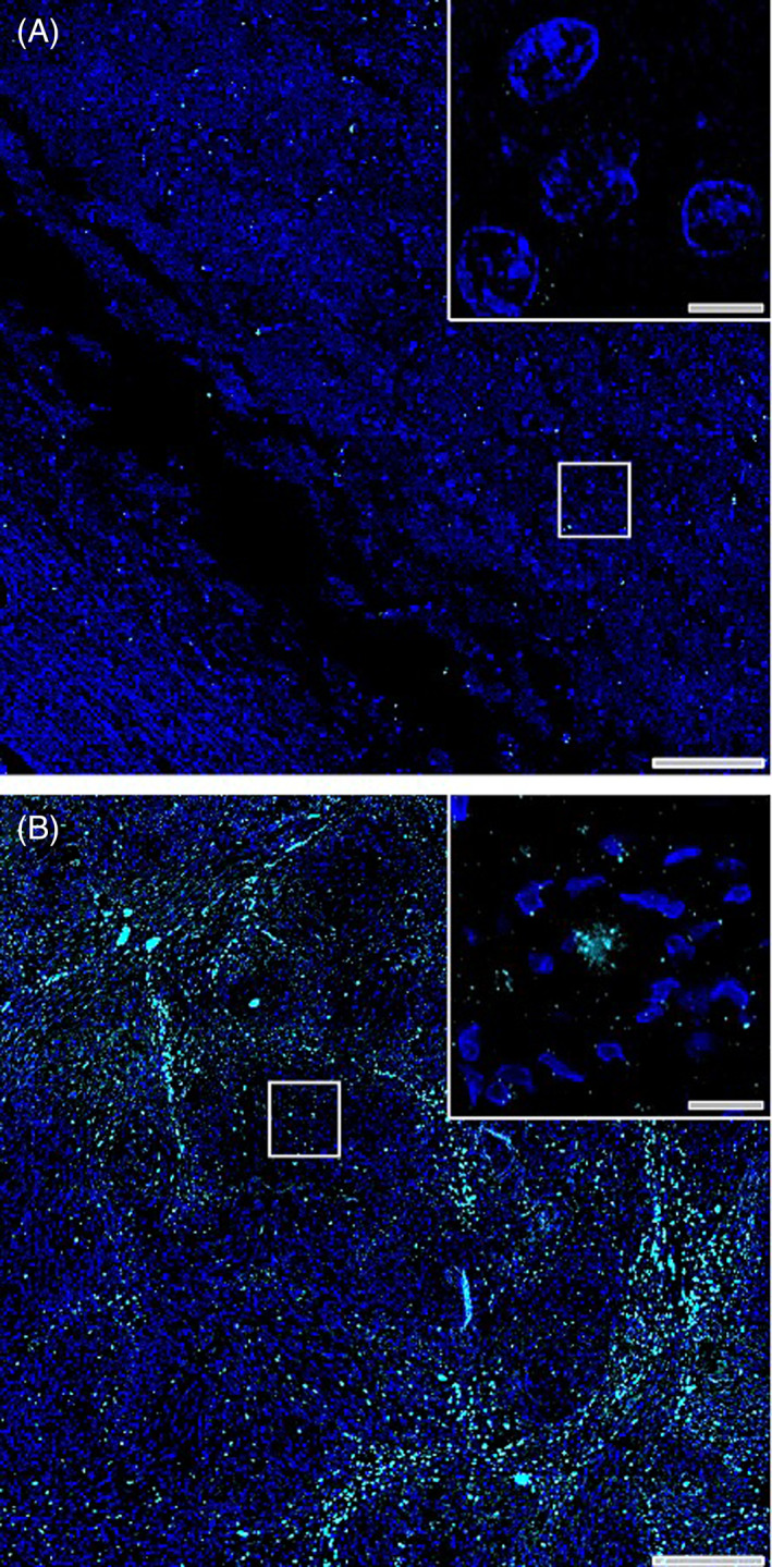
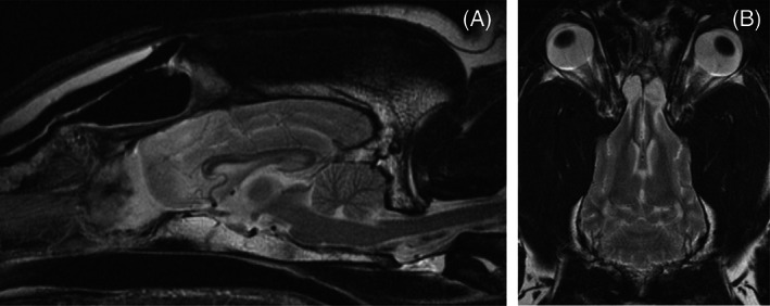
A 6-year-old female spayed Great Dane was evaluated for acute onset cluster seizures. Magnetic resonance imaging (MRI) identified a mass in the olfactory bulbs with a large mucoid component caudal to the primary mass. The mass was removed via transfrontal craniotomy and histopathology revealed a tyrosine crystalline-rich, fibrous meningioma with a high mitotic index. Repeat MRI at 6 months showed no detectable tumor regrowth. The dog is clinically normal with no seizures at the time of publication 10 months after surgery. This meningioma subtype is rare in humans. This unique meningioma occurred in a dog of younger age and uncommon breed for intracranial meningioma. Biological progression of this tumor subtype is unknown; however, growth rate might be slow despite the high mitotic index.
Keywords: atypical; brain tumor; crystal; dog; frontal; neoplasia; olfactory.
© 2023 The Authors. Journal of Veterinary Internal Medicine published by Wiley Periodicals LLC. on behalf of the American College of Veterinary Internal Medicine.
Conflict of interest statement
Authors declare no conflict of interest.
Figures


Similar articles
-
A modified bilateral transfrontal sinus approach to the canine frontal lobe and olfactory bulb: surgical technique and five cases.J Am Anim Hosp Assoc. 2000 Jan-Feb;36(1):43-50. doi: 10.5326/15473317-36-1-43. J Am Anim Hosp Assoc. 2000. PMID: 10667405
-
MRI and CT characteristics of a grade I meningioma with concurrent cribriform plate lysis in a dog.Vet Radiol Ultrasound. 2023 May;64(3):E23-E26. doi: 10.1111/vru.13196. Epub 2022 Nov 28. Vet Radiol Ultrasound. 2023. PMID: 36440542
-
A surgical approach to the canine olfactory bulb for meningioma removal.Vet Surg. 1987 Jul-Aug;16(4):273-7. doi: 10.1111/j.1532-950x.1987.tb00952.x. Vet Surg. 1987. PMID: 3507155
-
External beam radiation therapy for canine intracranial meningioma.Can Vet J. 2009 Jan;50(1):97-100. Can Vet J. 2009. PMID: 19337624 Free PMC article. Review. No abstract available.
-
Orbital Compartment Syndrome without Evidence of Orbital Mass or Ocular Compression After Pterional Craniotomy for Removal of Meningioma of the Frontal Lobe: A Case Report and Literature Review.World Neurosurg. 2020 Jul;139:588-591. doi: 10.1016/j.wneu.2020.04.094. Epub 2020 Apr 25. World Neurosurg. 2020. PMID: 32344145 Review.
References
-
- Foster ES, Carrillo JM, Patnaik AK. Clinical signs of tumors affecting the rostral cerebrum in 43 dogs. J Vet Intern Med. 1988;2:71‐74. - PubMed
-
- Sturges BK, Dickinson PJ, Bollen AW, et al. Magnetic resonance imaging and histological classification of intracranial meningiomas in 112 dogs. J Vet Intern Med. 2008;22:586‐595. - PubMed
-
- Belluco S, Marano G, Baiker K, et al. Standardization of canine meningioma grading: inter‐observer agreement and recommendations for reproducible histopathologic criteria. Vet Comp Oncol. 2022;20:509‐520. - PubMed
-
- Song RB, Vite CH, Bradley CW, Cross JR. Postmortem evaluation of 435 cases of intracranial neoplasia in dogs and relationship of neoplasm with breed, age, and body weight. J Vet Intern Med. 2013;27:1143‐1152. - PubMed
Publication types
MeSH terms
Substances
Grants and funding
LinkOut - more resources
Full Text Sources
