Case Reports
. 2023 Aug 2;31(8):2309-2325.
doi: 10.1016/j.ymthe.2023.06.006. Epub 2023 Jun 12.

Two cases of severe pulmonary toxicity from highly active mesothelin-directed CAR T cells

Affiliations
Case Reports

Two cases of severe pulmonary toxicity from highly active mesothelin-directed CAR T cells

Andrew R Haas et al. Mol Ther. .

Abstract

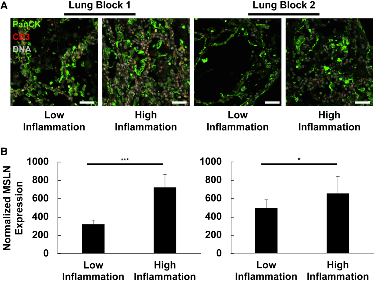
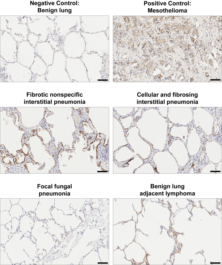
Multiple clinical studies have treated mesothelin (MSLN)-positive solid tumors by administering MSLN-directed chimeric antigen receptor (CAR) T cells. Although these products are generally safe, efficacy is limited. Therefore, we generated and characterized a potent, fully human anti-MSLN CAR. In a phase 1 dose-escalation study of patients with solid tumors, we observed two cases of severe pulmonary toxicity following intravenous infusion of this product in the high-dose cohort (1-3 × 10⁸ T cells per m2). Both patients demonstrated progressive hypoxemia within 48 h of infusion with clinical and laboratory findings consistent with cytokine release syndrome. One patient ultimately progressed to grade 5 respiratory failure. An autopsy revealed acute lung injury, extensive T cell infiltration, and accumulation of CAR T cells in the lungs. RNA and protein detection techniques confirmed low levels of MSLN expression by benign pulmonary epithelial cells in affected lung and lung samples obtained from other inflammatory or fibrotic conditions, indicating that pulmonary pneumocyte and not pleural expression of mesothelin may lead to dose-limiting toxicity. We suggest patient enrollment criteria and dosing regimens of MSLN-directed therapies consider the possibility of dynamic expression of mesothelin in benign lung with a special concern for patients with underlying inflammatory or fibrotic conditions.

Keywords: MSLN; cancer; cell transfer therapy; chimeric antigen receptor (CAR) T cells; immunotherapy.


Conflict of interest statement

Declaration of interests C.H.J., R.M.Y., and M.M.D. are inventors on patents and/or patent applications licensed to Novartis Institutes of Biomedical Research and receive license revenue from such licenses. J.L.B., B.E., K.M., and L.Z. are holders of stock options and patents with Novartis Institutes for Biomedical Research. R.M.Y. and M.M.D. are inventors on patents and/or patent applications licensed to Tmunity Therapeutics and receive license revenue from such licenses. C.H.J. and A.C. are scientific cofounders of Tmunity Therapeutics. C.H.J. is a scientific cofounder of Capstan Therapeutics and is a member of the scientific advisory boards of AC Immune, Alaunos, BluesphereBio, Cabaletta, Carisma, Cartography, Cellares, Celldex, Decheng, Poseida, Verismo, and WIRB-Copernicus. B.E. and R.J.G. completed work on this study before becoming employees at Miltenyi Biotec and AstraZeneca, respectively. R.J.G. holds or may hold AstraZeneca stock. N.C.S. holds equity in Fate Therapeutics and Pfizer. J.A.F. has received grants and personal fees from Cartography Bio., grants from Tmunity Therapeutics, and personal fees from Retro Bio and Shennon Bio outside the submitted work. Additionally, J.A.F. holds patents related to CAR T cells for cancer that are licensed and associated with royalties. S.F.L. is an inventor on patents in the areas of CAR T and biomarkers at Penn that were assigned to Novartis; received research funding from Novartis, Tmunity, and Cabaletta; and consults for Kite/Gilead. M.M.D. is a consultant for Tmunity Therapeutics and is a Consultant and Member on the Scientific Advisory Board of Cellares Corporation.

Figures

None
Graphical abstract
Figure 1
Figure 1
Characterization of novel anti-MSLN CAR constructs in vitro and in vivo (A) Cancer cell lines with varying MSLN expression levels were selected to test CAR T cell reactivity. The ovarian cancer cell line OVCAR8 and the pancreatic cancer cell line Panc02.03 were stained with soluble SS1 scFv to quantify MSLN surface expression (numbers indicate MFI of entire cell population). (B) Cytokine secretion was assessed by co-culturing 25,000 CAR T cells with either OVCAR8, Panc02.03, or Nalm6 cells for 18 h at E:T = 1. Error bars reflect standard deviation. Supernatant was harvested, and the IFNγ concentration measured. (C and D) Impedance-based killing assays were performed by culturing CAR T cells with OVCAR8 (C) and Panc02.03 (D) for 16 h at defined E:T ratios. (E and F) Xenograft studies were performed using representative gastric cancer (E) and lung cancer (F) cells to assess in vivo CAR T cell reactivity. Inserts show MSLN expression by flow cytometry (red histogram: K1 α-MSLN Ab, blue: secondary Ab only). (E) A total of 2 × 106 cells of the intermediate MSLN-expressing gastric cancer line N87 were injected subcutaneously. On day 7 post cancer cell inoculation, 5 × 106 CAR-positive transduced T cells were injected i.v. (see arrow). Tumor growth was monitored by caliper measurements and plotted as tumor volume over time. Activated, but untransduced (UTD) cells served as negative control. (F) A total of 2.5 × 106 cells of the low MSLN-expressing lung cancer line A549 were injected i.v. Five days later, 5 × 106 CAR-positive T cells were injected i.v. (see arrow). Bioluminescence imaging (BLI) was conducted at the points indicated and plotted over time. UTD cells and no treatment served as negative controls. (G) Human plasma membrane protein binding cell array was used to assess MSLN binder specificity. SS1, MSLN-5, and MSLN-11 scFvs with AVI-HIS were applied to cell microarrays expressing 3,559 unique transcripts. Binding was detected using a secondary antibody conjugated to Alexa Fluor 647 and detection of ZsGreen served as transfection/expression control. The array contained three isoforms of MSLN, which were detected as medium to strong binders with all three scFvs. TAOK3, TAO kinase 3; PPAP2B, lipid phosphate phosphohydrolase; P2Ry1, purinergic receptor P2Y1.
Figure 2
Figure 2
Serial chest radiologic studies captured the evolution of pulmonary toxicity in subjects 09 and 13 Both patients received serial chest radiologic studies following admission for complications arising in the setting of huCART-meso infusion. (A) Serial chest X-ray images document the evolution of pulmonary alveolar opacities consistent with non-cardiogenic pulmonary edema seen in subject 13. (B) Subject 09 received a chest CT approximately 2 weeks prior to huCART-meso infusion. After being admitted 2 days following huCART-meso infusion, she received multiple chest X-ray studies that documented new pulmonary alveolar opacities with notable air bronchograms (arrow) seen 2 days into her admission.
Figure 3
Figure 3
Engraftment of huCART-meso cells and systemic cytokine analysis (A) qPCR studies targeting the lentiviral DNA sequence of the huCART-meso cells were used to quantify the expansion kinetics of the mesoCART cells in peripheral blood specimens of subjects 13 and 09. (B) Non-cardiac C-reactive protein levels were assessed in pre- and post-infusion serum specimen. Serum cytokine levels were determined by multiplex Luminex assays on IFNγ (C), GM-CSF (D), TNFα (E), IL-6 (F), IL-10 (G), and IL-1RA (H). For specimens with cytokine values falling below the lowest control in the standard curve, a dashed line is included to denote the lowest concentration that can be reliably quantitated. Values below this line are plotted as reported, except for values reported as “out of range” that are plotted at zero. For both huCART-meso engraftment data and systemic cytokine measurements, pre-infusion specimens were designated day 0, and post-infusion specimens on the same day were designated day 0.5.
Figure 4
Figure 4
Autopsy findings within the lungs for subject 09 are notable for acute lung injury, extensive T cell infiltrates, and abundant CAR T cells (A) H&E-stained sections of lung tissue demonstrate acute lung injury including pulmonary edema, fibrin leakage, and focal hyaline membrane formation associated with an extensive polymorphous inflammatory infiltrate (left, middle). Scattered foci morphologically compatible with MPM are seen surrounded by lymphocytes that poorly infiltrate the tumor (right). Scale bars represent 250 μm (left), 50 μm (middle), and 500 μm (right). (B) Immunohistochemical studies reveal extensive T cell infiltrates in benign lung, with accumulation around vessels (arrow) and at the border of tumor deposits (arrowhead, left image). CD4+ T cells greatly outnumber CD8+ T cells (middle and right, respectively). Scale bars of the low-magnification images represent 500 μm. High-magnification images (30x) include scale bars corresponding to 30 μm. (C) qPCR studies targeting the WPRE sequence of the CAR T cells identify substantial enrichment of CAR T cells in lung and spleen at autopsy.
Figure 5
Figure 5
Inflammation correlates with enhanced MSLN protein expression in pulmonary epithelial cells from subject 09 (A) Representative immunofluorescence micrographs of independent autopsy specimens of lung collected from subject 09 are depicted. Heterogeneous levels of inflammatory infiltrates allow for direct comparison of MSLN protein expression from pulmonary epithelial cells (PanCK+ cells) from regions of relatively high vs. relatively low inflammation. Antibodies: PanCK (1:40, Cy3, green), CD3 (1:75, Alex Fluor 594, red). DNA was labeled using SYTO13 (gray). Scale bars represent approximately 50 μm. (B) MSLN protein expression was measured by digital counts from segments of PanCK+ cells. Normalized counts were generated by scaling data to the surface area of the illuminated segments. n = 10 segments per condition for block 1 (left) and n = 8 segments per condition for block 2 (right). ∗p < 0.05; ∗∗∗p < 0.001, two-tailed Student’s t test.
Figure 6
Figure 6
Immunohistochemical studies of human lung from a spectrum of inflammatory and/or fibrosing disease processes reveal variable expression of MSLN protein in alveolar cells Immunohistochemical staining for MSLN was performed on either human biopsies or autopsy specimens across a spectrum of lung diseases. Mesothelioma was included as a positive control for the study. Images were obtained at ×10 magnification, and scale bars represent 100 μm.

References

    1. Maude S.L., Frey N., Shaw P.A., Aplenc R., Barrett D.M., Bunin N.J., Chew A., Gonzalez V.E., Zheng Z., Lacey S.F., et al. Chimeric antigen receptor T cells for sustained remissions in leukemia. N. Engl. J. Med. 2014;371:1507–1517. doi: 10.1056/NEJMoa1407222. - DOI - PMC - PubMed
    1. Turtle C.J., Hanafi L.A., Berger C., Hudecek M., Pender B., Robinson E., Hawkins R., Chaney C., Cherian S., Chen X., et al. Immunotherapy of non-Hodgkin's lymphoma with a defined ratio of CD8+ and CD4+ CD19-specific chimeric antigen receptor-modified T cells. Sci. Transl. Med. 2016;8:355ra116. doi: 10.1126/scitranslmed.aaf8621. - DOI - PMC - PubMed
    1. Johnson L.A., June C.H. Driving gene-engineered T cell immunotherapy of cancer. Cell Res. 2017;27:38–58. doi: 10.1038/cr.2016.154. - DOI - PMC - PubMed
    1. Yong C.S.M., Dardalhon V., Devaud C., Taylor N., Darcy P.K., Kershaw M.H. CAR T-cell therapy of solid tumors. Immunol. Cell Biol. 2017;95:356–363. doi: 10.1038/icb.2016.128. - DOI - PubMed
    1. Hassan R., Thomas A., Alewine C., Le D.T., Jaffee E.M., Pastan I. Mesothelin Immunotherapy for Cancer: Ready for Prime Time? J. Clin. Oncol. 2016;34:4171–4179. doi: 10.1200/JCO.2016.68.3672. - DOI - PMC - PubMed

Publication types

LinkOut - more resources