. 2023 Jun 12;9(3):00662-2022.
doi: 10.1183/23120541.00662-2022. eCollection 2023 May.

Lung microhaemorrhage drives oxidative/inflammatory damage in α1-antitrypsin deficiency

Affiliations

Lung microhaemorrhage drives oxidative/inflammatory damage in α1-antitrypsin deficiency

Cesare Saltini et al. ERJ Open Res. .

Abstract

Background: Animal models using intratracheal instillation show that elastase, unopposed by α1-antitrypsin (AAT), causes alveolar damage and haemorrhage associated with emphysematous changes. The aim of the present study was to characterise any relationship between alveolar haemorrhage and human AAT deficiency (AATD) using bronchoalveolar lavage (BAL) and lung explant samples from AATD subjects.

Methods: BAL samples (17 patients, 15 controls) were evaluated for free haem (iron protoporphyrin IX) and total iron concentrations. Alveolar macrophage activation patterns were assessed using RNA sequencing and validated in vitro using haem-stimulated, monocyte-derived macrophages. Lung explants (seven patients, four controls) were assessed for iron sequestration protein expression patterns using Prussian blue stain and ferritin immunohistochemistry, as well as ferritin iron imaging and elemental analysis by transmission electron microscopy. Tissue oxidative damage was assessed using 8-hydroxy-2'-deoxyguanosine immunohistochemistry.

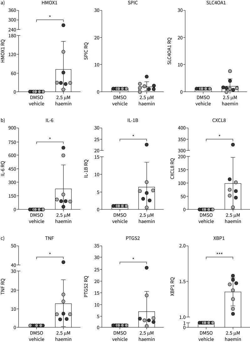
Results: BAL collected from AATD patients showed significantly elevated free haem and total iron concentrations. Alveolar and interstitial macrophages in AATD explants showed elevated iron and ferritin accumulation in large lysosomes packed by iron oxide cores with degraded ferritin protein cages. BAL macrophage RNA sequencing showed innate pro-inflammatory activation, replicated in vitro by haemin exposure, which also triggered reactive oxygen species generation. AATD explants showed massive oxidative DNA damage in both lung epithelial cells and macrophages.

Conclusions: BAL and tissue markers of alveolar haemorrhage, together with molecular and cellular evidence of macrophage innate pro-inflammatory activation and oxidative damage, are consistent with free haem stimulation. Overall, this initial study provides evidence for a pathogenetic role of elastase-induced alveolar haemorrhage in AATD emphysema.


Conflict of interest statement

Conflict of interest: Y. Xin declares funding for the present work from the National Science Foundation (Cooperative Agreement number DMR-1644779) and the State of Florida. M. Brantly declares funding for the present work from the Alpha-1 Foundation, of which they are Scientific Director. All other authors declare no competing interests.

Figures

FIGURE 1
FIGURE 1
Epithelial lining fluid (ELF) levels of a) haemin (control 0.2±0.1 μM, α1-antitrypsin deficiency (AATD) 72.0±108.7 μM; p=0.0149) and b) haem+nonencapsulated haemoglobin (control 21.5±25.0 μM, AATD 115.0±144.5 μM; p=0.00004). c) Total (haem) iron ELF levels are shown as inductively coupled plasma optical emission spectroscopy (ICPOES) total (non-haem and haem) iron (control 970.9±1015.1 ppb, AATD 8412.5±7997.5 ppb; p=0.0015). Haem+nonencapsulated haemoglobin was correlated with d) neutrophil elastase (NE), e) interleukin (IL)-6 and f) IL-8 (Pearson coefficient r=0.516, p=0.0002 and Spearman coefficient rs=0.397, p=0.005, r=0.408, p=0.004; and rs=0.320, p=0.028, r=0.330, p=0.022 and rs=0.373, p=0.009, respectively). ELF iron levels are shown as g) ICPOES non-haem iron (control 0.086±0.025 ppb, AATD 0.162±0.090 ppb; p=0.0043); and as h) ferritin (control 563.8±373.2 ng·mL−1, AATD 4162.4±4112.3 ng·mL−1; p=0.0024) and i) transferrin (control 4458.7±2283.6 ng·mL−1, AATD 7669.2±5464.7 ng·mL−1; p=0.0614) protein concentrations. Data are shown as ELF concentrations, normalised to bronchoalveolar lavage fluid urea concentrations, as described previously [17]. *: p<0.05; **: p<0.01; ***: p<0.001 (t-test).
FIGURE 2
FIGURE 2
a) Diff-Quik stained cytopreps of α1-antitrypsin deficiency (AATD) bronchoalveolar lavage cells show alveolar macrophages, with numerous adherent red blood cells and erythrophagocytic figures; b) Prussian Blue stained cytopreps demonstrate significantly greater iron accumulation in AATD macrophages (control 24.4±12.4 pixels2, AATD 75.6±90.8 pixels2; p=0.00015) c) compared to control. Arrows indicate examples of erythrocyte-like inclusions in a) and areas of iron accumulation in b). Scale bars=20 μm. d) Prussian blue iron stain, e) ferritin light chain (FTL) and f) and ferritin heavy chain (FTH) in AATD lung explanted tissue. Scale bars=50 μm. ImageJ quantitation of staining intensity for g) Prussian blue (control 99.0±88.5 pixels2, AATD 615.5±521.1 pixels2; p=0.020), h) FTL (control 2949.0±775.4 pixels2, AATD 7079.7±4111.2 pixels2; p=0.019) and i) FTH (control 575.6±228.6 pixels2, AATD 1584.2±1085.5 pixels2; p=0.025); all significantly higher in AATD compared to controls. *: p<0.05; ***: p<0.001 (t-test).
FIGURE 3
FIGURE 3
a) Control alveolar macrophages (AlvMacs) contain possibly numerous vesicles or remnants of lipid bodies; b) at higher magnification (inset), ferritin cores appear dispersed throughout the cytoplasm. AlvMacs seen between c) alveolar walls and d) alveolar interstitial cells did not show iron core accumulation. e, f) α1-Antitrypsin deficiency AlvMacs are characterised by numerous, densely iron-repleted lysosomal vesicles (arrows). g, h) Lysosomal and phagolysosomal content are seen similarly in interstitial macrophages (arrows).
FIGURE 4
FIGURE 4
Control alveolar macrophages (AlvMacs) (a, bright-field transmission electron microscopy (BF-TEM)) contain loosely packed ferritin cores in the cytoplasm (b, d, arrowheads) and/or arranged at the periphery of lysosomes/vesicles (b, c, arrows) by high-angle annular dark-field scanning transmission electron microscopy (HAADF-STEM) imaging (b–d). At atomic resolution, where the bright dots represent single iron atoms of a ferritin core (d, inset), control AlvMac ferritins are characterised as partially loaded with iron and have a crystalline structure of ferrihydrite. α1-Antitrypsin deficiency (AATD) AlvMacs (e, BF-TEM) contain large lysosomal vesicles (f–h, HAADF-STEM) repleted with iron at varying densities (f, arrows). In g) densely packed ferritin core vesicles (arrowhead), ferritin cores appear to be fully loaded with iron (inset) and show h) the oxidised crystalline structure of haematite α-Fe2O3 (inset). Most AATD AlvMac ferritins packed in lysosomal and phagolysosomal vesicles lack the 1–2-nm halo representing their protein cages (h, stars). The absence of the ferritin protein cage suggests ferritin degradation, as seen in haemosiderin. Immunoelectron microscopy assessment of ferritin light chain (FTL) expression in AATD AlvMacs. Ferritin iron cores (8 nm dense particles, arrowhead) and FTL antibody-bound with colloidal gold particles (12 nm dense particles, circles) are shown in the i) cytoplasm, j) lysosome and k) densely repleted phagolysosomes. The negative correlation between ferritin iron cores and FTL immunogold binding, indicating the presence of undegraded FTL protein, suggests ferritin protein cage degradation in denser lysosomes (l) (Pearson coefficient r= −0.79, p=0.000034). m) Elemental mapping and spectrometry validate iron density of AlvMac lysosomes as assessed by HAADF-STEM analysis. Energy-dispersive X-ray spectroscopy (EDS) maps show n) iron and o) oxygen in lysosomes, with p) individual lysosomes loaded with iron confirmed by EDS.
FIGURE 5
FIGURE 5
a) Scatter plot of the z-scores for the significantly enriched canonical pathways (α1-antitrypsin deficiency (AATD) versus AATD2). The most significantly activated pathways are highlighted red (positively enriched) or green (negatively enriched). b) Gene set enrichment analysis of the AATD2 full differential gene expression dataset with nine gene sets representing classic activation patterns M1 and M2, and the TPP gene lists of innate immune activation patterns [18]. The volcano plot shows positive (TPP-30, activated by tumour necrosis factor, PGE2 and P3C) and negative (M1-8, activated by interferon-γ) enrichment. The full list of gene sets is shown in supplementary table S1, sheet 1. HMGB1: high mobility group box 1; IL: interleukin; EIF2: eukaryotic initiation factor 2; FDR: false discovery rate.
FIGURE 6
FIGURE 6
In vitro activation (2.5 μM haemin) of iron regulation and M-TPP genes identified by differential gene expression and gene set enrichment analysis analysis of α1-antitrypsin deficiency (AATD) bronchoalveolar lavage-obtained alveolar macrophages, as assessed by reverse transcriptase quantitative PCR. a) Relative quantities (RQ) of the iron regulatory genes haem oxygenase (HMOX)1 showed significant activation (dimethyl sulfoxide (DMSO) 1.0±0.0 RQ, haemin 72.0±87.5 RQ; p=0.028) in AATD and control monocyte-derived macrophages (MDMs), but no activation of the Spi-C transcription factor (SPIC) (DMSO 1.0±0.0 RQ, haemin 2.0±1.7 RQ; p=0.072) and SLC40A1 (ferroportin 1) (DMSO 1.0±0.0 RQ, haemin 2.2±2.5 RQ; p=0.109) iron efflux regulatory genes by haemin. Relative quantities of b) the M-TPP leading edge genes interleukin (IL)-6 (DMSO 1.0±0.0 RQ, haemin 209.5±269.2 RQ; p=0.032), IL-1B (DMSO 1.0±0.0 RQ, haemin 6.5±6.9 RQ; p=0.029), C-X-C motif chemokine ligand (CXCL)8/IL-8 (DMSO 1.0±0.0 RQ, haemin 98.7±97.7 RQ; p=0.013) and c) tumour necrosis factor (TNF) (DMSO 1.0±0.0 RQ, haemin 13.0±12.4 RQ; p=0.032), prostaglandin-endoperoxide synthase (PTGS)2 (DMSO 1.0±0.0 RQ, haemin 7.2±8.5 RQ; p=0.039) and X-box binding protein (XBP)1 (DMSO 1.0±0.0 RQ, haemin 1.4±0.2 RQ; p=0.0007), all showing significant activation or RNA splicing (XBP1). *: p<0.05; ***: p<0.001 (t-test).
FIGURE 7
FIGURE 7
a) In vitro reactive oxygen species (ROS) generation by α1-antitrypsin deficiency (AATD) and control monocyte-derived macrophages (MDMs) induced by 2.5 µM haemin (dimethyl sulfoxide (DMSO) 1.3±0.2 dichlorodihydrofluorescein diacetate (DCFDA) fluorescence intensity, 2.5 µM haemin 2.5±0.5 intensity; p=0.0001) and 10 µM haemin (DMSO 1.3±0.2 intensity, 10 µM haemin 8.5±2.3 intensity; p=0.000014) was significantly higher than vehicle control both in AATD and control. ROS generation by AATD or control MDMs was not inhibited by α1-antitrypsin (AAT) at the normal epithelial lining fluid concentration (3 μM). b) Ex vivo assessment of oxidative nuclear damage by 8-hydroxy-2′-deoxyguanosine (8-OHdG) immunohistochemistry in a control lung tissue section. The “no primary” antibody, or control, stain is shown on the left and the 8-OHdG antibody stain on the right image. c) As above, control and 8-OHdG antibody stain of an AATD lung tissue section. The number of 8-OHdG stained nuclei was obtained by subtracting the numbers of brown stained nuclei in primary antibody slides from the number of counterstained (blue) nuclei on the “no primary” slide using ImageJ. Compared to control, numerous epithelial cells and macrophages (arrows) showing 8-OHdG positive nuclei were seen in AATD. d) Compared to control (178.1±282.1 pixels2 total stained area), significantly higher numbers of 8-OHdG-positive nuclei are seen in AATD sections (1231.1±866.2 pixels2 total stained area, p=0.009). ns: nonsignificant. **: p<0.01; ***: p<0.001 (t-test). e and f) Bright-field transmission electron microscopy imaging shows a type II epithelial cell e) of control tissue, and at higher magnification of the cytoplasm, iron cores are absent (f, inset). g and h) In AATD lung tissue, g) a type II epithelial cell shows sparse iron cores in the cytoplasmic reticulum (h, inset, arrows).
FIGURE 8
FIGURE 8
a) Haemorrhage. 1) Alveolar macrophage activation by pathogen-associated or damage-associated molecular patterns or damage-associated molecular patterns induces expression of interleukin (IL)-8 and auto-activation of α1-antitrypsin (AAT) expression to 2) induce neutrophil chemotaxis with 3) release of AAT-unmatched elastase. 4) Excess elastase causes alveolar membrane damage with red blood cell (RBC) diapedesis into the alveolar spaces. 5) Erythrophagocytosis leads to intracellular accumulation of ferritin and release into the alveolar spaces. 6) RBC lysis leads to haemoglobin and haem release in the alveolar fluid. b) Iron accumulation, inflammation and damage. In the presence of alveolar haemorrhage, 1) the levels of haemin increase beyond the normal alveolar concentration of haemopexin and 2) activate the M-TPP cytokine response (including IL-8, tumour necrosis factor and IL-6) and 3) oxygen radical production by alveolar macrophages. In the presence of unopposed elastase, 4) alveolar fluid ferritin is digested, with the liberation of high levels of iron. Haemin-induced generation of oxidative stress causes severe nuclear damage in iron-loaded 5) alveolar epithelial cells (brown nuclei) and 6) alveolar macrophages (brown nuclei).

References

    1. Greene CM, Marciniak SJ, Teckman J, et al. α1-Antitrypsin deficiency. Nat Rev Dis Primers 2016; 2: 16051. doi:10.1038/nrdp.2016.51 - DOI - PubMed
    1. Brantly ML, Paul LD, Miller BH, et al. . Clinical features and history of the destructive lung disease associated with alpha-1-antitrypsin deficiency of adults with pulmonary symptoms. Am Rev Respir Dis 1988; 138: 327–336. doi:10.1164/ajrccm/138.2.327 - DOI - PubMed
    1. Kaplan PD, Kuhn C, Pierce JA. The induction of emphysema with elastase. I. The evolution of the lesion and the influence of serum. J Lab Clin Med 1973; 82: 349–356. - PubMed
    1. Cosgrove S, Chotirmall SH, Greene CM, et al. . Pulmonary proteases in the cystic fibrosis lung induce interleukin 8 expression from bronchial epithelial cells via a heme/meprin/epidermal growth factor receptor/Toll-like receptor pathway. J Biol Chem 2011; 286: 7692–7704. doi:10.1074/jbc.M110.183863 - DOI - PMC - PubMed
    1. Lucey EC, Keane J, Kuang PP, et al. . Severity of elastase-induced emphysema is decreased in tumor necrosis factor-α and interleukin-1β receptor-deficient mice. Lab Invest 2002; 82: 79–85. doi:10.1038/labinvest.3780397 - DOI - PubMed